Introduction
Endobronchial ultrasound–guided transbronchial needle aspiration (EBUS-TBNA) is a minimally invasive diagnostic method for mediastinal and hilar lymphadenopathy
1. The main use of this technique is in the nodal staging of patients with lung cancer. EBUS-TBNA has also been performed for patients with sarcoidosis, tuberculosis, and lymphoma, and in the workup of mediastinal lymphadenopathy of unknown cause. The cumulative sensitivity of EBUS-TBNA in the lymph node staging of lung cancer is 88%–93%
2,3. Hence, EBUS-TB- NA has been replacing mediastinoscopy because of its high diagnostic yields and minimal invasiveness.
Incidence of Fever Following Endobronchial Ultrasound–Guided Transbronchial Needle Aspiration
Seo Yun Kim, M.D.
1, Jin woo Lee, M.D.
2, Young Sik Park, M.D.
2, Chang-Hoon Lee, M.D.
2, Sang-Min Lee, M.D., Ph.D.
2, Jae-Joon Yim, M.D., Ph.D.
2, Young Whan Kim, M.D., Ph.D.
2, Sung Koo Han, M.D., Ph.D.
2and Chul-Gyu Yoo, M.D., Ph.D.
21
Division of Pulmonology, Department of Internal Medicine, Korea Cancer Center Hospital, Seoul,
2Division of Pulmonary and Critical Care Medicine, Department of Internal Medicine and Lung Institute of Medical Research Center, Seoul National University College of Medicine, Seoul, Korea
Background: Endobronchial ultrasound–guided transbronchial needle aspiration (EBUS-TBNA) is a minimally invasive diagnostic method for mediastinal and hilar lymphadenopathy. This study aimed to investigate the incidence of fever following EBUS-TBNA.
Methods: A total of 684 patients who underwent EBUS-TBNA from May 2010 to July 2012 at Seoul National University Hospital were retrospectively reviewed. The patients were evaluated for fever by a physician every 6–8 hours during the first 24 hours following EBUS-TBNA. Fever was defined as an increase in axillary body temperature over 37.8
oC.
Results: Fever after EBUS-TBNA developed in 110 of 552 patients (20%). The median onset time and duration of fever was 7 hours (range, 0.5–32 hours) after EBUS-TBNA and 7 hours (range, 1–52 hours), respectively, and the median peak body temperature was 38.3
oC (range, 37.8–39.9
oC). In most patients, fever subsided within 24 hours; however, six cases (1.1%) developed fever lasting longer than 24 hours. Infectious complications developed in three cases (0.54%) (pneumonia, 2; mediastinal abscess, 1), and all three patients had diabetes mellitus. The number or location of sampled lymph nodes and necrosis of lymph node were not associated with fever after EBUS-TBNA. Multiple logistic regression analysis did not reveal any risk factors for developing fever after EBUS-TBNA.
Conclusion: Fever is relatively common after EBUS-TBNA, but is transient in most patients. However, clinicians should be aware of the possibility of infectious complications among patients with diabetes mellitus.
Keywords: Endoscopic Ultrasound-Guided Fine Needle Aspiration; Fever; Infection
Address for correspondence: Chul-Gyu Yoo, M.D., Ph.D.
Division of Pulmonary and Critical Care Medicine, Department of Internal Medicine and Lung Institute of Medical Research Center, Seoul National University College of Medicine, 101 Daehak-ro, Jongno-gu, Seoul 03080, Korea
Phone: 82-2-2072-2210, Fax: 82-2-762-9662 E-mail: [email protected]
Received: Aug. 2, 2016 Revised: Sep. 12, 2016 Accepted: Oct. 4, 2016
cc
It is identical to the Creative Commons Attribution Non-Commercial License (http://creativecommons.org/licenses/by-nc/4.0/).
Copyright © 2017
The Korean Academy of Tuberculosis and Respiratory Diseases.
EBUS-TBNA also has an extremely low complication rate.
According to a Japanese nationwide survey, the infectious complication rate of EBUS-TBNA was 0.19%
4. A recent study of 3,123 cases undergoing the procedure reported infectious complications in 0.16%
5. Serious complications such as medi- astinal abscess, pneumomediastinum, empyema, pericarditis, sepsis, and intramural hematoma have been described in case reports
6-8. However, with the increasing use of this method, significant and critical complications may increase.
Fever following bronchoscopy has frequently been re- ported, with a range of 5–16%
9-11, and incidence of fever after conventional TBNA was 10–13%
12,13. However, few studies have investigated fever following EBUS-TBNA. Therefore, in this study, we assessed the incidence of fever following EBUS- TBNA.
Materials and Methods
1. Patients
A total of 684 patients who underwent EBUS-TBNA from
May 2010 to July 2012 at Seoul National University Hospital were retrospectively reviewed. Patients were excluded if they had at least one of the following criteria: body temperature
>37.5
oC during the 48 hours prior to EBUS-TBNA (n=56), therapeutic bronchoscopy (n=6), conventional bronchoscopy within 24 hours of EBUS-TBNA (n=6), and discharge within 24 hours of EBUS-TBNA (n=64). Finally, 552 patients were in- cluded in the analysis.
2. EBUS-TBNA procedures
All EBUS-TBNA was performed using a real-time linear probe (BF-UC260F-OL8 and BF-UC260FW; Olympus, Tokyo, Japan). We used a 22-guage needle (NA-201SX-4022; Olym- pus) for transbronchial aspiration, and conventional flexible bronchoscopy was performed before the EBUS-TBNA ex- amination. If the patient had already taken the bronchoscopy examination, the EBUS-TBNA was conducted immediately after. We used a flexible bronchoscope with a 5.9 mm diam- eter (model BF-200 or BF-1T240; Olympus Optical Co., Tokyo, Japan). Of 552 patients, 532 (96.4%) underwent conventional bronchoscopy before the EBUS-TBNA examination. EBUS-
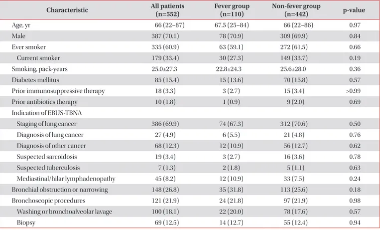
Table 1. Comparison of baseline characteristics and bronchoscopic findings and procedures between fever and non-fever group
Characteristic All patients
(n=552)
Fever group (n=110)
Non-fever group
(n=442) p-value
Age, yr 66 (22–87) 67.5 (25–84) 66 (22–86) 0.97
Male 387 (70.1) 78 (70.9) 309 (69.9) 0.84
Ever smoker 335 (60.9) 63 (59.1) 272 (61.5) 0.66
Current smoker 179 (33.4) 30 (27.3) 149 (33.7) 0.19
Smoking, pack-years 25.0±27.3 22.8±24.3 25.6±28.0 0.36
Diabetes mellitus 85 (15.4) 15 (13.6) 70 (15.8) 0.57
Prior immunosuppressive therapy 18 (3.3) 3 (2.7) 15 (3.4) >0.99
Prior antibiotics therapy 10 (1.8) 1 (0.9) 9 (2.0) 0.69
Indication of EBUS-TBNA
Staging of lung cancer 386 (69.9) 74 (67.3) 312 (70.6) 0.50
Diagnosis of lung cancer 27 (4.9) 6 (5.5) 21 (4.8) 0.76
Diagnosis of other cancer 68 (12.3) 12 (10.9) 56 (12.7) 0.62
Suspected sarcoidosis 19 (3.4) 3 (2.7) 16 (3.6) 0.78
Suspected tuberculosis 7 (1.3) 2 (1.8) 5 (1.1) 0.63
Mediastinal/hilar lymphadenopathy 45 (8.2) 12 (10.9) 33 (7.5) 0.24
Bronchial obstruction or narrowing 148 (26.8) 35 (31.8) 113 (25.6) 0.18
Bronchoscopic procedures 121 (21.9) 24 (21.8) 97 (21.9) 0.98
Washing or bronchoalveolar lavage 100 (18.1) 22 (20.0) 78 (17.6) 0.57
Biopsy 69 (12.5) 14 (12.7) 55 (12.4) 0.94
Values are presented as median (range) or number (%).
EBUS-TBNA: endobronchial ultrasound–guided transbronchial needle aspiration.
TBNA was performed with the patient under moderate seda- tion using fentanyl and midazolam, as well as local anesthesia using lidocaine.
3. Assessment of fever
The patients were evaluated for fever by a physician every 6–8 hours during the first 24 hours following EBUS-TBNA.
Fever was defined as an increase in axillary body temperature over 37.8
oC.
4. Laboratory evaluations
White blood cells (WBC) and neutrophil counts were ob- tained before EBUS-TBNA in and at the time of fever. When fever developed after EBUS-TBNA, two sets of blood cultures (both aerobic and anaerobic) were performed.
5. Statistics
A chi-square test was used to compare categorical variables, and a t test was used to compare continuous variables. The odds ratios for the risk of fever following EBUS-TBNA were analyzed using a multivariate logistic regression model, in- cluding age, sex, any variables with a p<0.20, and prior immu- nosuppressive or antibiotic therapy that could impact fever after EBUS-TBNA. A p<0.05 was considered to indicate sig-
nificance. All statistical analyses were performed using SPSS version 19.0 (IBM Corp., Armonk, NY, USA).
6. Ethics statement
The study was approved by the Institutional Review Board of Seoul National University Hospital (IRB No. 1212-112-453) and conducted in accordance with the Declaration of Hel- sinki. The informed consent requirement was waived due to the retrospective study design.
Results
1. Incidence of fever following EBUS-TBNA
The records of a total of 552 patients who underwent EBUS- TBNA were reviewed. The median age was 66 years (range, 22–87 years), and 387 patients were male (70.1%). The base- line clinical characteristics of the study population are sum- marized in Table 1. The indications of EBUS-TBNA were for the staging of lung cancer in 386 cases, diagnosis of cancer (lung cancer in 27 cases, and others in 68 cases, suspected of sarcoidosis in 19 cases, tuberculosis in seven cases, and me- diastinal or hilar lymphadenopathy in 45 cases). Fever after EBUS-TBNA developed in 110 of 552 patients (20%).
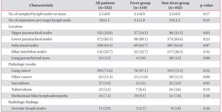
Table 2. Comparisons of characteristics of lymph nodes, final diagnosis, and radiologic findings between fever and non- fever group
Characteristic All patients
(n=552) Fever group
(n=110) Non-fever group
(n=442) p-value
No. of sampled lymph nodes or mass 2.3±0.9 2.4±0.9 2.2±0.9 0.17
No. of aspirations per target lymph node 3.0±1.1 3.2±1.0 2.9±1.2 0.51
Location
Upper paratracheal nodes 125 (22.6) 27 (24.5) 98 (22.3) 0.62
Lower paratracheal nodes 472 (85.5) 98 (89.1) 374 (84.6) 0.23
Subcarinal nodes 350 (63.4) 69 (62.7) 281 (63.6) 0.87
Hilar/interlobar nodes 142 (25.7) 25 (22.7) 117 (26.5) 0.42
Lung parenchymal mass 24 (4.5) 4 (3.6) 20 (4.5) 0.80
Pathologic results
Lung cancer 395 (71.6) 76 (67.1) 319 (72.2) 0.52
Other cancer 63 (11.4) 13 (11.8) 50 (11.3) 0.88
Sarcoidosis 27 (4.9) 5 (4.5) 22 (5.0) 0.85
Tuberculosis 23 (4.2) 7 (6.4) 16 (3.6) 0.19
Mediastinal/hilar lymphadenopathy 41 (7.4) 10 (9.1) 31 (7.0) 0.46
Radiologic findings
Necrotic lymph nodes 11 (2.0) 3 (2.7) 8 (1.8) 0.46
2. Risk factors of fever following EBUS-TBNA
One hundred twenty one patients (21.9%) underwent bron- choscopic procedure including biopsy, washing and bron- choalveolar lavage (BAL). There were no differences in abnor- mal bronchoscopic findings such as bronchial obstruction or narrowing and bronchoscopic procedures between the fever and non-fever group.
The total number of sampled lymph nodes or lung masses was 2.3±0.9. The most frequently punctured lymph node was the lower paratracheal lymph node in 472 cases (85.5%), fol- lowed by the subcarinal lymph node in 350 cases (63.4%).
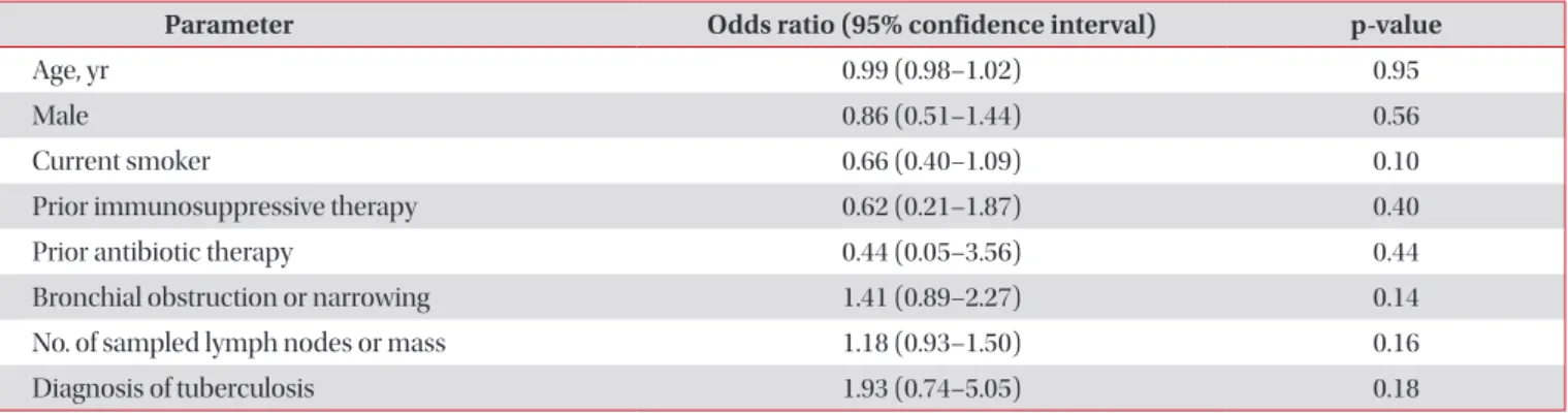
There were no differences in the number or location of sampled lymph nodes and necrosis of lymph node on com- puted tomography (CT) between groups (Table 2). When we evaluated factors for developing fever after EBUS-TBNA using a multivariate model including age, smoking history, prior im- munosuppressant therapy, prior antibiotic therapy, bronchial obstruction or narrowing, number of sampled lymph nodes or mass and diagnosis of tuberculosis, no risk factors for fever following EBUS-TBNA were observed (Table 3).
3. Clinical characteristics of febrile patients after EBUS- TBNA
The median onset and duration of fever after EBUS-TBNA was 7 hours (range, 0.5–32 hours) and 7 hours (range, 1–52 hours), respectively, and the median peak body temperature was 38.3
oC (range, 37.8
oC–39.9
oC). Of 110 febrile patients, 43 patients (39%) received antibiotic treatment due to fever after EBUS-TBNA. Fever in most patients (94.5%) resolved within 24 hours. Accompanying symptoms were chill in 46.4%, fe- brile sense in 30.9%, general ache in 10.9%, and dyspnea in 5.5% patients. Thirty-four patients (30.4%) had no symptoms.
Blood culture samples at the time of fever were taken in 56 (50.9%) patients and positive in two (1.8%). Streptococcus hominis was identified in only a single positive culture of the
patients and is part of the normal skin flora thereby able to
contaminate blood cultures. None of the bacteraemic patients showed clinical features suggestive of infection. Therefore, no true bacteremia was found. Peripheral WBC and neutrophil counts increased significantly at the time of fever than the pre–EBUS-TBNA values (n=57) (Figure 1).
4. Infectious complications following EBUS-TBNA Six of 110 febrile patients developed prolonged fever lasting longer than 24 hours, and pneumonia developed in two of the six patients. Three of six patients (50%) with prolonged fever had diabetes mellitus and 12 of 104 patients (11.5%) with fever that subsided within 24 hours had diabetes mellitus. Diabetes mellitus was more common in patients with prolonged fever than in patients with non-prolonged fever (p=0.032). Both pa- tients who developed pneumonia had diabetes.
We experienced one case of mediastinal abscess developing after EBUS-TBNA. A 69-year-old female patient with underly-
Table 3. Predictors of fever after EBUS-TBNA (multivariable analysis)
Parameter Odds ratio (95% confidence interval) p-value
Age, yr 0.99 (0.98–1.02) 0.95
Male 0.86 (0.51–1.44) 0.56
Current smoker 0.66 (0.40–1.09) 0.10
Prior immunosuppressive therapy 0.62 (0.21–1.87) 0.40
Prior antibiotic therapy 0.44 (0.05–3.56) 0.44
Bronchial obstruction or narrowing 1.41 (0.89–2.27) 0.14
No. of sampled lymph nodes or mass 1.18 (0.93–1.50) 0.16
Diagnosis of tuberculosis 1.93 (0.74–5.05) 0.18
EBUS-TBNA: endobronchial ultrasound–guided transbronchial needle aspiration.
Figure 1. Changes in peripheral blood white blood cell (WBC) and neutrophil counts before endobronchial ultrasound (EBUS)–
guided transbronchial needle aspiration and at the time of fever in the fever group (n=57). *p=0.030.
†p<0.001.
0
WBC Neutrophil
12,000 10,000 8,000 6,000 4,000 2,000
*
Pre-EBUS At the time of fever (x10 / L)3