• 검색 결과가 없습니다.

Successful Rechallenge with Imatinib in a Patient with Chronic Myeloid Leukemia Who Previously Experienced Imatinib Mesylate Induced Pneumonitis

N/A
N/A
Protected

Academic year: 2021

Share "Successful Rechallenge with Imatinib in a Patient with Chronic Myeloid Leukemia Who Previously Experienced Imatinib Mesylate Induced Pneumonitis"

Copied!
4
0
0

로드 중.... (전체 텍스트 보기)

전체 글

(1)

Successful Rechallenge with Imatinib in a Patient with Chronic Myeloid Leukemia Who Previously Experienced Imatinib Mesylate Induced Pneumonitis

Seong Woo Go, M.D.

1

, Boo Kyeong Kim, M.D.

1

, Sung Hak Lee, M.D.

2

, Tae-Jung Kim, M.D.

2

, Joo Yeon Huh, M.D.

1

, Jong Min Lee, M.D.

1

, Jick Hwan Hah, M.D.

1

, Dong Whi Kim, M.D.

1

, Min Jung Cho, M.D.

1

, Tae Wan Kim, M.D.

1

and Ji Young Kang, M.D.

1

Departments of

1

Internal Medicine and

2

Pathology, The Catholic University of Korea College of Medicine, Seoul, Korea

Imatinib mesylate is a targeted therapy that acts by inhibiting tyrosine kinase of the bcr-abl fusion oncoprotein, which is specific to chronic myeloid leukemia (CML), and the c-transmembrane receptor, which is specific to gastrointestinal stromal tumors. Interstitial pneumonitis is a rare adverse event of imatinib therapy. It is clinically difficult to distinguish from infectious pneumonia, which can frequently occur due to the underlying disease. The standard treatment for imatinib-induced pneumonitis is to discontinue the medication and optionally administer corticosteroids. However, there are a few cases of successful retrial with imatinib. We describe a case of successful rechallenge of imatinib in a patient with imatinib-induced interstitial pneumonitis and CML without a recurrence of the underlying disease after 3 months of follow-up.

Keywords: Leukemia, Myelogenous, Chronic, BCR-ABL Positive; Imatinib; Lung Diseases, Interstitial

Philadelphia-positive acute lymphocytic leukemia and gastro- intestinal stromal tumors (GIST) to inhibit c-transmembrane receptor (c-kit), and platelet derived growth factor (PDGF) tyrosine kinase

1

. However, imatinib has various side effects and commonly causes pleural effusion and general edema.

Additionally, nausea, muscle cramps, abdominal pain, skin rash, and diarrhea can occur. The incidence of severe respira- tory adverse events is very rare and, in particular, interstitial pneumonitis is extremely uncommon, with a incidence of 0.2% to 1.3%

2

. In Korea, to our knowledge, only three cases of imatinib-induced pneumonitis have been reported, which developed in two patients with CML and one with GIST
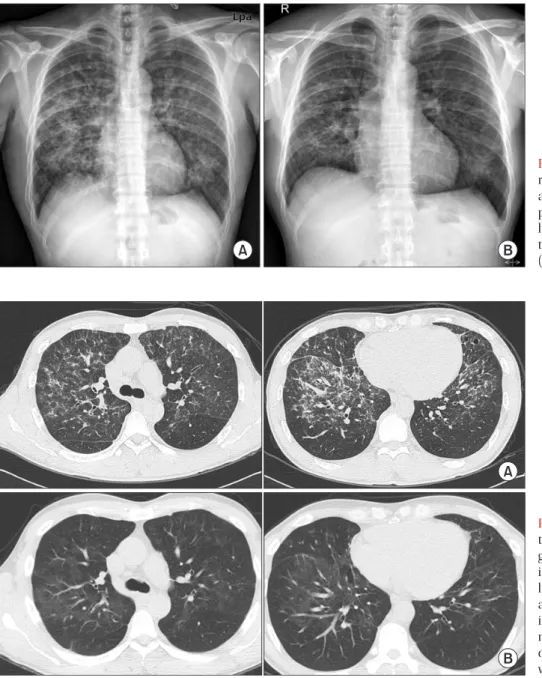
3,4

. Moreover, there has been no reports of successful rechallenge with imatinib. Herein, we describe a case of imatinib-induced interstitial pneumonitis in a patient with CML, who improved by temporarily stopping the drug and starting steroid therapy.

The pateint was restarted on imatinib to control the underly- ing disease without recurrence of the pneumonitis at the 3 month follow-up.

Copyright © 2013

The Korean Academy of Tuberculosis and Respiratory Diseases.

All rights reserved.

Introduction

Imatinib is a protein tyrosine kinase inhibitor that acts on the bcr-abl gene of the abnormal Philadelphia chromo- some in chronic myeloid leukemia (CML). It is also used in

CASE REPORT

http://dx.doi.org/10.4046/trd.2013.75.6.256

ISSN: 1738-3536(Print)/2005-6184(Online) • Tuberc Respir Dis 2013;75:256-259

256

Address for correspondence: Ji Young Kang, M.D.

Department of Internal Medicine, Seoul St. Mary’s Hospital, The Catholic University of Korea College of Medicine, 222 Banpo-daero, Seocho-gu, Seoul 137-701, Korea

Phone: 82-2-2258-6060, Fax: 82-2-596-2158 E-mail: [email protected]

Received: Apr. 29, 2013 Revised: Jun. 24, 2013 Accepted: Sep. 16, 2013

cc

It is identical to the Creative Commons Attribution Non-Commercial

License (http://creativecommons.org/licenses/by-nc/3.0/).

(2)

Successful rechallenge with imatinib induced pneumonitis in CML

http://dx.doi.org/10.4046/trd.2013.75.6.256 257

www.e-trd.org

Case Report

A 43-year-old man was admitted to the hospital for 2 weeks of dyspnea. He was diagnosed with CML 3 months ago and took 400 mg of imatinib/day for 2 months. His vital signs were blood pressure, 110/80 mm Hg; pulse rate, 72/min; respiratory rate, 20/min; and body temperature, 36.4°C. He presented with an acute ill-looking appearance but no specific findings were noted on neck and abdominal examinations. Heart beat was regular and breathing sounds were clear. The laboratory findings were as follows: white blood cells, 3,630/mL (segment- ed neutrophils, 54%; lymphocytes, 25%; eosinophils, 12%); he- moglobin, 12.4 g/dL; platelets, 136,000/mL, C-reactive protein, 0.31 mg/dL (normal range, 0.01–0.47 mg/dL); total protein, 5.7

g/dL; albumin, 3.4 g/dL, aspartate aminotransferase/alanine aminotransferase, 27/28 U/L; lactate dehydrogenase, 853 IU/

L; and blood urea nitrogen/creatinine 13.4/0.99 mg/dL. His arterial blood gas analysis on room air revealed pH, 7.447;

PaO

2

, 31.2 mm Hg; PaCO

2

, 79.0 mm Hg; and HCO

3

, 21.2 mEq/

L with oxygen saturation of 96.6%. A chest X-ray showed dif- fuse ground glass opacities and reticular nodules on both lung fields (Figure 1). A chest computed tomography (CT) scan revealed patchy ground glass opacity and septal thickening in the middle and lower zones of both lungs (Figure 2A). A pulmonary function test showed a forced vital capacity (FVC) of 3.85 L (83.6% of predicted value), forced expiratory volume in 1 second (FEV1) of 2.81 L (79.4% of predicted value), FEV1/

FVC of 78.36%, and a carbon monoxide diffusing capacity of

Figure 1. Chest X-ray shows bilateral reticulonodular infiltration in both lungs at admission (A) and slight regression of peribronchial patchy opacities in both lungs at 2 weeks after discontinuing ima- tinib and commencing steroid treatment (B).

Figure 2. Chest high resolution computed

tomography scan reveals patchy ground

glass opacities with some interlobar and

intralobular septal thickening in both

lungs, predominantly seen in the central

and upper lung zones (A), and interval

improvement of the interstitial pneumo-

nia with some remaining ground glass

opacity after 12 weeks of rechallenge

with imatinib (B).

(3)

SW Go et al.

258 Tuberc Respir Dis 2013;75:256-259 www.e-trd.org

50.2%. He underwent a bronchoscopy with bronchoalveolar lavage (BAL) and transbronchial lung biopsy (TBLB). The differential counts in the BAL fluid revealed macrophages, 15%; lymphocytes, 75%; neutrophils, 5%; and eosinophils, 2%. A microbiologic study including the culture of bacteria, fungus with Pneumocystis jirovecii, and Mycobacterium in bronchial specimens was all negative. The polymerase chain reaction assays of mycoplasma pneumonia, nine respiratory viruses such as influenza virus A and B, respiratory syncytial virus, metapneumo virus, rhino virus, parainfluenza virus, adeno virus, corona virus, bocavirus, and cytomegalovirus were all negative. Histological findings for the TBLB at the lingular segment demonstrated fibroblastic plug formations in the alveoli with infiltrations of chronic inflammatory cells and type II pneumocyte proliferation, mild fibrous thickening in the interstitium, which suggested organizing pattern of in- terstitial pneumonia (Figure 3). No evidence suggested infec- tious pneumonia; thus, we strongly suspected drug-induced interstitial pneumonitis caused by imatinib. We stopped the imatinib and started 1 mg/kg prednisolone daily. After 2 weeks of treatment, the dyspnea and abnormal X-ray findings improved, and he was discharged with 30 mg prednisolone (Figure 1B). After 1 month of treatment, he restarted 300 mg imatinib to control the underlying disease concomitant with 20 mg prednisolone. No recurrence of respiratory symptoms was observed on a chest CT after 12 weeks of imatinib rechal- lenge (Figure 2B).

Discussion

Recently, many targeted anti-neoplastic agents such as gefitinb, erlotinib, imatinib, bevacitumab, and rituximab has been commonly used to treat various malignancies. Although the incidence of pulmonary toxicity is rare with these targeted therapies, these drugs can induce severe respiratory events

such as interstitial pneumonitis and pulmonary eombolism

5

. Imatinib can causes pulmonary complications such as pleural effusion and pneumonitis. The largest study about imatinib treatment by Ohnishi et al.

2

reported that among 5,500 pa- tients treated by the drug, there were 27 cases of interstitial lung disease of the 3,023 adverse events. The median period to develop interstitial pneumonitis from starting the drug was 49 days and the median daily dose of the drug at the time of the diagnosis was 400 mg. However, no significant correlation was observed between development of the disease and the dose or duration of imatinib. But, the incidence of the disease seemed higher in patients who had pre-existing pulmonary diseases. In the current case, it took 70 days for the interstitial pneumonitis to occur, and the dosage of imatinib was 400 mg/

day. The current patient had no previous lung disease.

Although the pathophysiology of imatinib-induced inter-

stitial pneumonitis has not been identified clearly until now,

the noncytotoxic or cytotoxic types are suggested as possible

mechanisms

6

. The noncytotoxic type manifests as eosino-

philic pneumonia, hypersensitive pneumonitis, or obstruc-

tive bronchiolitis, whereas the cytotoxic pattern manifests as

interstitial edema, lymphocyte infiltration, and destruction

of alveolar epithelial cells. In addition, imatinib aggravates

interstitial pneumonitis by inhibiting PDGF tyrosine kinase,

which leads to acute lung injury

7,8

. The diagnosis is made

based on history, clinical symptoms, radiological findings, and

pathological results. Chest radiographs show diverse patterns

such as a hypersensitivity reaction, interstitial pneumonitis,

cryptogenic-organizing pneumonia, nodular or peribron-

chovascular lesions, and diffuse alveolar damage

2

. Pathologic

manifestations also vary and include diffuse alveolar damage,

nonspecific interstitial pneumonia, bronchiolitis obliterans

organizing pneumonia, eosinophilic pneumonia, and pulmo-

nary hemorrhage

9

. The current case was strongly suggested

initially by lung histology, showing fibrous plugs in the alveoli

with infiltration of inflammatory cells in the interstitium, and

Figure 3. (A, B) Transbronchial lung

biopsy specimen reveals organizing pat-

tern of interstitial pneumonia, showing

fibroblastic plug formation in the alveoli

with infiltration of chronic inflammatory

cells and mild fibrous thickening in the

interstitium (H&E stain; A, ×40; B, ×200).

(4)

Successful rechallenge with imatinib induced pneumonitis in CML

http://dx.doi.org/10.4046/trd.2013.75.6.256 259

www.e-trd.org

by exclusion of other infectious causes via microbiologic and clinical studies. Finally, he was confirmed to have drug- induced interstitial pneumonitis due to improvement of the pneumonitis by discontinuing the causative drug and initiat- ing steroid therapy.

The standard treatment is to stop the drug, observe the course of the disease, and optionally administer steroids

5

. Al- though the prognoses are diverse, mostly the disease is mild and resolves promptly with treatment

5

. It can rarely cause fatal or irreversible results such as respiratory failure or permanent structural distortion of the lung

10

. In addition, the prognosis might be poor if a patient has a history of lung disease. Some other options for managing the underlying disease of patients with imatinib-induced pneumonitis have been reported. Oh- nishi et al.

2

found that 23 of 27 patients with imatinib-induced interstitial pneumonitis recovered with corticosteroid treat- ment and discontinuation of the drug. They reported that four of 11 patients who were readministered 100–400 mg imatinib daily relapsed with interstitial pneumonitis. Delomas et al.

11

reported a case of a patient with CML with pneumonitis who did not relapse after switching to nilotinb. Three cases of ima- tinib-induced interstitial pneumonia have been reported in Korea; two CML and one patient with GIST

3,4

. The patient with CML stopped imatinib treatment and took steroid therapy, but died. The other patient with CML recovered with steroid therapy, changed from imatinib to nilotinib, and survived. The other patient with GIST improved by discontinuing the ima- tinib and commencing steroid therapy. The present case is the first domestic report of successful imatinib rechallenge in a patient with CML after recovery from interstitial pneumonitis without recurrence at a 3 month follow-up. However, symp- toms and chest radiographs should be monitored because a pneumonitis relapse can occur during readministration of the drug. In addition, further studies about duration of discontinu- ing imatinib and starting steroid therapy, optimal timing for imatinib rechallenge, and the superiority between an imatinib retrial or switching to another drug are required.

In conclusion, as imatinib-induced pneumonitis can be fatal with irreversible results and can be confused with more common infectious diseases, it is important for clinicians to consider the possibility of imatinib-induced pneumonitis when patients develop respiratory symptoms or abnormal

radiologic features during imatinib treatment. In addition, it should be kept in mind that susseccful rechallenge with ima- tinib coud be another option for a underlying disease after a recovery from interstitial pneumonitis related with imatinib mesylate.

References

1. Deininger MW, Goldman JM, Melo JV. The molecular biology of chronic myeloid leukemia. Blood 2000;96:3343-56.

2. Ohnishi K, Sakai F, Kudoh S, Ohno R. Twenty-seven cases of drug-induced interstitial lung disease associated with ima- tinib mesylate. Leukemia 2006;20:1162-4.

3. Kim TH, Kim BG, Cho SW, Cho SK, Kim HJ, Yuh YJ, et al. Ima- tinib-mesylate induced interstitial pneumonitis in two CML patients. Tuberc Respir Dis 2011;71:210-5.

4. Lee JW, Kim HJ, Kim KJ, Shin KC, Hong YH, Chung JH, et al. A case of imatinib-mesylate associated hypersensitivity pneu- monitis. Tuberc Respir Dis 2005;59:423-6.

5. Barber NA, Ganti AK. Pulmonary toxicities from targeted therapies: a review. Target Oncol 2011;6:235-43.

6. Yokoyama T, Miyazawa K, Kurakawa E, Nagate A, Shimamoto T, Iwaya K, et al. Interstitial pneumonia induced by imatinib mesylate: pathologic study demonstrates alveolar destruc- tion and fibrosis with eosinophilic infiltration. Leukemia 2004;18:645-6.

7. Snyder LS, Hertz MI, Peterson MS, Harmon KR, Marinelli WA, Henke CA, et al. Acute lung injury: pathogenesis of intraal- veolar fibrosis. J Clin Invest 1991;88:663-73.

8. Walsh J, Absher M, Kelley J. Variable expression of platelet- derived growth factor family proteins in acute lung injury.

Am J Respir Cell Mol Biol 1993;9:637-44.

9. Rossi SE, Erasmus JJ, McAdams HP, Sporn TA, Goodman PC.

Pulmonary drug toxicity: radiologic and pathologic manifes- tations. Radiographics 2000;20:1245-59.

10. Seki N, Ito A, Watanabe K, Shibakuki R, Seto T, Uematsu K, et al. Irreversible imatinib-induced pneumonitis following long- term imatinib administration. Intern Med 2007;46:1941-2.

11. Delomas T, Darne C, Besson C. Lack of recurrence of ima-

tinib-induced interstitial lung disease with nilotinib. Leuk

Lymphoma 2012;53:332-3.

수치

Figure  1.  Chest  X-ray  shows  bilateral  reticulonodular infiltration in both lungs  at admission (A) and slight regression of  peribronchial patchy opacities in both  lungs at 2 weeks after discontinuing  ima-tinib and commencing steroid treatment  (B)

참조

관련 문서

This book contains hundreds of complete, working examples illustrating many common Java programming tasks using the core, enterprise, and foun- dation classes APIs.. Java Examples

1 John Owen, Justification by Faith Alone, in The Works of John Owen, ed. John Bolt, trans. Scott Clark, "Do This and Live: Christ's Active Obedience as the

The participants were asked to describe the practices of special classes at middle schools in terms of the relationships with a variety of

Therefore, in this paper, the solar array of HST is modeled as a simple model with beams and membranes, and the phenomenon of thermal induced vibration is verified and

Frequency and clinical significance of the expression of the multidrug resistance proteins MDR1/P-glycoprotein, MRP1, and LRP in acute myeloid leukemia: a

In this study, we found that Pin1 plays a pivotal role in insulin- induced AP-1 activation through its interaction with p70S6K and prolongs activity of

Activating mutation in the tyrosine kinase JAK2 in polycythemia vera, essential thrombocythemia and myeloid metaplasia with myelofibrosis.. A gain-of-function

The feed is commonly a solution in a solvent like ethanol or t-butanol, and the nonsolvent is water..