경기지역 학교 단체급식소 식품 및 환경 중 식중독균 분석
오태영1․백승엽1․구민선1,2․이종경3․김승민1․박경민2․황대근2․김현정1,2
1한국식품연구원
2과학기술연합대학원대학교
3한양여자대학 식품영양과
Analysis of Foodborne Pathogens in Food and Environmental Samples from Foodservice Establishments at Schools in Gyeonggi Province
Tae Young Oh1, Seung-Youb Baek1, Minseon Koo1,2, Jong-Kyung Lee3, Seung Min Kim1, Kyung-Min Park2, Daekeun Hwang2, and Hyun Jung Kim1,2
1Food Safety Research Group, Korea Food Research Institute
2Department of Food Biotechnology, University of Science and Technology
3Department of Food and Nutrition, Hanyang Women's University
ABSTRACT Foodborne illness associated with food service establishments is an important food safety issue in Korea.
In this study, foodborne pathogens (Bacillus cereus, Clostridium perfringens, Escherichia coli, pathogenic Escherichia coli, Listeria monocytogenes, Salmonella spp., Staphylococcus aureus, and Vibrio parahaemolyticus) and hygiene in- dicator organisms [total viable cell counts (TVC), coliforms] were analyzed for food and environmental samples from foodservice establishments at schools in Gyeonggi province. Virulence factors and antimicrobial resistance of detected foodborne pathogens were also characterized. A total of 179 samples, including food (n=66), utensil (n=68), and environ- mental samples (n=45), were collected from eight food service establishments at schools in Gyeonggi province. Average contamination levels of TVC for foods (including raw materials) and environmental samples were 4.7 and 4.0 log CFU/g, respectively. Average contamination levels of coliforms were 2.7 and 4.0 log CFU/g for foods and environmental swab samples, respectively. B. cereus contamination was detected in food samples with an average of 2.1 log CFU/g.
E. coli was detected only in raw materials, and S. aureus was positive in raw materials as well as environmental swab samples. Other foodborne pathogens were not detected in all samples. The entire B. cereus isolates possessed at least one of the diarrheal toxin genes (hblACD, nheABC, entFM, and cytK enterotoxin gene). However, ces gene encoding emetic toxin was not detected in B. cereus isolates. S. aureus isolates (n=16) contained at least one or more of the tested enterotoxin genes, except for tst gene. For E. coli and S. aureus, 92.7% and 37.5% of the isolates were susceptible against 16 and 19 antimicrobials, respectively. The analyzed microbial hazards could provide useful information for quantitative microbial risk assessment and food safety management system to control foodborne illness outbreaks in food service establishments.
Key words: foodservice, foodborne pathogen, toxigenic genes, antibiotics susceptibility
Received 24 September 2015; Accepted 12 November 2015 Corresponding author: Hyun Jung Kim, Food Safety Research Group, Korea Food Research Institute, Sungnam, Gyeonggi-do 13539, Korea
E-mail: [email protected], Phone: +82-31-780-9271
서 론
식중독으로 인한 우리나라의 사회경제적 손실비용은 2005년 1조 6천억 원으로, 주요 식품안전 이슈 중 하나이다 (1). 이 중 단체급식 발생 식중독은 건당 발생 환자수가 많아 이에 대한 관심이 집중되고 있다. 단체급식 이용자수는 2010년 기준 국민의 25%가 넘는 1,390만 명으로 추정되며, 최근 5년간 학교(직영 및 위탁) 및 기업의 단체급식에서 발
생된 식중독 환자수는 연간 2,521~4,515명이었다. 2014년 기준 건당 평균 식중독 환자수는 68.4명, 전체 환자수는 4,515명이었는데 이 중 학교 급식(직영)에서 전체 식중독 환자의 89.9%가 발생하여 단체급식 중 안전관리 필요성이 높은 것으로 조사되었고 다음으로 기업의 단체급식과 학교 급식(위탁)의 순이었다(2). 2014년 학교 단체급식소의 원인 물질별 식중독 통계에 의하면 pathogenic Escherichia coli 가 17건(환자수 1,441명)으로 가장 높게 나타났으며 그 뒤 로 Clostridium perfringens가 10건(환자수 944명), nor- ovirus가 8건(환자수 333명), Staphylococcus aureus가 3 건(환자수 99명), Salmonella spp.가 2건(환자수 852명)으 로 나타났다(2). 이들 병원성 미생물은 조리, 가공과정 중
가열을 하지 않고 그대로 소비되는 신선편의 식품(3) 및 불 충분한 가열 또는 조리로 식품에 교차 오염되는 경우의 주요 식중독 유발요인으로 알려져 있다(4). 단체급식소 식중독 발 생의 주요 원인으로는 급식시설의 노후화 및 환경의 열악함, 오염된 식재료와 조리기구의 사용, 식품의 저장, 보관 등 관 리 및 조리단계에서의 온도관리 미흡, 조리 종사자의 개인위 생 불량, 교차오염 그리고 위생관리체계 미비 등이 제시된 바 있다(5). 아울러 식중독 발생을 감소시키기 위한 위생관 리 영역은 크게 식품 원자재의 위생, 조리 종사자의 개인위 생, 급식소의 조리 기구나 조리환경 위생 등으로 분류할 수 있다(6). 단체급식소 식중독 제어와 안전관리 기술을 개발하 기 위해서는 무엇보다 주요 공정 중 식품, 도구와 환경 시료 중의 미생물학적 위해인자에 대한 체계적인 분석에 근거한 위험분석이 요구된다. 국내외에서 식품안전관리를 위해 사 전 예방적 안전관리 시스템을 도입하는 추세로 이와 관련하 여 식품안전 분야에서 위험분석의 중요성이 크게 인식되고 있다(7,8). 위험분석은 식품 등에 존재하는 위해요인이 인체 에 노출되었을 때 발생할 수 있는 유해 영향과 발생확률을 과학적으로 예측하는 일련의 과정으로 국내의 경우 2005년 도 식품위생법에 관련 규정이 신설되면서 식품안전의 측면 에서 위험분석이 강조되어 왔다. 기존의 정부 주도 식품안전 관리는 물론이고 산업적 식품안전관리의 영역까지 널리 이 용되는 추세로(9), 단체급식소 식중독 안전관리에도 활용 가 능성이 높다. 이에 본 연구에서는 위험분석에 기반을 둔 단 체급식소 식중독 사전 예방 시스템 개발을 위하여 식중독 환자 발생 건수가 가장 많아 안전관리 우선순위가 높은 학교 급식(직영)에서 식품, 조리 도구 및 환경 중 식중독균을 분석 하고 이들 미생물의 병원성 인자 및 항생제 내성을 확인하여 단체급식소 미생물 위험분석을 위한 정보를 제공하고자 하 였다.
재료 및 방법
시료수집
2014년 5월부터 8월까지 경기도 소재 총 8개 학교의 급 식소를 방문하여 전처리 및 조리 단계의 식품, 음용수, 조리 도구 및 환경 swab 시료를 무균적으로 멸균백에 채취하였 다. 채취 후 즉시 냉장상태로 실험실에 운반하여 미생물 분 석을 수행하였다. 모든 분석은 시료를 채취한 후 6시간 이내 에 진행되었으며, 시료는 분석 진행 전까지 4°C로 유지하였 다.
위생지표균 분석
단체급식소의 위생지표를 조사하기 위하여 일반세균수와 대장균군을 정량 분석하였다. 일반세균수는 식품공전(10) 에 따라 시료 25 g에 멸균 인산 완충 희석액 225 mL를 가하 여 균질기(Stomacher®400 Circulator, Seward Labora- tory Systems Inc., Davie, FL, USA)를 이용하여 260 rpm
에서 2분간 균질화시킨 후 시험액 1 mL를 취하여 멸균 인산 완충 희석액 9 mL에 단계 희석하였다. 각 단계 희석액 1 mL를 평판에 분주하고 Plate Count Agar(PCA, Merck, Darmstadt, Germany)를 약 15 mL씩 부어 고르게 혼합한 후 37°C에서 24~48시간 배양하여 성장한 집락 수를 측정 하였다.
대장균군(coliforms)은 일반세균수와 동일한 시험액 10, 1, 0.1 mL씩 3개를 듀람관을 넣은 Brilliant-green bile lactose broth(BGLB, Merck)에 접종하고 37°C에서 48시 간 배양하여 가스의 발생이 확인되면 Endo agar(Merck)에 획선 배양하였다. 전형적인 집락이 확인되면 Nutrient agar (Merck)에 획선 배양하여 성장한 집락이 그람 음성, 무아포 성 간균으로 확인되면 대장균군 양성으로 확정하고, 최확수 표에 따라 대장균군 수를 산출하였다.
식중독균 분석
Bacillus cereus의 정량분석은 식품공전(10)에 따라 실 시하였다. 일반세균수 분석과 동일한 시험액 1 mL를 단계 희석한 후, Mannitol Eggyolk Polymyxine agar(MYEP, Merck)에 각 단계 희석액을 0.2 mL씩 5장에 도말하여 총 접종액이 1 mL가 되게 한 다음 30°C에서 24시간 배양하였 다. 성장한 집락 주변에 lecithinase를 생성하는 혼탁한 환 이 있는 분홍색 집락을 계수하였다. 계수한 평판에서 5개 이상의 전형적인 집락을 선별하여 TSA(Merck)에 획선 도 말하고 VITEK2®compact(BioMerieux, Marcy l'Etoile, France)로 확인 후 식품공전에 따라 B. cereus를 최종 확인 하고 정량 결과값에 반영하였다.
C. perfringens, E. coli,pathogenic E. coli, L. mono- cytogenes, Salmonella spp., S. aureus, Vibrio para- haemolyticus와 같은 병원성 미생물 정성시험은 식품공전 (10)에 따라 진행되었다. 즉 시료 25 g에 분석할 병원성 미 생물에 따른 225 mL의 배양액을 각각 가한 후 균질기를 이용하여 1분간 균질화시킨 다음 L. monocytogenes는 30°C, 나머지 균주는 35~37°C에서 18~24시간 배양하였 다. 병원성 미생물별 증균 배양액을 10 μL 취하여 각 균별 선택배지에 접종한 후 37°C에서 배양하였다. 이때 L. mon- ocytogenes는 30°C에서 24시간 배양하였고, C. per- fringens는 37°C에서 혐기배양 하였다. 의심되는 집락을 TSA(Merck)에 획선 도말한 후 37°C에서 24시간 배양한 다음 VITEK2®compact(BioMerieux)로 최종 확인하였다.
병원성 인자 분석
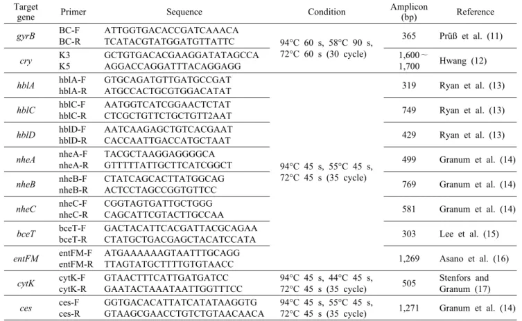
분리 동정된 B. cereus와 S. aureus의 독소유전자는 PCR 로 확인 동정하였다. 각 분리주의 genomic DNA는 DNeasy Blood and Tissue Kit(Qiagen GmbH, Hilden, Germany) 을 사용하여 추출한 후 template DNA로 사용하였으며, S.
aureus는 Lysozyme(Sigma-Aldrich Co., St. Louis, MO, USA)을 포함한 enzymatic lysis buffer를 사용하였다. PCR
Table 1. List of primers used in the PCR for the detection of virulence associated genes in B. cereus Target
gene Primer Sequence Condition Amplicon
(bp) Reference
gyrB BC-F
BC-R
ATTGGTGACACCGATCAAACA
TCATACGTATGGATGTTATTC 94°C 60 s, 58°C 90 s, 72°C 60 s (30 cycle)
365 Prüß et al. (11)
cry K3
K5
GCTGTGACACGAAGGATATAGCCA AGGACCAGGATTTACAGGAGG
1,600∼
1,700 Hwang (12) hblA hblA-F
hblA-R
GTGCAGATGTTGATGCCGAT ATGCCACTGCGTGGACATAT
94°C 45 s, 55°C 45 s, 72°C 45 s (35 cycle)
319 Ryan et al. (13)
hblC hblC-F hblC-R
AATGGTCATCGGAACTCTAT
CTCGCTGTTCTGCTGTT2AAT 749 Ryan et al. (13)
hblD hblD-F hblD-R
AATCAAGAGCTGTCACGAAT
CACCAATTGACCATGCTAAT 429 Ryan et al. (13)
nheA nheA-F nheA-R
TACGCTAAGGAGGGGCA
GTTTTTATTGCTTCATCGGCT 499 Granum et al. (14)
nheB nheB-F nheB-R
CTATCAGCACTTATGGCAG
ACTCCTAGCCGGTGTTCC 769 Granum et al. (14)
nheC nheC-F nheC-R
CGGTAGTGATTGCTGGG
CAGCATTCGTACTTGCCAA 581 Granum et al. (14)
bceT bceT-F bceT-R
GACTACATTCACGATTACGCAGAA
CTATGCTGACGAGCTACATCCATA 303 Lee et al. (15)
entFM entFM-F entFM-R
ATGAAAAAAGTAATTTGCAGG
TTAGTATGCTTTTGTGTAACC 1,269 Asano et al. (16)
cytK cytK-F cytK-R
GTAACTTTCATTGATGATCC GAATACTAAATAATTGGTTTCC
94°C 45 s, 44°C 45 s,
72°C 45 s (35 cycle) 505 Stenfors and Granum (17) ces ces-F
ces-R
GGTGACACATTATCATATAAGGTG GTAAGCGAACCTGTCTGTAACAACA
94°C 45 s, 55°C 45 s,
72°C 45 s (35 cycle) 1,271 Granum et al. (14)
반응액은 AccuPowerTM premix(Bioneer, Daejeon, Korea) 에 template DNA 1 μL와 각각의 specific primer 1 μL를 넣고 증류수로 전체 용량이 20 μL가 되게 첨가하였다. B.
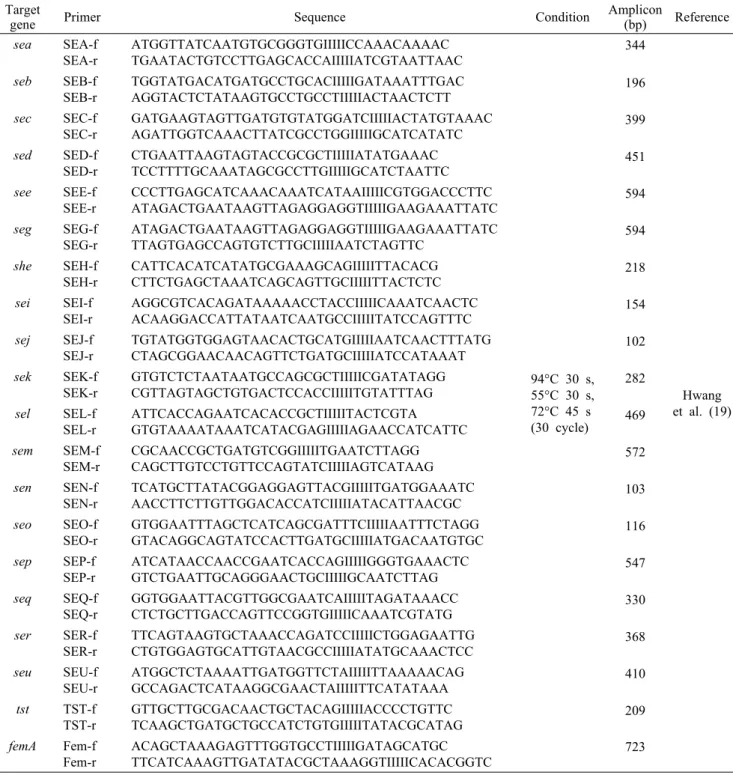
cereus와 S. aureus의 enterotoxin gene을 분석하기 위한 primer 염기서열과 PCR 반응 조건은 Table 1, 2와 같다.
PCR cycler(Eppendorf, Hamburg, Germany)의 반응조건 은 94°C에서 7분간 predenaturation 시킨 후 Table 1과 2의 조건에 따라 annealing 시키고(11-18), 72°C에서 7분 간 extension을 진행하였다. 증폭된 DNA는 2.0% agarose gel을 사용하여 TBE buffer 하에서 100 V로 50분간 전기 영동을 실시한 후 EtBr로 15분간 염색한 다음 UV로 관찰하 였다. 독소유전자 확인을 위해 사용된 표준균주는 B. cer- eus ATCC 14579(diarrheagenic), B. cereus NCCP 14796 (emetic), S. aureus ATCC 13565(sea), ATCC 14458 (seb), S. aureus ATCC 19095(sec, seg, seh, sei, seg, sel, sem, sen, seo, seu), S. aureus ATCC 23235(sed, ser), S. aureus FRI913(see, sek, seq), S. aureus NRS 110(sej), S. aureus NRS111(tst) 등이었다(19,20).
항생제 내성
학교 급식소에서 분리 동정된 E. coli와 S. aureus의항생 제 내성을 VITEK2®system(BioMerieux)의 AST-N224 test Card와 AST-P601 test Card를 이용하여 broth dilu-
tion법으로 시험하였다. 시험한 항생제로는 E. coli의 경우 amikacin, ampicillin, amoxicillin/clavulanic acid, aztre- onam, cefazolin, cefepime, cefotaxime, cefoxitin, cef- tazidime, ciprofloxacin, ertapenem, gentamicin, imipe- nem, piperacillin/tazobactam, tigercycline, trimetho- prim/sulfamethoxazole과 같고, S. aureus는 benzylpeni- cillin, clindamycin, ciprofloxacin, erythromycin, fusidic acid, gentamicin, habekacin, linezolid, mupirocin, ni- trofurantoin, oxacillin, quinupristin/dalfopristin, rifam- picin, teicoplanin, telithromycin, tetracycline, tigecy- cline, trimethoprim/sulfamethoxazole, vancomycin과 같 다. Clinical and Laboratory Standard Institute(CLSI)의 기준을 따라 항생제 내성 여부를 판별하였으며(21), E. coli ATCC 10536과 S. aureus ATCC 23235를 표준균주로 사 용하였다.
급식소 환경미생물 분포
미생물학적 위해인자를 분석한 총 8개 급식소 중 두 곳을 선정하여 급식소 환경의 미생물 분포를 분석하였다. 선정된 급식소에서 절단기, 칼, 세척대, 앞치마, 고무장갑, 화장실 세면대 손잡이, 전처리용 건조대, 냉장고 손잡이 등의 표면 을 멸균 면봉으로 획득하여 0.85% 생리식염수에 현탁 후, 현탁액을 Tryptic Soy Agar(TSA, Merck)에 도말하여 생
Table 2. List of primers used in the PCR for the detection of virulence associated genes in S. aureus Target
gene Primer Sequence Condition Amplicon
(bp) Reference sea SEA-f
SEA-r
ATGGTTATCAATGTGCGGGTGIIIIICCAAACAAAAC TGAATACTGTCCTTGAGCACCAIIIIIATCGTAATTAAC
94°C 30 s, 55°C 30 s, 72°C 45 s (30 cycle)
344
Hwang et al. (19) seb SEB-f
SEB-r
TGGTATGACATGATGCCTGCACIIIIIGATAAATTTGAC
AGGTACTCTATAAGTGCCTGCCTIIIIIACTAACTCTT 196 sec SEC-f
SEC-r
GATGAAGTAGTTGATGTGTATGGATCIIIIIACTATGTAAAC
AGATTGGTCAAACTTATCGCCTGGIIIIIGCATCATATC 399 sed SED-f
SED-r
CTGAATTAAGTAGTACCGCGCTIIIIIATATGAAAC
TCCTTTTGCAAATAGCGCCTTGIIIIIGCATCTAATTC 451 see SEE-f
SEE-r
CCCTTGAGCATCAAACAAATCATAAIIIIICGTGGACCCTTC
ATAGACTGAATAAGTTAGAGGAGGTIIIIIGAAGAAATTATC 594 seg SEG-f
SEG-r
ATAGACTGAATAAGTTAGAGGAGGTIIIIIGAAGAAATTATC
TTAGTGAGCCAGTGTCTTGCIIIIIAATCTAGTTC 594
she SEH-f SEH-r
CATTCACATCATATGCGAAAGCAGIIIIITTACACG
CTTCTGAGCTAAATCAGCAGTTGCIIIIITTACTCTC 218 sei SEI-f
SEI-r
AGGCGTCACAGATAAAAACCTACCIIIIICAAATCAACTC
ACAAGGACCATTATAATCAATGCCIIIIITATCCAGTTTC 154 sej SEJ-f
SEJ-r
TGTATGGTGGAGTAACACTGCATGIIIIIAATCAACTTTATG
CTAGCGGAACAACAGTTCTGATGCIIIIIATCCATAAAT 102 sek SEK-f
SEK-r
GTGTCTCTAATAATGCCAGCGCTIIIIICGATATAGG
CGTTAGTAGCTGTGACTCCACCIIIIITGTATTTAG 282
sel SEL-f SEL-r
ATTCACCAGAATCACACCGCTIIIIITACTCGTA
GTGTAAAATAAATCATACGAGIIIIIAGAACCATCATTC 469 sem SEM-f
SEM-r
CGCAACCGCTGATGTCGGIIIIITGAATCTTAGG
CAGCTTGTCCTGTTCCAGTATCIIIIIAGTCATAAG 572
sen SEN-f SEN-r
TCATGCTTATACGGAGGAGTTACGIIIIITGATGGAAATC
AACCTTCTTGTTGGACACCATCIIIIIATACATTAACGC 103 seo SEO-f
SEO-r
GTGGAATTTAGCTCATCAGCGATTTCIIIIIAATTTCTAGG
GTACAGGCAGTATCCACTTGATGCIIIIIATGACAATGTGC 116 sep SEP-f
SEP-r
ATCATAACCAACCGAATCACCAGIIIIIGGGTGAAACTC
GTCTGAATTGCAGGGAACTGCIIIIIGCAATCTTAG 547
seq SEQ-f SEQ-r
GGTGGAATTACGTTGGCGAATCAIIIIITAGATAAACC
CTCTGCTTGACCAGTTCCGGTGIIIIICAAATCGTATG 330 ser SER-f
SER-r
TTCAGTAAGTGCTAAACCAGATCCIIIIICTGGAGAATTG
CTGTGGAGTGCATTGTAACGCCIIIIIATATGCAAACTCC 368 seu SEU-f
SEU-r
ATGGCTCTAAAATTGATGGTTCTAIIIIITTAAAAACAG
GCCAGACTCATAAGGCGAACTAIIIIITTCATATAAA 410 tst TST-f
TST-r
GTTGCTTGCGACAACTGCTACAGIIIIIACCCCTGTTC
TCAAGCTGATGCTGCCATCTGTGIIIIITATACGCATAG 209 femA Fem-f
Fem-r
ACAGCTAAAGAGTTTGGTGCCTIIIIIGATAGCATGC
TTCATCAAAGTTGATATACGCTAAAGGTIIIIICACACGGTC 723
존한 균주를 대상으로 MS system(VITEK MS, BioMe- rieux)을 이용하여 분석하였다. 배양한 미생물을 1 μL ma- trix solution과 함께 Target Slide에 접종하고 분석한 후 VITEK MS system(BioMerieux)의 고급 스펙트럼 분류기 소프트웨어(Myla v.3.2, BioMerieux)를 이용하여 결과를 분석하였다. 시험 결과의 보정을 위해 사용한 표준균주는 E. coli ATCC 8739였다.
결과 및 고찰
위생지표균 오염도
위생지표균 및 식중독균을 분석한 총 8개 경기도 소재 학교 급식소는 농촌 3곳, 도시 4곳 및 벽지 1곳으로 구성되 어 있으며 8개 학교 전체의 1일 식수 인원은 14,573명이었 다. 학교 급식소에서 채취한 식품, 조리도구 및 환경 시료의 위생 실태를 살펴보기 위하여 지표균으로 총균수와 대장균
Fig. 1. Contamination of hygiene indicator microorganisms and B. cereus in food, equipment and environmental sample from foodservice establishments of schools. A, total viable counts of foods (n=66); B, total viable counts of equipment and environ- ments (n=56); C, coliform of foods (n=66); D, coliform of equip- ment and environments (n=56); E, B. cereus of foods (n=66).
군을 분석하였다. 총 66개 식품 시료의 총균수는 평균 4.7 log CFU/g, 최대 8.1 log CFU/g이었고, 대장균군은 평균 3.1 log CFU/g, 4.0 log CFU/g으로 총균수에 비해 대장균 군의 오염 수준이 낮았다. Cho와 Park(22)의 연구에서는 식재료의 총균수가 5.5 log CFU/g, 최대 6.8 log CFU/g으 로 평균값은 더 높은 수준이었으나 편차가 크지 않았으며, 대장균군은 2.7 log CFU/g, 최대 3.6 log CFU/g으로 본 연구 결과보다 약간 낮은 수준으로 검출되었다. 한편 총 56 개 환경 시료의 경우 총균수는 평균 2.7 log CFU/g, 최대 4.1 log CFU/g으로 식품 시료보다 낮은 수준으로 분석되었 으나 대장균군의 경우 식품 시료와는 달리 환경 시료에서 평균 4.0 log CFU/g, 최대 5.4 log CFU/g으로 총균수에 비해 높은 수준으로 분석되었다(Fig. 1). 식품공전(10)에서 즉석섭취, 편의식품류의 세균수 기준은 5 log CFU/g으로 제시되었으나, 대장균군은 따로 명시되지 않았다. Solberg 등(23)도 세균수 기준을 5 log CFU/g 이하로 제안하였고, 대장균군은 3 log MPN/g 이하로 검출되지 않아야 한다고 제안한 바 있다. 대장균군에 대한 기준은 현재 정해져 있지 않으나 분변지표균인 대장균군의 급식소 환경 시료에서 높 은 수준으로 검출된 결과는 환경에 의한 교차오염의 가능성 을 배제할 수 없음을 나타내고 있어 이에 대한 추가 연구가 요구된다.
식중독균 분석
학교 급식소의 식품, 조리도구 및 환경 시료를 채취하여 B. cereus의 정량분석 및 C. perfringens, E. coli,patho- genic E. coli, L. monocytogenes, Salmonella spp., S.
aureus, V. parahaemolyticus의 정성분석을 실시하였다.
B. cereus는 토양에서 유래된 미생물로 토양, 물, 먼지와
같은 자연환경에 널리 분포되어 있으며 원재료를 통해 식품 에 오염된다. 일반적으로 B. cereus는 열에 안정성이 있는 포자를 형성하며 이는 135°C의 온도에서도 4시간 동안 생 존이 가능하므로(24), 식품에 이미 오염되었을 경우 일반적 인 열처리로는 포자를 제거할 수 없다. 식품 내 B. cereus의 수준이 3~6 log CFU/g 이상일 경우 식중독을 야기할 수 있음이 보고된 바 있다(24-27). 본 연구에서는 B. cereus의 정량분석 결과 식품 시료(n=66)에서 평균 2.1 log CFU/g, 최대 4.1 log CFU/g으로 분석되었으며, 이 중 조리된 식품 의 오염도는 4 log CFU/g 이하로 식품공전의 기준 이하였다 (10)(Fig. 1). 식품 시료 중 E. coli와 S. aureus는 조리 전 시료(n=14)의 각각 35.7%, 21.4%에서 검출되었으며 검출 된 시료는 E. coli의 경우 숙주나물, 참나물, 쑥갓, 감자, S.
aureus의 경우 다진 당근, 쑥갓, 감자였다. 조리단계 식품 시료(n=52)에서는 모두 음성으로 분석되었다. 조리도구 (n=68)의 경우 모두 음성이었으며 환경 시료에서는 S. aur- eus가 냉장고 손잡이(n=14)에서 1개가 양성으로 검출되었 고 그 외의 다른 환경 시료는 모든 항목에서 음성으로 분석 되었다(Table 3). 그 외 C. perfringens, L. monocyto- genes, Salmonella spp., V. parahaemolyticus는 분석된 식품, 조리도구 및 환경 시료에서 모두 음성이었다. E. coli는 조리 전 시료에서 35.7% 양성으로 나타났으나 병원성 인자 는 검출되지 않았다. S. aureus는 건강한 사람의 25~50%
가 보균자이며 이 중 15~20%는 enterotoxin gene을 생성 할 수 있기 때문에 조리 종사자에 의해 오염될 가능성이 있 다(28). 말레이시아의 초등학교 급식 종사자의 손을 대상으 로 S. aureus를 조사한 결과 조리 전, 조리 중, 조리 후에 각각 74.1%, 65.9%, 70.6%로 검출되어(29) 조리 종사자가 오염의 매개가 될 수 있으며, 본 연구팀이 조리 전 시료와 환경에서 분리된 S. aureus의 rep-PCR 기반 유전적 상동성 을 분석한 결과 동일 균주임이 확인되었으며(data not shown), 급식소 내 종사자-환경-식품 간의 교차오염에 대 한 안전관리가 필요한 것으로 사료된다.
병원성 인자 분석
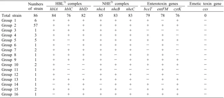
학교 급식소의 식품에서 분리한 B. cereus 분리주 86주 를 대상으로 설사유발독소인 HBL(hbl A, C, D), NHE(nhe A, B, C), enterotoxin gene(bceT, entFM, cytK) 그리고 구토 유발 독소인 cereulide(ces) 독소유전자를 분석하였 다. PCR 분석 결과 모든 B. cereus 분리주에서 독소유전자 가 분석되었으며 독소유전자 분포에 따라 여러 그룹으로 구 분할 수 있었다(Table 4). HBL complex는 모든 분리주에 서 양성으로 나타났으며, 모든 hbl gene을 보유하는 분리주 는 전체 86주 중 74주(86.0%)였다. NHE complex에서는 82주가 nhe의 모든 gene에 양성으로 나타났다(95.3%).
Enterotoxin gene 3종류를 모두 보유하는 분리주는 68주 (79%)였으며 bceT는91.8%, entFM은 90.6%, 그리고 cytK 는 88.3%가 양성으로 검출되었다. 기존 연구 결과 임상과
Table 4. Profiles of toxin-encoding genes in B. cereus isolates obtained from foods and environmental samples from foodservice establishments of schools
Numbers of strain
HBL1) complex NHE2) complex Enterotoxin genes Emetic toxin gene
hblA hblC hblD nheA nheB nheC bceT entFM cytK ces
Total strain Group 1 Group 2 Group 3 Group 4 Group 5 Group 6 Group 7 Group 8 Group 9 Group 10 Group 11 Group 12 Group 13 Group 14 Group 15 Group 16
86 6 57
1 3 5 1 2 1 1 2 1 1 1 1 2 1
84 + + + + + + + + + + + + +
- +
- 76
+ + + +
-
- + + +
-
-
- + + + +
82 + + + + + + + + +
-
-
- + + + +
85 + + + + + + + + + + + + +
- + +
83 + + + + + + +
-
- +
- + + + + +
83 + + + + + + + + + +
- + + +
- +
79 + +
- + +
-
- + + +
-
-
- + + +
78
- +
- + + + + + + + + + +
- + +
76 + +
-
- +
-
-
- + +
-
- + + + +
0
-
-
-
-
-
-
-
-
-
-
-
-
-
-
-
-
1)HBL, hemolysin. 2)NHE, nonhemolytic enterotoxin.
Table 5. Profiles of toxin-encoding genes in S. aureus isolates obtained foods and environmental samples from foodservice establish- ments of schools
Numbers
of strain femA sea1) seb1) sec1) sed1) see1) seg seh sei sej sek sel sem sen seo sep seq ser seu tst2) Total strain
Group 1 Group 2 Group 3 Group 4
16 7 3 5 1
16 + + + +
11 + +
- +
0
-
-
-
- 0
-
-
-
- 0
-
-
-
- 0
-
-
-
- 6
-
- + +
7 +
-
-
- 6
-
- + +
0
-
-
-
- 10
+ +
-
- 6
-
- + +
6
-
- + +
6
-
- + +
6
-
- + +
0
-
-
-
- 10
+ +
-
- 0
-
-
-
- 6
-
- + +
0
-
-
-
-
1)Classical enterotoxin gene, sea, seb, sec, sed, see.
2)tst, toxic-shock syndrome toxin.
식품 및 곡물시료에서 분리한 120주의 B. cereus의 독소유 전자는 HBL complex가 94.2%로 본 연구 결과보다 더 높게 분석되었으나 NHE complex에서 90.0%, enterotoxin 중 entFM은 65.8%, 그리고 cytK는 52.5%가 양성으로 본 연 구 결과에 비해 낮게 나타났다(30). 한편 단체급식소 식품에 서 분리된 B. cereus에서 구토 유발 독소유전자인 ces는 모든 분리주에서 음성이었고 Kim 등(30)의 연구에서도 음 성으로 나타나 유사한 경향을 보였다. 급식소에서 분리된 S. aureus 16주에 대하여 enterotoxin 생성유전자를 분석 한 결과 4개의 서로 다른 group으로 분류되었다(Table 5).
Group 1은 sea, seh, sek, seq 유전자를 동시에 보유하는 group으로 전체 분리주의 43.7%를 차지하였다. Group 2는 sea, sek, seq 유전자를 동시에 보유하였고 전체 분리주의 18.7%를 차지하였으며, group 3은 seg, sei, sel, sem, sen, seo, seu 유전자를 보유하였으며 전체 분리주의 31.2%에 해당되었다. Group 4는 전체 분리주의 6.3%를 차지하였으 나 독소유전자를 가장 많이 보유(sea, seg, sei, sel, sem, sen, seo, seu)하였다. Group 2와 3은 식품에서만 분리되었 고, group 1의 경우 식품과 환경에서 발견되어 S. aureus가
독소 생성에 유리한 환경에 노출되는 경우 식중독을 유발할 수 있음을 보여주었다. 모든 분리주에서 독소유전자가 검출 되었으며 전형적인 장독소유전자(sea~see) 중에서 sea를 제외한 나머지 유전자는 분석되지 않았으며, seg와 sei 유전 자는 같은 시료에서 검출되었다. seg와 sei toxin gene은 항상 결합되어 있다고 알려져 있으며(31) 인간 독소충격증 후군과 포도상구균성 열상 피부증후군의 원인이 될 수 있다 (32). Aydin 등(33)의 연구에서 육제품, 유제품 및 제과류, 그리고 즉석식품 등에서 분리한 S. aureus 147주 중 62%에 서 S. aureus enterotoxin gene, exfoliative toxin gene, toxic-shock syndrome toxin gene이 검출되었고, 전형적 인 장독소유전자보다 sei gene이 더 많이 검출되어 본 연구 결과와는 차이를 보였다. 한편 toxic-shock syndrome toxin(tst) 유전자는 모든 분리주에서 검출되지 않았다.
항생제 내성
항생제는 보건 위생뿐만 아니라 각종 농축수산물 생산 공 정에서도 사용되고 있다. 항생제의 오남용으로 인한 내성균 주의 출현은 집단 식중독 발생 시 초기 진료에 결정적 영향
Table 6. Antimicrobial resistance of E. coli and S. aureus iso- lated from food and environmental samples of foodservice estab- lishments of schools
Bacteria Resistance phenotype Number (%) of isolates
E. coli
CFZ1) FOX SXT CFZ-FOX
Susceptible to all tested antibiotics2)
2 (4.9) 1 (2.4) 1 (2.4) 1 (2.4) 38 (92.7)
S. aureus P GM P-GM
Susceptible to all tested antibiotics3)
5 (31.3) 10 (62.5) 5 (31.3) 6 (37.5)
1)CFZ, cefazolin; FOX, cefoxitin; SXT, trimethoprim/sulfame- thoxazole; P, benzylpenicillin; GM, gentamicin
2)92.7% of the E. coli strains were susceptible against ampicillin, amoxicillin/clavulanic acid, piperacillin/tazobactam, cefotax- ime, ceftazidime, cefepime, aztreonam, ertapenem, imipenem, amikacin, gentamicin, ciprofloxacin and tigecycline.
3)37.5% of the S. aureus strains were susceptible against ox- acillin, habekacin, ciprofloxacin, erythromycin, telithromycin, clindamycin, quinupristin/dalfopristin, linezolid, teicoplanin, vancomycin, tetracycline, tigecycline, nitrofurantoin, fusidic acid, mupirocin, rifampicin, trimethoprim/sulfamethoxazole.
0%
20%
40%
60%
80%
100%
Cutting machine
Apron Knives Sink Refrigerator handles Pectobacterium spp. Pantoea spp. Micrococcus spp.
Erwinia spp. Enterobacter spp. Burkholderia spp.
Bacillus spp.
A
spp.
spp.
spp. spp.
spp.
spp.
spp.
0%
20%
40%
60%
80%
100%
Knives Sink Glove Knob
(restroom) Table
Streptococcus spp. Staphylococcus spp. Raoultella spp.
Rahnella spp. Pseudomonas spp. Pantoea spp.
Lactococcus spp. Klebsiella spp. Enterobacter spp.
Acinetobacter spp.
B
spp.
spp.
spp.
spp.
spp.
spp.
spp.
spp.
spp.
spp.
Fig. 2. Profiles of culturable microorganisms in environmental samples of foodservice establishments. A, foodservice establishment
Ⅰ; B, foodservice establishment Ⅱ.
을 주기 때문에 분리균주에 대한 항생제 내성 여부를 분석할 필요가 있다(28). E. coli 41주를 16종의 항생제를 이용하여 내성을 분석한 결과 92.7%의 분리주에서 모든 항생제에 대 한 감수성이 있었으나 cefazolin에 2개 균주(4.9%)가 내성 을 보였고, cefoxitin, trimethoprim/sulfamethoxazole에 각각 1개 균주(2.4%)가 내성을 보였다. 1개 균주는 cefazo- lin과 cefoxitin에 대해 동시에 내성이 있었다(Table 6).
Yoo 등(34)이 유통식품에서 분리된 대장균 50주를 대상으 로 항생제 감수성을 시험한 결과 50%의 분리주에서 모든 항생제에 대한 감수성을 보였으며, ampicillin과 amox- icillin/clavulanic acid 그리고 tetracycline에서 각각 36%, 32%, 22%의 내성을 확인하였고, 다제내성 분리주가 전체 의 40%를 차지하였다. Kim 등(35)의 연구에서는 샐러드,
새싹 등 국내 신선편이 식품에서 분리된 대장균 120주의 항 생제 내성을 분석한 결과 ampicillin에 대한 내성이 14.2%
로 가장 높았고 piperacillin 11.7%, cefalotin 10.0%의 순 으로 분석되었으며, 2개 이상의 항생제에 다제내성을 보이 는 균주는 전체 신선편이 식품 분리주의 14.1%로 본 연구에 비해 높았다. 본 연구에서 S. aureus의 19종의 항생제에 대 한 내성을 분석한 결과 benzylpenicillin에서 5개 균주(31.3
%), gentamicin에서 10개 균주(62.5%)가 내성을 보였고, 항생제 2종(benzylpenicillin, gentamicin)에 대해 다제내 성을 보인 분리주가 5개 분석되었다. 전체 분리주의 37.5%
는 시험한 모든 항생제에 감수성이 있었다(Table 6). 국내 식품에서 분리된 S. aureus의 항생제 내성을 살펴보면 돈육 제품 124점에서 분리한 S. aureus 30주의 항생제 내성 결과 penicillin과 ampicillin에 대해 76.7%, 70.0%의 높은 내성 을 보였고(36), Kim 등(28)의 연구에서 들깻잎 재배단지에 서 분리한 S. aureus 31주를 대상으로 항생제 감수성을 확 인한 결과 모든 분리주에서 내성이 나타났고 penicillin과 ampicillin의 내성이 각각 96.8%, 96.8%로 매우 높게 나타 나 본 연구 결과와는 차이를 보였다. 이와 같이 학교 급식소 에서 분리된 E. coli와 S. aureus의 경우 항생제 내성률이 낮았으며, 다제내성률도 높지 않아 국내 학교 급식에서 분리 된 주요 균주의 경우 항생제 내성에 대한 위험은 크지 않은 것으로 사료된다.
급식소 환경미생물 분포
급식소 중 두 곳을 선정해 급식소 환경미생물 군집의 구성 을 분석하여 환경 중 교차오염 가능성을 확인하고자 하였다.
급식소 Ⅰ의 절단기, 칼, 세척대, 앞치마, 냉장고 손잡이를 대상으로 미생물 분포를 분석한 결과 Bacillus spp., Burkholderia spp., Enterobacter spp., Erwinia spp., Micrococcus spp., Pantoea spp., Pectobacterium spp.가 분석되었으며, Pantoea spp.가 가장 높은 비율로 분석되었 고 Bacillus spp.가 다음으로 분석되었다. 또 다른 급식소(급 식소 Ⅱ)에서는 칼, 세척대, 고무장갑, 화장실 세면대 손잡 이, 건조대(전처리용)를 대상으로 분석했을 때 Acineto-
bacter spp., Enterobacter spp., Klebsiella spp., Lacto- coccus spp., Pantoea spp., Pseudomonas spp., Rahnella spp., Raoultella spp., Staphylococcus spp., Strepto- coccus spp.가 분석되었고, 그중 Raoultella spp.가 가장 높은 비율로 분석되었으며 뒤를 이어 Streptococcus spp.
가 다음으로 높은 비율로 분석되었다(Fig. 2). 급식소 Ⅰ의 Pantoea spp.의 경우 도마, 앞치마, 칼에서 공통적으로 분석 되었고, 급식소 Ⅱ의 경우 칼, 개수대, 그리고 장갑에서 Enterobacter spp.가 검출되어 이를 공통적으로 사용하는 급식소 조리원이 교차오염의 매개가 되었을 가능성을 보여 주고 있다.
요 약
본 연구에서는 건당 환자수가 높아 식품안전관리 우선순위 가 높은 단체급식소의 식품, 조리도구 및 환경에서 식중독균 을 분석하고 이들 미생물의 병원성 인자 및 항생제 내성을 확인하여 미생물 위험분석을 위한 기본정보를 제공하고자 하였다. 경기도 소재 총 8개(농촌 3, 도시 4 및 벽지 1) 학교 급식소에서 식품 시료(n=66), 조리도구(n=44) 및 환경 시료 (n=56) 등 총 179점의 시료를 채취하여 지표세균 및 식중독 균을 분석하였다. 식품 시료에서 총균수는 평균 4.7 log CFU/g, 최대 8.1 log CFU/g으로 대장균군의 평균 오염도 3.1 log CFU/g, 최대 오염도 4.0 log CFU/g으로 높았다.
선반 및 개수대 등 환경 시료의 총균수는 평균 2.7 log CFU/
g, 최대 4.1 log CFU/g으로 식품 시료보다 낮은 수준으로 분석되었으나 대장균의 경우 평균 4.0 log CFU/g, 최대 5.4 log CFU/g으로 식품 시료보다 오염 수준이 높아 환경으로 부터의 교차오염 가능성을 배제할 수 없었다. 병원성 미생물 중 Bacillus cereus의 정량분석 결과 식품(원료, 조리단계 및 조리식품 포함) 시료에서 평균 2.1 log CFU/g, 최대 4.1 log CFU/g으로 분석되었으나, 이 중 조리된 식품의 오염도 는 10,000 CFU/g 이하로 식품공전의 기준 이하로 오염되어 있었다. Escherichia coli는 식품 중 조리 전 시료(n=14)에 서만 검출율 35.7%로 분석되었으며 조리단계의 식품, 조리 도구 및 환경 시료에서는 검출되지 않았다. Staphylococcus aureus의 경우 조리 전 식품 원료(n=14)의 21.4%에서 검출 되었으며 환경 시료(냉장고 손잡이)에서 1건 양성으로 검출 되었고, 조리단계의 식품, 조리도구 및 환경 시료에서는 검 출되지 않았다. 그 외 Clostridium perfringens, Listeria monocytogenes, Salmonella spp., Vibrio parahaemoly- ticus는 분석된 모든 시료에서 모두 음성이었다. 분리된 B.
cereus의 독소유전자(hblACD, nheABC, entFM, cytK enterotoxin gene)를 분석한 결과 구토 유발 독소인 ces는 모두 음성이었으나 분석된 86주 모두 적어도 1종 이상의 설사 유발 독소유전자가 검출되었으며 66.2%의 균주는 설 사 유발 독소유전자를 모두 보유하고 있었다. 식품과 환경에 서 분리한 S. aureus(n=16)의 장독소 생성 유전자를 분석한
결과 모두 1종 이상의 독소유전자가 검출되었다. 전형적인 장독소유전자 중에서는 sea만 검출되었으며, 독소충격증후 군 toxin(tst) 유전자는 모든 분리주에서 검출되지 않았다.
집단 식중독 발생 시 초기 진료에 결정적 영향을 주는 항생 제 내성 여부를 분석한 결과 E. coli(n=41)의 92.7%는 분석 한 항생제 16종에 대해 내성을 보이지 않았고 cefazolin에 대한 내성률이 4.9%로 가장 높았으며, 1개 균주에서만 2개 항생제에 대해 다제내성을 보여 국내외 항생제 내성률보다 낮았다. S. aureus(n=16)는 시험한 19종 항생제 중 genta- micin에 대한 내성률이 62.5%로 가장 높았으며 일부 균주 에서 2주 항생제에 대해 다제내성이 관찰되었다. 한편 단체 급식소 2개소의 조리도구와 환경 중 미생물 군집을 분석한 결과 특정균이 도구와 환경에서 중복 검출되어 도구와 환경 중 교차오염 가능성을 간접적으로 시사하였다. 이와 같이 본 연구에서 단체급식소 식품, 조리도구 및 환경 중 위생지 표균과 병원성 미생물의 오염패턴을 분석하고 분리된 균주 의 독성인자와 항생제 내성 정보를 분석하였다. 관련 정보는 단체급식소 미생물 위험분석과 이를 바탕으로 사전적, 정량 적 안전관리 기술 개발에 활용 가능할 것으로 사료된다. 한 편 식중독 유발의 다른 원인인 바이러스류와 기타 원인에 대한 연구는 진행되지 않아 추가 연구가 필요하다.
감사의 글
본 연구는 한국식품연구원의 연구비 지원으로 수행되었으 며, 이에 감사드립니다.
REFERENCES
1. Shin H, Lee S, Kim JS, Kim J, Han KH. 2010. Socioecono- mic costs of food-borne disease using the cost-of-illness model: applying the QALY method. J Prev Med Public Health 43: 352-361.
2. Food Safety Korea. Available from: http://www.foodsafety- korea.go.kr/portal/healthyfoodlife/foodPoisoningStat.do?
menu_no=519&menu_grp=MENU_GRP02 (accessed Sep 2015).
3. Beuchat LR. 1996. Listeria monocytogenes incidence on vegetable. Food Control 7: 223-228.
4. Heiman KE, Garalde VB, Gronostaj M, Jackson KA, Beam S, Joseph L, Saupe A, Ricotta E, Waechter H, Wellman A, Adams-Cameron M, Ray G, Fields A, Chen Y, Datta A, Burall L, Sabol A, Kucerova Z, Trees E, Metz M, Leblanc P, Lance S, Griffin PM, Tauxe RV, Silk BJ. 2015. Multistate outbreak of listeriosis caused by imported cheese and evi- dence of cross-contamination of other cheeses, USA, 2012.
Epidemiol Infect 30: 1-11.
5. Hong SH. 2014. The microbiological assessment and identi- fication of food utensils and foodservice facilities in school.
J Fd Hyg Safety 29: 189-194.
6. Bae HJ. 2006. Analysis of contamination of bacteria from raw materials, utensils, and worker's hands to prepared foods in foodservice operations. J Korean Soc Food Sci Nutr 35:
655-660.
7. Lee JK, Kwak NS. 2004. Microbiological risk assessment
for food safety control. Food Science and Industry 37: 61- 71.
8. Lee SH. 2011. Quantitative microbial risk assessment. Safe Food 6: 13-16.
9. Kim HJ, Kim SM, Ok G, Koo M. 2015. Microbiological risk assessment for industry. Safe Food 10: 13-22.
10. MFDS. 2014. Food Code. Ministry of Food And Drug Safe- ty, Seoul, Korea. 10-3-1_1.
11. Prüβ BM, Dietrich R, Nibler B, Mrtlbauer E, Scherer S.
1999. The hemolytic enterotoxin HBL is broadly distributed among species of the Bacillus cereus group. Appl Environ Microbiol 65: 5436-5442.
12. Hwang JH. 2009. Biochemical characteristics and enterotoxin gene distribution of food-borne Bacillus cereus. MS Thesis.
Kyungwon University, Gyeonggi, Korea.
13. Ryan PA, Macmillan JD, Zilinskas BA. 1997. Molecular cloning and characterization of the genes encoding the L1 and L2 components of hemolysin BL from Bacillus cereus.
J Bacteriol 179: 2551-2556.
14. Granum PE, O'Sullivan K, Lund T. 1999. The sequence of the non-haemolytic enterotoxin operon from Bacillus cereus.
FEMS Microbiol Lett 177: 225-229.
15. Lee DS, Kim KS, Kwon KS, Hong KW. 2008. A multiplex PCR assay for the detection and differentiation of enter- otoxin-producing and emetic toxin-producing Bacillus cer- eus strains. Food Sci Biotechnol 17: 761-765.
16. Asano SI, Nukumizu Y, Bando H, Iizuka T, Yamamoto T.
1997. Cloning of novel enterotoxin genes from Bacillus cer- eus and Bacillus thuringiensis. Appl Environ Microbiol 63:
1054-1057.
17. Stenfors LP, Granum PE. 2001. Psychrotolerant species from the Bacillus cereus group are not necessarily Bacillus weihenstephanensis. FEMS Microbiol Lett 197: 223-228.
18. Ehling-Schulz M, Svensson B, Guinebretiere MH, Lindbäck T, Andersson M, Schulz A, Fricker M, Christiansson A, Granum PE, Märtlbauer E, Nguyen-The C, Salkinoja-Salonen M, Scherer S. 2005. Emetic toxin formation of Bacillus cer- eus is restricted to a single evolutionary lineage of closely related strains. Microbiology 151: 183-197.
19. Hwang SY, Kim SH, Jang EJ, Kwon NH, Park YK, Koo HC, Jung WK, Kim JM, Park YH. 2007. Novel multiplex PCR for the detection of the Staphylococcus aureus super- antigen and its application to raw meat isolates in Korea.
Int J Food Microbiol 117: 99-105.
20. Oh SK, Koo M, Lee N, Kim HJ, Oh SW, Choi SY. 2011.
Distribution of newly described enterotoxin-like genes in Staphylococcus aureus isolated from ready-to-eat foods in Korea. Food Sci Biotechnol 20: 579-584.
21. Clinical and Laboratory Standards Institute. 2014. Perform- ance standards for antimicrobial susceptibility testing; 24th informational supplement (M100-S24). Clinical and Labora- tory Standards Institute, Wayne, PA, USA. p 68-75.
22. Cho SK, Park JH. 2012. Microbial contamination analysis for drinking water, foodstuff, and cooked food for foodser- vice operation. Korean J Food Sci Technol 44: 478-483.
23. Solberg M, Buckalew JJ, Chen CM, Schaffner DW, O'Neill K, McDowell J, Post LS, Bodeck M. 1990. Microbiological
safety assurance system for foodservice facilities. J Food Technol 44: 68,70-73.
24. Granum PE, Lund T. 1997. Bacillus cereus and its food poisoning toxins. FEMS Microbiol Lett 157: 223-228.
25. Andersson A, Ronner U, Granum PE. 1995. What problems does the food industry have with the spore-forming patho- gens Bacillus cereus and Clostridium perfringens?. Int J Food Microbiol 28: 145-155.
26. U.S. Department of Agriculture/Food Science & Inspection Service. 2004. Examination of meat and poultry products for Bacillus cereus. U.S. Department of Agriculture/Food science & Inspection Service Microbiology Guidebook.
Available from http://www.fsis.usda.gov/wps/wcm/connect/
7aa41946-bd89-4ba9-91cf-7ea72e15e677/Mlgchp12.pdf?
MOD=AJPERES (accessed Sep 2015).
27. U.S. Food & Drug Administration. 2004. Bacteriological an- alytical manual. U.S. Food & Drug Administration Center for Food Safety & Applied Nutrition. Available from http://
www.fda.gov/Food/FoodScienceResearch/LaboratoryMeth ods/ucm070875.htm (accessed Sep 2015).
28. Kim SR, Cha MH, Chung DH, Shim WB. 2015. Profiles of toxin genes and antibiotic susceptibility of Staphylococcus aureus isolated from perilla leaf cultivation area. J Fd Hyg Safety 30: 51-58.
29. Tan SL, Lee HY, Mahyudin NA. 2014. Antimicrobial resist- ance of Escherichia coli and Staphylococcus aureus isolated from food handler's hands. Food Control 44: 203-207.
30. Kim JB, Kim JM, Cho SH, Oh HS, Choi NJ, Oh DH. 2011.
Toxin genes profiles and toxin production ability of Bacillus cereus isolated from clinical and food samples. J Food Sci 76: 25-29.
31. Chen TR, Chiou CS, Tsen HY. 2004. Use of novel PCR primers specific to the genes of staphylococcal enterotoxin G, H, I for the survey of Staphylococcus aureus strains iso- lated from food-poisoning cases and food samples in Tai- wan. Int J Food Microbiol 92: 189-197.
32. Jarraud S, Cozon G, Vandenesch F, Bes M, Etienne J, Lina G. 1999. Involvement of enterotoxins G and I in staph- ylococcal toxic shock syndrome and staphylococcal scarlet fever. J Clin Microbiol 37: 2446-2449.
33. Aydin A, Sudagidan M, Muratoglu K. 2011. Prevalence of staphylococcal enterotoxins, toxin genes and genetic-relat- edness of foodborne Staphylococcus aureus strains isolated in the Marmara Region of Turkey. Int J Food Microbiol 148: 99-106.
34. Yoo YA, Kim MS, Kim KS, Park SH, Jung SK. 2010.
Antimicrobial resistance and implicated genes of E. coli iso- lated from commercial and cooked foods in Seoul. J Fd Hyg Safety 25: 220-225.
35. Kim SM, Oh T, Kim HJ. 2015. Antimicrobial resistance, molecular, and phenotypic diversity of Escherichia coli iso- lates from fresh vegetable products in Korea. J Korean Soc Appl Biol Chem 58: 745-750.
36. Kim HJ, Koo M. 2012. Estimation of dietary exposure to antimicrobial resistant Staphylococcus aureus from pork- based food dishes. Korean J Food Sci Ani Resour 32: 91-97.