주박(酒粕)에서 분리된 다당류의 대식세포 면역조절 활성
⁃연구노트⁃
박우용1․성낙윤1․변의홍1․오광훈2․변명우3․유영춘4
1공주대학교 식품공학과, 2공주대학교 사범대학 체육교육과
3우송대학교 외식조리영양학부, 4건양대학교 의과대학 미생물학교실
Immuno-Modulatory Activities of Polysaccharides Separated from Jubak in Macrophage Cells
Woo-Young Park1, Nak-Yun Sung1, Eui-Hong Byun1, Kwang-Hoon Oh2, Myung-Woo Byun3, and Yung Choon Yoo4
1Department of Food Science and Technology and 2Department of Physical Education, College of Education, Kongju National University
3Department of Culinary Nutrition, Woosong University
4Department of Microbiology, College of Medicine, Konyang University
ABSTRACT Activating macrophage cells play an important role in the host immune defense system. In this paper, immuno-modulatory activities of polysaccharides separated from Jubak (JPS) in macrophage cells were investigated.
Immuno-modulatory activities were estimated based on cell proliferation, nitric oxide (NO) and cytokine production, degree of mitogen-activated protein kinases (MAPKs), and nuclear factor (NF)-κB phosphorylation in RAW264.7 macro- phage cells. JPS (62.5 to 250 μg/mL) did not induce a cytotoxic event. Additionally, NO and proinflammatory cytokines (tumor necrosis factor-α and interleukin-6) production significantly increased in a dose-dependent manner. Similarly, phosphorylation of MAPKs and NF-κB increased upon JPS treatment. Therefore, our results suggest that polysaccharides separated from Jubak can induce macrophage activation through MAPK and NF-κB signaling and induction of Th1 polarization.
Key words: Jubak polysaccharide, immuno-modulatory activity, macrophage activation, nitric oxide production, cytokine production
Received 29 January 2015; Accepted 2 June 2015
Corresponding author: Yung Choon Yoo, Department of Microbiol- ogy, College of Medicine, Konyang University, Daejeon 302-631, Korea
E-mail: [email protected], Phone: +82-42-600-6495
서 론
주박(酒粕)은 곡류와 효모, 누룩을 주원료로 막걸리나 청 주를 양조하는 과정에서 생산되는 부산물로 흔히 술지게미 라고 한다(1). 영양학적으로 당질, 알코올, 유기산 및 효모 등을 함유하며, 그중 다당류와 단백질이 풍부하게 함유되어 있다고 보고된다(2). Saccharomyces cerevisiae 및 Asper- gillus oryzae는 양조과정에서 주로 사용되는 미생물로 전 통주의 품질을 결정하는 매우 중요한 인자이고, 이들 자체에 도 다양한 영양성분을 함유하고 있다고 보고된다(3). 최근 전통주의 생산 및 소비가 증가하면서 부산물인 주박 또한 그 양이 증가하고 있어 환경적인 문제를 야기할 수 있으므로 부산물인 주박의 재활용에 대한 관심이 지속적으로 증가되 는 추세이다. 그러나 현재 양조과정에서 얻어지는 대부분의
주박은 단순히 가축의 사료로 이용되거나 전량 폐기되고 있 는 실정이다(4). 기존의 연구에서 주박 추출물의 항암, 항염 증, 항균 활성, 항산화 및 항당뇨 등의 기능성이 보고되었으 나(5-7), 주박의 주성분인 다당체의 면역 활성 효과에 대한 연구는 아직 미흡한 실정이다.
대식세포는 선천 및 적응 면역시스템에서 방어와 조절을 담당하는 면역세포로 외부로부터 유입되는 항원을 직접적 으로 제거하거나 무력화시키는 기능을 수행한다. 항원제시 세포로서 대식세포는 pro-inflammatory cytokine인 tumor necrosis factor(TNF)-α, interleukin(IL)-6와 nitric ox- ide(NO) 등과 같은 다양한 물질을 분비하여 자기 자신 또는 적응면역체계를 담당하는 T림프구를 활성화시킨다(8).
다당체는 생체 내에서 세포매개 조절작용 등의 다양한 생 물학적 현상의 매개체로써 중요한 역할을 수행하는 것으로 알려져 있다. 최근 천연소재로부터 분리한 기능성 다당체 (biological response modifier)의 연구가 활발하게 수행되 고 있으며, 항종양, 면역증진, 항염증, 미백 효과 등의 다양 한 기능성이 입증되고 있다(9-11). 대표적인 전통 발효식품 인 재래식 조선간장으로부터 추출한 다당체는 항보체 활성
과 쥐의 복강에서 분리한 peritoneal macrophage의 pro- inflammatory cytokine(IL-6, IL-12)의 분비를 증가시켜 면역체계를 활성화시키는 것으로 보고되었다(12).
본 연구는 주박에서 추출한 다당체의 대식세포 면역 활성 능에 대해 알아보기 위하여 마우스 대식세포 유래의 세포주 인 RAW264.7 세포주에서 면역증진 매개인자인 NO, pro- inflammatory cytokine의 분비능과 cytokine의 분비능을 조절하는 조절인자인 mitogen-activated protein kinase (MAPK) 인산화 및 nuclear factor(NF)-κB의 핵 내로의 이동에 미치는 영향에 관하여 알아보았다.
재료 및 방법
주박 조다당(JPS) 추출
본 연구에서 사용된 주박은 경상북도 김천시 해인주조에 서 제공받아 사용하였다. 건조된 주박을 실험실용 분쇄기 (NSG-1002SS, Hanil, Seoul, Korea)로 분쇄하고 주박 분 말 50 g에 400 mL의 distilled water를 가하여 100°C에서 2시간 동안 열수추출 하였다. 추출물을 Filter paper(No.4, Whatman, Kent, UK)로 여과하고 여과액에 70% 에탄올을 가하여 4°C에서 12시간 동안 방치한 후, 원심분리(3,200 rpm, 20 min) 하여 침지된 조다당(JPS)을 분리하고 이를 동결건조 하여 실험에 사용하였고 이때 얻어진 조다당의 수 율은 2.2%(g/g)였다.
세포배양
마우스의 대식세포주인 RAW264.7 cell은 한국세포주은 행(Seoul, Korea)에서 분양받아 사용하였으며, 세포배양을 위해 100 unit/mL penicillin 및 100 unit/mL streptomycin 과 10% fetal bovine serum을 포함하는 Roswell Park Memorial Institute(RPMI) 1640 배지(Life Technology, Carlsbad, CA, USA)를 사용하였으며, 세포는 37°C, 5%
CO2 incubator(Thermo Fisher Scientific, Carlsbad, CA, USA)에서 배양하였다.
내독소 함량 평가
JPS의 내독소 함량은 Limulus Amebocyte Lysate assay kit(GenScript, Piscataway, NJ, USA)을 구입하여 측정하 였으며, JPS의 내독소 함량은 15 pg/mL(0.1 EU/mL) 이하 로 측정되었다.
세포 생존율 평가
RAW264.7 cell을 96 well plate에 3×10⁴ cell/well의 농 도로 분주한 후 37°C, 5% CO2 incubator에서 12시간 동안 배양하면서 세포를 완전히 부착시키고 JPS를 phosphate buffered saline(PBS; WelGene, Daegu, Korea)에 용해하 여 62.5, 125 및 250 μg/mL의 농도로 24시간 동안 처리하 였다. Well당 30 μL의 3-(4,5-dimethylthiazol-2-yl)-2,5-
diphenyltetrazolium bromide; thiazolyl blue(MTT; Sigma- Aldrich Co., St. Louis, MO, USA) 용액(5 mg/mL)을 첨가 하여 4시간 동안 반응시켰다. MTT 시약의 첨가로 생긴 formazan을 녹이기 위해서 dimethyl sulfoxide(DMSO, Sigma-Aldrich Co.)를 100 μL씩 첨가하고 1시간 후 mi- croplate reader를 이용하여 517 nm에서 흡광도를 측정하 였고, Control(medium only)의 흡광도 값을 기준으로 세포 생존율을 비교하였다.
Cytokine 분비 유도능 평가
48 well plate에 RAW264.7 cell을 5×10⁴ cell/well로 분 주한 후 37°C, 5% CO2 incubator에서 12시간 동안 배양하 면서 세포를 완전히 부착시키고, PBS에 용해된 JPS(62.5, 125 및 250 μg/mL) 또는 양성대조구인 lipopolysaccharide (LPS, 200 ng/mL) 농도로 처리하여 24시간 동안 배양한 후 배양 상등액을 분리하였다. 분리된 배양 상등액에서 IL-6, TNF-α 및 NO의 함량을 측정하였다. Cytokine 함량은 ELISA kit(eBioscience Co., San Diege, CA, USA)을 사용 하여 측정하였으며, 이때 cytokine의 농도는 kit에 포함되어 있는 표준용액으로부터 산출된 표준곡선으로부터 계산되었 다.
Nitric oxide(NO) 유도능 평가
분리된 배양 상등액 100 μL에 동량의 Griess(Sigma- Aldrich Co.) 시약을 처리하여 10분 동안 반응시킨 후 mi- croplate reader를 이용하여 517 nm에서 흡광도를 측정하 였다. NO의 농도는 sodium nitrite(NaNO2, Sigma-Aldrich Co.)를 사용하여 얻은 표준직선과 비교하여 산출하였다.
Western blot analysis
RAW264.7 대식세포를 6 well plate에 2×10⁶ cell/well 의 농도로 분주하여 12시간 동안 완전히 부착시키고 JPS를 62.5 및 125 μg/mL의 농도로 처리하였다. 배양이 끝난 세포 를 수집하여 PBS로 3회 세척한 다음, NP40 Cell lysis buf- fer(Biosource, Seoul, Korea)를 첨가한 후 13,000 rpm에 서 15분간 원심분리 해서 cell lysate를 분리하였다. 핵 내의 단백질을 분리하기 위하여 상기 수집된 세포에 저장성 완충 액[10 mM N-(2-hydroxyethyl)piperazine-N'-(2-eth- anesulfonic acid)(HEPES), pH 7.9, 1.5 mM magnesium chloride(MgCl2), 10 mM potassium chloride(KCl), 0.5 mM dithiothreitol(DTT), 1 μM leupeptin과 0.2 mM phe- nyl methyl sulfonyl fluoride(PMSF)]을 20분간 처리한 후 12,000×g에서 1분간 원심분리 하여 세포질과 핵을 분리하 였으며, 분리된 핵을 고장성 완충액[20 mM HEPES, pH 7.9, 25% glycerol, 420 mM ethylene diamine tetra ace- tic acid 0.5 mM DTT, 1 μM leupeptin, 0.2 mM PMSF]을 처리하여 10,000×g에서 20분간 원심분리 하여 핵 단백질 을 추출하였다. 분리된 cell lysate는 BCA protein de-
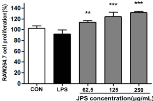
Fig. 1. Cell proliferation activity of Jubak polysaccharide (JPS) in RAW264.7 macrophage cell. JPS were treated at the concen- tration of 62.5, 125, and 250 μg/mL. Cell proliferation was con- ducted in triplicates (n=3), and the results are expressed as mean
±SD. Statistical analysis was performed using the Student's two tailed t-test with a significance level of **P<0.01, ***P<0.001.
tection kit(Thermo Fisher Scientific Inc., Rockford, IL, USA)을 사용하여 단백질 정량을 실시하였고, well당 20 μg 의 cell lysate를 10% polyacrylamide gel에 각각 loading 하여 SDS-PAGE로 변성 분리하였다. 이를 polyvinylidene difluoride membrane(Millipore, Merck KGaA, Darmstadt, Germany)으로 transfer 하였고, membrane은 anti-body 의 비특이적 결합을 방지하기 위해 blocking solution(skim milk 5%) 20 mL에서 1시간 동안 방치하였다. 이후 TBST (20 nM tris-HCl, 150 mM NaCl, 0.05% Tween-20, pH 7.5)로 10분씩 3회 세척하였으며, p-p38, p-38, p-ERK, ERK, p-JNK, JNK 및 NF-κB의 발현량을 측정하기 위해 1차 항체(Cell Signaling Technology, Danvers, MN, USA) 를 1:2,000으로 희석하여 4시간 동안 반응시키고 TBST로 5분간 3회 세척하였다. 이후 2차 항체(goat-anti rabbit lgG, Calbiochem, La Jolla, CA, USA)를 1:5,000으로 희석 하여 2시간 동안 반응시키고 현상을 위하여 electrochemil- uminescence(Millipore Merck KGaA) reagent를 사용하 여 인화하였다.
통계분석
이상의 실험에서 얻어진 결과는 Statistical Package for Social Sciences(SPSS, 10.0, IBM, Chicago, IL, USA) software를 이용하여 one way ANOVA test로 분석하였으 며, 시료 간의 유의성은 Student's two tailed t-test로 *P<
0.05, **P<0.01 및 ***P<0.001 수준에서 비교하였다.
결과 및 고찰
JPS의 세포 생존율 평가
JPS가 대식세포의 세포독성에 미치는 영향을 평가하기 위하여 대식세포 RAW264.7 cell에 농도별 JPS를 처리하여 JPS에 대한 세포의 생존율을 MTT 방법을 통하여 평가하였 다(Fig. 1). JPS를 62.5, 125 및 250 μg/mL의 농도로 처리
하였을 때 모든 농도에서 세포독성이 나타나지 않았고 농도 의존적으로 세포 생존율이 증가되는 것으로 관찰되었다. 따 라서 JPS는 대식세포의 세포독성에 영향을 미치지 않았으 며, 추후 JPS의 처리가 cytokine 및 NO의 생성능에 어떠한 영향을 미치는지 알아보기 위하여 JPS의 농도를 62.5, 125 및 250 μg/mL로 고정하여 실험하였다
JPS의 NO 및 cytokine 분비능
대식세포는 숙주의 면역반응에 중추적인 역할을 수행한 다. 병원성 항원이 생체 내에 침입하게 되면 대식세포가 이 를 포식하여 항원에 대한 정보를 T 세포에 전달하여 세포매 개면역반응이 일어난다. 일반적으로 활성화되지 않은 대식 세포는 외래 항원에 대한 면역기능을 하는 데 효과적이지 않다(13). 반면에 활성화된 대식세포는 생체 내 미생물 및 바이러스에 감염된 세포를 제거하는 데 매우 효과적이다 (14). 이처럼 대식세포의 활성화 여부는 외부로부터 유입된 미생물의 침입에 있어서 초기면역반응의 증강을 나타내는 지표로서 다양한 물질들의 면역 활성능을 평가할 때 주로 이 용된다(15). 대식세포 활성화에 미치는 영향을 평가하기 위 해서 RAW264.7 대식세포에 JPS를 처리한 후 세포 상등액 에서 pro-inflammatory cytokine인 TNF-α, IL-6와 NO의 생성에 대하여 알아보았다. 앞선 결과에서 세포독성에 영향 을 미치지 않는 농도인 62.5, 125 및 250 μg/mL의 농도로 JPS를 처리하여 대식세포 활성화 인자인 cytokine(TNF-α, IL-6)과 NO의 생성을 관찰한 결과 JPS의 처리는 농도 의존 적으로 TNF-α, IL-6와 NO의 분비능을 증가시키는 것으로 관찰되었다(Fig. 2A, B). 대식세포 활성화에 중요한 역할을 수행하는 NO는 nitric oxide synthase(NOS)와 산소가 결 합하여 L-arginine을 산화시켜 생성되는데, 미생물에 감염 된 세포나 암세포를 제어하는 것으로 보고되고 있다(16).
하지만 과량 분비된 NO는 혈관 투과성, 부종 등의 염증반응 을 악화시키고 염증매개물질의 생합성을 촉진하여 염증을 심화시키는 것으로 알려져 있다(17). 따라서 세포독성이 없 는 농도에서 NO의 증가는 면역기능을 활성화시키는 것으로 판단할 수 있다. 본 연구에서도 62.5, 125 및 250 μg/mL의 JPS를 처리하였을 때 세포독성을 확인할 수 없었으며, NO 분비능이 농도 의존적으로 증가하는 것으로 관찰되어 JPS 의 처리는 대식세포의 면역 활성능을 증강시키는 것으로 사 료된다.
활성화된 대식세포가 분비하는 cytokine은 광범위한 면 역신호 전달인자로서 특정 수용체를 통해 세포들의 성숙 및 성장을 유도하고 체액성 및 세포매개성 면역반응을 조절함 으로써 면역시스템에서 중요한 역할을 수행한다. 대식세포 이외에 cytokine을 분비하는 세포는 혈관 내피세포(endo- thelial cell)와 비만세포(mast cell)가 있으나 이들이 분비 하는 cytokine은 면역조절작용 외 다른 역할을 수행하며, 면역조절에 관여하는 cytokine인 IL-1, IL-6, TNF-α는 주 로 활성화된 대식세포에 의해 생성된다(18,19). 따라서 본
A
B
Fig. 2. Nitric oxide (NO) and cytokine (TNF-α and IL-6) production activity of Jubak polysaccharide (JPS) in RAW264.7 macrophage cell. JPS were treated at the concentration of 62.5, 125, and 250 μg/mL. NO (A) and cytokine (B: TNF-α and IL-6) production were conducted in triplicates (n=3), and the results are expressed as mean±SD. Statistical analysis was performed using the Student's two tailed t-test with a significance level of *P<0.05, **P<0.01, and ***P<0.001.
A B
Fig. 3. Phosphorylation activity of Jubak poly- saccharide (JPS) on MAPKs and NF-κB in RAW264.7 macrophage cells. JPS were treated at the concentration of 62.5 and 125 µg/mL for 45 min. Cells lysates were subjected to SDS- PAGE and immunoblotting analysis was per- formed using each specific antibody to MAPKs (phospho-p38, phospho-ERK1/2, and phospho- JNK1/2) (A) and NF-κB (p65) (B).
실험에서는 대식세포의 활성 지표인 cytokine의 분비량을 통해서 대식세포의 활성화 정도를 알아본 결과 JPS를 대식 세포에 세포독성이 없는 62.5, 125 및 250 μg/mL의 농도로 처리하였을 때 TNF-α 및 IL-6의 함량이 농도 의존적으로 증가하는 것으로 보아 JPS의 처리는 대식세포의 활성화를 유도하는 것으로 사료된다(Fig. 2B)
JPS의 MAPKs 및 NF-κB의 인산화에 미치는 영향 상기 실험에서 JPS의 처리가 cytokine 및 NO의 분비를 증가시킴으로써 대식세포 활성화를 유도하는 것을 확인할
수 있었다. 초기 면역반응이 시작되면 면역세포 내 신호전달 에 관여하는 MAPKs(ERK, JNK, p-38) 및 NF-κB의 인산 화가 이루어지고, NF-κB가 최종 활성화되어 면역 방어기작 을 나타내는 cytokine 및 NO의 분비에 영향을 미치는 것으 로 보고된다(13). 따라서 JPS의 처리에 의한 대식세포 활성 화의 정확한 면역기전을 알아보기 위해서 대식세포에 JPS 의 농도를 62.5 및 125 μg/mL로 처리하고 MAPKs와 NF-κB 의 인산화 정도를 관찰했을 때 JPS의 처리가 MAPK(ERK, JNK, p-38) 및 NF-κB의 인산화를 증가시키는 것으로 확인 되었다(Fig. 3). NF-κB는 세포 내에서 비활성상태로 존재
하며 NF-κB가 면역 활성인자들에 의해 자극을 받으면 IκB- α가 인산화되고 NF-κB는 핵 안으로 이동하여 NO 및 cyto- kine 등 다양한 면역 활성 매개체의 유전자 발현을 유도시킨 다(20,21). 본 연구에서 JPS의 처리에 의하여 NO 및 cyto- kine의 분비가 증가된 것은 MPAKs의 인산화로 NF-κB의 활성이 증가되었기 때문인 것으로 사료된다.
요 약
본 연구는 양조과정의 부산물인 주박으로부터 분리한 다당 류(JPS)가 초기 면역반응에 중추적인 역할을 수행하는 대식 세포에서 활성화를 유도하는지에 관한 여부를 알아보기 위 해서 수행되었다. 주박에서 분리한 다당류를 마우스 유래 대식세포인 RAW264.7 cell에 처리하였을 때 대식세포의 활성화의 지표인 NO와 cytokine(IL-6, TNF-α)의 분비가 증가되었다. 또한 이러한 NO와 cytokine의 증가의 원인에 관한 면역기전에 관하여 알아본 결과 JPS의 처리는 MAPKs (ERK, JNK, p-38)의 인산화를 촉진시켜 NF-κB의 활성을 유도하여 면역세포의 활성인자들의 분비를 촉진시킨 것으 로 관찰되었다.
감사의 글
본 연구는 2014년도 미래창조과학부의 재원으로 한국연구 재단 기초연구사업(No. NRF-2012R1A1A2009507)과 농 림축산식품부 고부가가치식품기술개발사업(No. 112078- 3)의 지원으로 수행되었으며, 그 지원에 감사드립니다.
REFERENCES
1. Kim TY, Jeon TW, Yeo SH, Kim SB, Kim JS, Kwak JS.
2010. Antimicrobial, antioxidant and SOD-like activity ef- fect of Jubak extracts. Korean J Food & Nutr 23: 299-305.
2. Lee JH, Park SM, Park CD, Jung HJ, Kim HS, Yu TS.
2007. Characteristics of Ju-Back fertilizer on growth of crop plants. J Life Sci 17: 1562-1570.
3. Lee HS, Park CS, Choi JY. 2010. Quality characteristics of the mashes of Takju prepared using different yeasts. Korean J Food Sci Technol 42: 56-62.
4. Cho SY, Park JW, Rhee C. 1998. Edible films from protein concentrates of rice wine meal. Korean J Food Sci Technol 30: 1097-1106.
5. Kang HT, Lee SH, Kim SY, Kim MS, Shin WC, Sohn HY, Kim JS. 2014. Anti-proliferative activities of solvent frac- tions of lees extracts in human colorectal HCT116 cells.
J Life Sci 24: 967-972.
6. Park MJ, Kang HT, Kim JS. 2014. Anti-inflammatory ef-
fects of extracts and their solvent fractions of rice wine lees.
J Life Sci 24: 843-850.
7. Kim S, Cho W. 2006. Effects of Takju (Korean turbid rice wine) lees on the serum glucose levels in streptozotocin-in- duced diabetic rats. Korean J Food Culture 21: 638-643.
8. Jiang MH, Zhu L, Jiang JG. 2010. Immunoregulatory ac- tions of polysaccharides from Chinese herbal medicine.
Expert Opin Ther Targets 14: 1367-1402.
9. Kim DM, Kim KH, Sung NY, Jung PM, Kim JS, Kim JK, Kim JH, Choi J, Song BS, Lee JW, Kim JK, Yook HS.
2011. Effects of gamma irradiation on the extraction yield and whitening activity of polysaccharides from Undaria pinnatifida sporophyll. J Korean Soc Food Sci Nutr 40:
712-716.
10. Ha ES, Hwang SH, Yu KW, Shin KS, Cho HM, Kim CH, Park WM, Yoon TJ. 2003. Immunostimulation activity of the crude polysaccharides fractionated from Eleutherococcus senticosus, and its application to prevent of tumors by com- bination therapy with cisplatin. Yakhak Hoeji 47: 159-166.
11. Kim SB, Lee GW, Lee UY, Lee TS. 2007. Studies on im- muno-modulatory and antitumor effects of crude polysac- charides extracted from fruiting body of Oudemansiella radicata. Korean J Mycol 35: 109-114.
12. Lee MS, Shin KS. 2014. Intestinal immune-modulating ac- tivities of polysaccharide isolated from commercial and tra- ditional Korean soy sauces. J Korean Soc Food Sci Nutr 44: 228-234.
13. Brewer MS, Ikins WG, Harbers CAZ. 1992. TBA values, sensory characteristics, and volatiles in ground fork during long-term frozen storage: Effects of packaging. J Food Sci 57: 558-563.
14. Kim HI, Lee BM. 1996. Stevioside, a natural sweetener:
Is it safe? J Fd Hyg Safety 11: 323-327.
15. Bios MS. 1958. Antioxidant determinations by the use of a stable free radical. Nature 181: 1199-1200.
16. Benzie IF, Strain JJ. 1996. The ferric reducing ability of plasma (FRAP) as a measure of "antioxidant power": the FRAP assay. Anal Biochem 230: 70-76.
17. Ryu JH, Ahn H, Kim JY, Kim YK. 2003. Inhibitory activity of plant extracts on nitric oxide synthesis in LPS-activated macrophages. Phytother Res 17: 485-489.
18. Boyle JJ. 2005. Macrophage activation in atherosclerosis:
pathogenesis and pharmacology of plaque rupture. Curr Vasc Pharmacol 3: 63-68.
19. Olszewski MB, Groot AJ, Dastych J, Knol EF. 2007. TNF trafficking to human mast cell granules: mature chain-de- pendent endocytosis. J Immunol 178: 5701-5709.
20. Chandel NS, Trzyna WC, McClintock DS, Schumacker PT.
2000. Role of oxidants in NF-κB activation and TNF-α gene transcription induced by hypoxia and endotoxin. J Immunol 165: 1013-1021.
21. Kim NY, Kim YK, Bae KJ, Choi JH, Moon JH, Park GH, Oh DH. 2005. Free radical scavenging effect and extraction condition of ethanol extracts and fractions of wild grape seed (Vitis coignetiea). J Korean Soc Food Sci Nutr 34:
755-758.