스테비아로부터 분리한 Austroinulin의 RAW264.7 세포에 대한 항염증 효과
변 명 우
우송대학교 외식조리영양학부
Anti-Inflammatory Activity of Austroinulin from Stevia rebaudiana in LPS-induced RAW264.7 Cells
Myung-Woo Byun
Dept. of Culinary Netrition, Woosong University, Daejeon 300-718, Korea
Abstract
The leaves of Stevia rebaudiana are well-known in Japan, Korea, and China as a natural sweetener. Medicinal uses of this plant originated in Paraguay and Brazil in the form of aqueous decoctions of the leaves used as a contraceptive agent and for the treatment of hyperglycemia. In the present study, the antioxidant, anti-hyper- tension, and anti-inflammatory activities of S. rebaudiana extracts are investigated for their use in food. The biologically-active compound was isolated and purified from S. rebaudiana. The isolated compound was identified as austroinulin (C
20H
34O
3; molecular weight 322) by mass, IR spectrophotometry, 1D, and 2D-NMR. Austroinulin was characterized as a diterpenoid possessing a 3-methylpenta-2,4-dienyl at C-9. When subjected to an in- flammatory mediator inhibitory assay from lipopolysaccharide (LPS)-activated macrophages, the austroinulin in- hibited the enhanced production of nitric oxide (NO) and inducible nitric oxide synthase (iNOS) expression (10 μ g/mL=67.9 and 45.1%, respectively). This was significant and dose-dependent. The results suggest that aus- troinulin from S. rebaudiana inhibited the NO and iNOS in RAW 264.7 cells.
Key words: Stevia rebaudiana, austroinulin, RAW264.7, anti-inflammation
E-mail: [email protected]
Phone: 82-42-630-9864, Fax: 82-42-630-9389
서 론
스테비아(
Stevia rebaudiana)는 국화과(Asteraceae)에 속 하는 다년생 여러해살이풀로 감미료로 주로 잎을 사용하며, 건조한 것은 식품첨가물, 화장품원료 친환경 농자재 등으로 이용되고 있다(1). 스테비아는 남미의 파라과이, 아르헨티 나, 브라질 등의 국경산간지 하천, 습지대주변에서 약용을 목적으로 재배하여 왔으며, 1899년에 처음 연구되어 100여 개 이상의 terpenes과 flavonoids 등이 phytochemicals로 함 유되어 있는 것으로 알려져 있다(2).
스테비아의 주요성분으로는 diterpenoid glycosides의 steviolbioside, rebaudioside A~E, stevioside 등의 성분이 함유되어 있는 것으로 알려져 있으며, 그 밖에 jhanol, ster- ebin A-N, 6-O-acetyl-austroinulin 등의 diterpenes 성분이 알려져 있다(3-5). 이러한 스테비아에 대한 연구는 대부분 잎으로부터 diterpenoid glycosides의 분리 및 성분연구이 며, 그중에서 stevioside는 생쥐에 대한 insulin-sensitive와 insulin-resistant 당뇨조절 효과 등이 보고되고 있다(6). 최 근 diterpene 화합물들은 항산화, 항박테리아, 항바이러스 등 에 효과가 확인되고 있으며 새로운 기작의 염증제로 확인되
고 있다(7-9).
염증 관련 질환은 물리적 충격이나 화학물질 및 세균감염
등의 자극에 대한 생체 방어 기작이며, 지속적인 염증반응은
점막손상을 촉진시켜 결과적으로 관절염, 당뇨병, 동맥경화
증 및 암 등의 발생을 이끈다(10). 생체 내 염증을 촉진하는
효소로는 cyclooxygenase-2(COX-2)와 lipoxygenase(LOX)
가 잘 알려져 있으며(11,12), 이들 효소를 저해하는 물질은
염증성 질환을 저해하는 항염증제로서 개발 가능성이 높다
(13). 지금가지 개발된 함염증제는 크게 스테로이드계와 비
스테로이드계로 나눌 수가 있으며, 모두 염증반응의 주요
매개체인 프로스타글란딘의 생합성을 억제함으로 항염증작
용을 나타내는 약제이다. 그러나 이들 함염증제는 위염, 신
장염 및 심장질환 등을 초래함으로써 인체 안전성면에서 문
제점을 안고 있어 그 사용이 일부 제한되고 있기에 현재 천
연으로부터 보다 안전한 항염증 물질을 검색하는 연구가 활
발하게 진행되고 있을 뿐 아니라 일상 식생활에서 섭취하는
식품중 항염증제로 이용하기 위한 물질이 계속 탐색되고 있
다(14,15). 특히 우리나라에서는 위령선, 하고초, 괄루근 등
의 한방 및 생약제가 섭취되고 있고, 그중 조인스정은 관절
염 증상 완화제로 천연물신약으로 사용되고 있다.
최근에는 국민 소득의 향상과 노령화 등 사회적 변화에 따라 건강에 대한 관심도가 고조되면서 각종 건강보조식품 이 개발되고 그 수요 또한 증가하고 있는 실정이다(16). 따라 서 본 연구에서는 스테비아는 앞에서 언급한 바와 같이 이용 성이 높은 식물임에도 불구하고 diterpene 성분에 대한 생리 활성이 과학적으로 주로 연구된 바 없어 항염증 효과를 규명 하기 위한 일환으로 스테비아로부터 diterpene 주요성분인 austroinulin 화합물을 분리 정제하고, 이를 이용한 RAW 246.7 세포주에 대한 염증 억제 효과를 규명하고자 하였다.
재료 및 방법
실험재료
본 실험에 사용한 스테비아는 2010년 10월 전라북도 정읍 시에서 수확한 것을 구입하여 사용하였으며, 건조된 상태로 저온(-5
oC)에서 보관하면서 실험재료로 사용하였다.
시약 및 크로마토그래피용 충진제
본 실험에 사용된 column chromatography 충진물로서 silica gel 60(70-230 mesh, Merck, Darmstadt, Germany)와 diaion HP-20(Sigma-Aldrich, St. Louis, MO, USA)을 사용 하였으며, 물질분리 및 확인을 위한 thin-layer chromatog- raphy(TLC)는 pre-coated silica gel 60(0.25 mm, Merck) plat를 사용하였다. 그 외 용매 및 시약류는 분석용 특급 또 는 1급 시약을 각각 사용하였다.
스테비아로부터 austroinulin의 분리 및 정제
음건 세절한 스테비아 시료 1 kg을 80% 에탄올(20 L×
3)로 실온에서 3일간 추출하였다. 추출액을 감압 농축시켜 340 g의 흑갈색의 조추출물을 얻었다. 추출물을 다이아이온 칼럼을 통해 H
2O, methanol, CHCl
3층으로 나누었다. 그 다 음 methanol 층을 H
2O와 ethyl acetate(1:1) 혼합용액 2,000 mL와 함께 분획여두에 넣고 실온에서 3시간, 3회 반복하여 분획한 후 ethyl acetate 분획물 50 g을 얻었다. 얻어진 추출 물을 실리카겔 칼럼에서(10×30 cm, 230~400 mesh, 700 g) hexane : acetone(50:1, 40:1, 20:1, 10:1, 8:1, 6:1, 3:1, 1:1)으로 순차적으로 용출시켜 7개의 subfraction(1-7)을 얻었다. 얻 어진 subfraction 5를 다시 실리카겔 칼럼에서(2.5×30 cm, 230~400 mesh, 200 g) hexane : ethyl acetate(20:1, 10:1, 8:1, 6:1, 4:1, 2:1, 1:1)로 정제하여 diterpenes 화합물 austro- inulin 385 mg을 순수 분리하였다.
세포 배양
본 실험에서 사용한 마우스 대식세포주 RAW264.7 세포 는 한국세포주은행(KCLB, Seoul, Korea)에서 분양받아 10% fetal bovine serum(FBS; Hyclone, Logan, UT, USA) 와 100 units/mL of penicillin, 100 mg/mL of streptomycin 을 첨가한 DMEM 배지(Invitrogen, Carlsbad, CA, USA)를
사용하여 37
oC, 5% CO
2incubator에서 배양하였으며, 세포 밀도가 높아지면 trypsin-EDTA를 처리하여 계대배양하면 서 실험에 사용하였다.
세포독성 측정
세포독성은 EZ-Cytox cell viability assay kit(DAEIL lab, Seoul, Korea)를 사용하여, 제조사의 실험방법에 따라서 측 정하였다. RAW264.7 세포를 최종농도 2×10
5cells/mL가 되도록 96 well plate에 분주한 후, 37°C, 5% CO
2incubator 에서 24시간 배양한 다음, austroinulin을 2.5, 5, 10, 20, 40 μ g/mL의 농도로 첨가하여 24시간 배양하였다. 배양 후, EZ-Cytox 시약 10 mL를 넣고 4시간 동안 배양한 후 micro- plate reader(Benchmark Plus, Bio-Rad, Hercules, CA, USA)를 이용하여 480 nm에서 흡광도를 측정하였으며, 대 조군에 대한 세포 생존율을 백분율로 나타내었다.
Nitric oxide 측정
RAW264.7 세포를 최종농도 2×10
5cells/mL가 되도록 96 well plate에 분주한 후, 37°C, 5% CO
2incubator에서 24시간 배양하였다. 배양 후, austroinulin을 2.5, 5, 10 μg/mL의 농도 로 전 처리하고 1시간 후에 LPS 1 μg/mL를 처리한 후, 20시 간 동안 배양하였다. 배양액에 동량의 Griess 시약(Sigma- Aldrich)을 가하고 실온에서 15분간 반응시킨 후 microplate reader(Bio-Rad)를 이용하여 570 nm에서 흡광도를 측정하 였다. NO 농도는 sodium nitrite의 표준곡선을 이용하여 결 정하였다.
Western blot을 이용한 iNOS 단백질 분석
RAW264.7 세포를 최종농도 2×10
5cells/mL가 되도록 100 mm dish에 분주한 후, 37°C, 5% CO
2incubator에서 24 시간 배양한 후, austroinulin을 2.5, 5, 10 μg/mL의 농도로 전 처리하고 1시간 후에 LPS 1 μg/mL를 처리한 후, 20시간 동안 배양하였다. 배양 후 dish에서 세포를 회수하여 PBS로 세척하고 NP40 cell lysis buffer(Sigma-Aldrich)를 첨가하 여 얼음에서 세포를 용해하였다. 세포 용해액에 sample buf- fer를 섞어 100
oC에서 5분간 끓인 후 8% SDS-PAGE를 수행 하였다. 전기영동 후 단백질을 nitrocellulose membrane으 로 transfer한 후 5% skim milk로 실온에서 1시간 blocking 후 iNOS 항체를 4°C에서 하루 밤 동안 반응시켰다. 반응 후 TBS-T buffer로 10분간 3번 세척한 후 2차 anti-rabbit IgG conjugated HRP를 실온에서 2시간 반응시켰다. TBS-T buf- fer로 10분간 3번 세척하고 enhanced chemiluminescence detection system(Amersham Pharmacia, Bucks, UK)를 사 용하여 iNOS 단백질을 확인하였다.
통계학적 검정
모든 실험은 3회 반복실험에 대한 평균(mean)±표준편차
(standard deviation; SD)로 나타내었다. 통계학적 분석은
SPSS Inc.(Chicago, IL, USA) version 18.0을 이용하였으며,
Fig. 1. Isolation of austroinulin from the leaves of Stevia rebaudiana.
(A) (B)
Austroinulin
OHOH
OH OH
OH OH
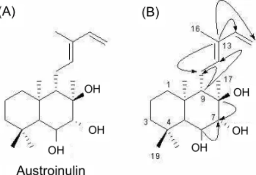
Fig. 2. (A) Structure of austroinulin isolated from the leaves of Stevia rebaudiana (B) HMBC correlation of austroinulin (CDCl
3, 125 MHz).
one-way analysis of variance(ANOVA)를 시행하여 p<
0.05인 경우를 유의적인 것으로 판정하였다.
결과 및 고찰
스테비아로부터 austroinulin의 분리 및 정제
농축한 스테비아 80% 에탄올 추출물로부터 diaion 칼럼 크로마토그래피 실시하여 클로로필을 제거한 methanol층을 얻었다. Methanol층을 H
2O/ethyl acetate로 분획하여 di- terpenoid glycoside가 완전히 제거된 ethyl acetate 분획물 을 얻었다. Ethyl acetate 분획물을 실리카겔 칼럼크로마토 그래피를 통해 active-guided fractionation하여 분리하였 다. 이때
n-hexane-acetone(50:1→1:1) 혼합용매를 이용하 여 용매의 극성을 변화시키면서 용출시키고, TLC pattern에 따라서 fraction 하였다. 그에 대한 활성 측정결과 용출액 중 subfraction 5에서 NO에 대한 활성 효과를 보였다. 용출 액 중 subfraction 5를
n-hexane-ethyl acetate 혼합용매에 서 극성을 변화시켜가며 2차 실리카겔 칼럼크로마토그래피 하여 분리한 결과 TLC pattern에서 용출액 중 fraction 8-12 에서 main point인 1개의 화합물을 분리하였고, UV spec- trum 254 nm에서 TLC 확인 결과 단일 화합물임을 알 수 있었다(Fig. 1).
분리한 austroinulin의 구조 동정
스테비아의 80% 에탄올 추출물로부터 active-guided fractionation하여 분리된 단일 화합물의 구조분석은 JNM- ECA 500 spectrometer(Jeol, Tokyo, Japan)를 사용하여 1D, 2D-NMR 스펙트럼과 LC/MS(Agilent 1100 series, Boe- blingen, Germany)를 이용하여 MS를 측정하였고, 기존문헌 의 측정값과 비교하여 구조를 분석하였다. 분리된 austro- inulin 화합물은 흰색의 분말로서 LC/MS를 통해 분자이온
[M]
+의 피크가
m/
z=322를 확인하였다.
1H-NMR 스펙트 럼의 양성자(proton) 개수와
13C-NMR 탄소 개수를 고려하 면 화합물은 C
20H
34O
3의 분자식을 갖는 화합물로 확인하였 다.
1H-NMR 스펙트럼에서는 quaternary methyls이
d0.87, 0.96, 1.11, 1.14(각각 3H)이고 cyclohexane ring의 carbinyl hydrogen이
d3.58(1H, d,
J=10 Hz), 3.39(1H, t,
J=10 Hz)이 었다. 전형적으로 3-methylpenta-2,4-dienyl에서 관찰되는 2개의 olefinic hydrogens
d5.43(1H, t,
J=6.6 Hz), 6.83(1H, d,
J=17 Hz)과 1개의 methylene
d5.08, 5.17(2H, br d,
J=17 Hz)을 관찰하였다.
13C-NMR 스펙트럼에서는 전형적으로 3-methylpenta-2,4-dienyl에서 관찰되는 6개의 탄소 피크 를 관찰하였다. 특히
d113.7의 피크는 3-methylpenta-2,4- dienyl의 methylene으로 확인되었다(Fig. 2). HMBC를 비롯 한 2D-NMR을 통해 화합물은 decalin system에서 C-9에 3-methylpenta-2,4-dienyl 치환체를 가지고 있음을 추론할 수 있었다(Fig. 3). 이상의 분광학적인 자료를 종합하면 화합 물은 austroinulin로 확인되었으며 문헌에 보고된 분광학적 인 자료와 일치하였다(17,18).
Austroinulin: white crystals; mp 75~78
oC; MS:
m/
z[M]
+322;
1H-NMR(500 MHz, CDCl
3)
dH(ppm): 0.87(3H, s, H-18), 0.96(3H, s, H-20), 1.11(3H, s, H-17), 1.14(3H, s, H- 19), 1.11~1.51(6H, m, H-1,2,3,5,9), 1.76(3H, s, H-16), 2.21 (1H, m, H-11), 2.40(1H, m, H-11), 3.39(1H, t,
J=10 Hz, H- 7), 3.58(1H, t,
J=10 Hz, H-6), 5.08, 5.17(2H, br d,
J=17 Hz, H-15), 5.43(1H, t,
J=6.6 Hz, H-12), 6.83(1H, d,
J=17 Hz, H-14);
13C-NMR(500 MHz, CDCl
3)
dc(ppm): 17.0(C-20), 18.2(C-2), 19.4(C-17), 19.9(C-16), 22.3(C-19), 22.8(C-11), 33.8(C-4), 36.4(C-18), 39.2(C-10), 39.9(C-10), 43.4(C-3), 57.3(C-5), 59.6(C-9), 71.7(C-6), 77.3(C-8), 85.0(C-7), 113.8 (C-15), 130.9(C-13), 133.6(C-12), 133.7(C-14).
RAW264.7 세포의 독성 확인
마우스 대식세포인 RAW264.7은 염증반응에서 중추적인
역할을 수행하며 항염증 효과를 평가하는 염증모델로 많이
사용된다. 이러한 RAW264.7 세포를 이용하여 스테비아에
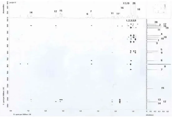
Fig. 3. HMBC spectral data of austroinulin from the Stevia rebaudiana (in CDCl
3).
0 20 40 60 80 100 120
Austroinulin (μg/mL)
Cell viability (% of control) .
0 2.5 5 10 20 40
Fig. 4. Effect of austroinulin on cell viability. RAW264.7 cells were treated with various concentrations of austroinulin for 24 hr and relative cell viability was assessed by WST-1 assay. Error bars represent the mean±SD.
0 2 4 6 8 10 12 14 16
NO (μM) .
LPS (1 μg/mL) - + + + + austroinulin (μg/mL) - - 2.5 5 10
#
*
**
**
Fig. 5. Effect of austroinulin on LPS-induced NO production.
RAW264.7 cells were treated with various concentrations of aus- troinulin for 1 hr prior to the addition of LPS (1 μg/mL) and the cells were further incubated for 20 hr. Error bars represent the mean±SD.
#p<0.01 vs. control,
*p<0.05 vs. LPS,
**p<0.01 vs. LPS.
서 분리한 austroinulin의 세포 독성을 cell viability assay에 의해 확인한 결과 Fig. 4와 같이 나타났다. 본 연구에서 처리 한 실험군의 농도 0∼40 μg/mL에서 95% 이상의 생존율을 보여 스테비아에서 분리한 austroinulin 화합물은 세포독성 을 가지지 않는 것으로 나타났다.
Nitric oxide 생성 저해활성 확인
내독소로 잘 알려진 lipopolysaccharide(LPS)는 그람 음 성균의 세포외막에 존재하며, RAW264.7 세포와 같은 mac- rophage 또는 monocyte에서 tumor necrosis actor-α(TNF- α ), interleukin-6(IL-6), interleukin-1β(IL-1β)와 같은 pro- inflammatory cytokine을 증가시키는 것으로 알려져 있다 (19). RAW264.7 cell의 NO 생성억제 정도를 측정하기 위하 여 스테비아에서 분리한 austroinulin 화합물을 농도별로 세
포에 처리하여 생성되는 NO양을 측정한 결과 Fig. 5와 같이 나타났다. LPS를 첨가하지 않은 군에서는 8~10%의 NO를 생성하였고, austroinulin 화합물을 2.5, 5, 10 μg/mL로 처리 한 결과에서 농도 의존적으로 NO의 생성을 억제하는 것으로 관찰되었으며, 10 μg/mL에서 NO의 생성을 67.9%를 억제하 였다. 이는 Park 등(20)의 인삼 ginsenoside Rb1 화합물과 Suh 등(21)의 인동덩굴 biflavonoil 화합물이 LPS로 유도된 RAW264.7 세포의 NO 생성 억제능에 미치는 결과와 비교하 여 보면 스테비아에서 분리된 austroinulin 화합물의 NO 생 성 억제능이 우수하다는 것을 확인할 수 있었다.
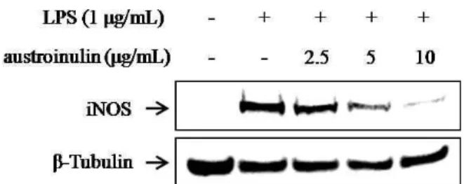
iNOS 단백질의 발현 측정 결과
자극에 유도된 iNOS는 오랜 기간 동안 다량의 NO를 생성
Fig. 6. Effect of austroinulin on LPS-induced iNOS expression levels. RAW264.7 cells were treated with various concentrations of austroinulin for 1 hr prior to the addition of LPS (1 μg/mL) and the cells were further incubated for 20 hr. Cells were har- vested and subjected to western blotting.
하게 되고, 생성된 NO는 guanylyl cyclase의 활성과 동시에 주위 조직에 세포독성을 나타낸다. 즉 LPS로 유도되어진 RAW264.7 세포에서 iNOS의 protein level의 감소에 의해 항염증 효과를 기대할 수 있다. 스테비아에서 분리한 aus- troinulin에 대해 iNOS를 저해하는 효과가 있는지 알아보기 위하여 RAW264.7 세포에 LPS(1 μg/mL)를 처리하고 1시간 뒤, 스테비아에서 분리한 austroinulin 화합물을 2.5, 5, 10 μ g/mL로 처리한 후 20시간 배양한 후 iNOS protein의 발현 율 변화를 western blotting으로 확인하였다. 이때 austro- inulin 화합물을 2.5, 5, 10 μg/mL 농도로 처리한 결과 iNOS protein 발현율은 각각 11.4, 26.8, 45.1%를 감소시켜 우수한 활성이 관찰되었다(Fig. 6). 이는 Zhao 등(22)이나 Metzger 등(23)의 보고에 의하면 RAW264.6 세포에 LPS로 자극한 후 생리활성물질에 의한 항염증 효과를 살펴본 결과 iNOS expression과 iNOS enzymatic activity의 down-regulation 에 따라 NO의 생성이 억제됨을 보고하였다.
이런 결과는 스테비아에서 분리된 astroinuline 화합물이 LPS에 의해 유도되는 염증효소인 iNOS 유전자의 발현을 효 과적으로 억제시켜 NO의 생성을 억제시킨 것으로 사료된다.
요 약
본 연구에서는 스테비아는 이용성이 높은 식물임에도 불 구하고 diterpene 성분에 대한 생리활성이 과학적으로 주로 연구된 바 없어 active-guided fractionation 방법을 이용하 여 항염증 효과를 나타내는 지표화합물을 규명하기 위해 실 리카겔 칼럼크로마토그래피 방법을 이용하여 분리 및 정제 한 후,
1H와
13C-NMR, COSY, DEPT, HMQC, HMBC spec- trum(500 MHz, CDCl
3), MS, IR 분석을 통하여 분자량 322 의 astroinulin임을 구조 동정하였다. 특히 HMBC spectrum 을 통해 분리된 화합물이 decalin system에서 C-9에 3-me- thylpenta-2,4-dienyl 치환체를 가지고 있음을 알 수 있었다.
분리된 astroinulin을 RAW264.7 세포에 2.5, 5, 10 μg/mL 처리한 결과 농도 의존적으로 NO의 생성을 억제하였다. 마 찬가지로 iNOS protein의 발현에서도 농도 의존적으로 저해 하였으며, 화합물을 2.5, 5, 10 μg/mL 처리한 결과 각각 11.4,
26.8, 45.1% 감소하였다. Astroinulin 화합물을 10 μg/mL의 낮은 농도에서 NO와 iNOS를 각각 67.9, 45.1%를 저해하는 결과를 나타내었다. 이와 같은 결과는 스테비아에서 분리된 astroinulin 화합물은 RAW264.7 세포에서 LPS에 의해 유도 된 NO와 iNOS 단백질을 유의적으로 억제하는 것으로 확인 되어 이를 이용한 기능성식품 및 의약품 개발 가능성을 지닌 약용 식물자원인 것으로 판단된다.
문 헌