66
한방안이비인후피부과학회지 제28권 제3호(2015년 8월)
J Korean Med Ophthalmol Otolaryngol Dermatol 2015;28(3):66-75 pISSN 1738-6640 eISSN 2234-4020
http://www.ood.or.kr http://dx.doi.org/10.6114/jkood.2015.28.3.066
Original Article / 원저
나이관련 황반변성에 대한 동서의학적 고찰
정유진·고우신·윤화정
동의대학교 한의과대학 안이비인후피부과학교실
The Study on the Korean and Western Medical Literatures for Age-Related Macular Degeneration
Yu-Jin Jung·Woo-Shin Ko·Hwa-Jung Yoon
Dept. of Oriental Medical Opthalmology & Otolaryngology & Dermatology, Dong-eui University
Abstract
Object : The purpose of this study is to understand age-related maculardegeneration(AMD) with both western and Korean medicine.
Methods : We investigated the comprehension of general AMD degenation in both western and Korean medicine through literature review.
Results : The results are as follows.
1. AMD prevalent increasing as the population ages; however, treatment options remain limited and incompletely defined.
2. Generally macular degeneration has been affected by aging and is associate with the function of kidney(腎) and liver(肝) in Korean medicine.
Conclusion : Further studies are needed to apply comprehension of AMD in Korean medicine to clinical stage.
Key words : macular degeneration; age-related macular degenation; Korean medicine; Review
ⓒ 2015 the Society of Korean Medicine Ophthalmology & Otolaryngology & Dermatology
This is an Open Access journal distributed under the terms of the Creative Commons Attribution Non-Commercial License (http://creativecommons.org/license/by-nc/3.0/) which permits unrestricted non-commercial use, distribution, and reproduction in any medium, provided the original work is properly cited.
정유진 외 2인 : 나이관련 황반변성에 대한 동서의학적 고찰
67
Ⅰ. 서 론
망막의 중심부에 위치하면서 중심 시력을 담당하고 있는 황반 세포의 모양과 성질이 변하여 중심시력의 손실을 가져오는 질환을 황반 변성이라고 한다1). 2004년 미국에서 조사한 결과, 중심시력 손실과 영구 적인 시각장애를 초래하는 후기 나이관련 황반변성 환자가 175만명이며, 중심부 또는 미만성의 드루젠 (Drusen)이 125㎛ 이상 축적된 중기 나이관련 황반변 성 환자는 730만명으로 추정되며, 매년 증가하는 추 세이다2).
한의학적으로 황반 변성은 중심시력이 손상되어 흐 릿하게 보이거나 굴절되어 보이고, 결국에는 시야 결 손으로 이른다는 점에서 眼昏, 眼盲 및 眼花 질환에 해당된다3).
황반 변성의 원인은 환경적 요인, 흡연 등의 생활 패턴, 자외선, 음주, 염증 등으로 다양하지만, 연령의 증가가 가장 주요한 위험 요인이다. 연령 관련 황반변 성(AMD)은 선진국의 50세 이상의 노령자들의 시력 장애에서 가장 흔한 원인으로 알려져 있다4). 현재까지 황반변성과 관련된 한의학 연구로는 서 등5)의 두통을 동반한 황반변성 환자의 치험례 논문 1 편에 불가하여, 황반변성에 대한 한방적 접근은 아직 까지 미비한 실정이다.
황반변성의 원인과 발생기전에 대하여 여러 가지 가설이 제기되고 있으나 명확하게 밝혀지지 않았으며, 진단 및 치료에 있어서도 현대의학의 연구 결과가 다 양하게 발표되고 있는 바, 본 논문에서는 최근 황반변 성 관련 해외 및 국내 논문을 분석하여 황반변성의 최신 지견을 모색하고 한의학적인 연구 방향을 발전 시키고자 하였다.
Ⅱ. 연구 방법
국내 서양의학 및 한의학 관련 잡지에 소개된 황반 변성에 관한 논문들과 해외 의학 관련 잡지에 수록된 황반변성에 대한 논문을 대상으로 조사, 연구하였다.
해외에 발표된 논문은 Pubmed를 활용하였고, 검 색어는 “Age-related macular degeneration”으로 하 였으며, 제한 조건은 Full text available로 검색하여 조사된 다수의 논문 중 15편을 참고하였다(Table 1).
국내에서 발표된 황반변성 관련 논문은 한국학술정 보(KISS, Korean studies Information Servixe System)와 전통의학정보 포털(OASIS, Oriental Medicine Advanced Searching Integrated System) 에서 황반변성을 검색하여 서양의학 관련 논문 11편, 한의학 관련 논문 1편을 재료로 삼았다(Table 2).
Ⅲ. 결과 및 고찰
1. 나이관련 황반변성에 대한 서양의학적 이해 1) 정의 및 역학
나이관련 황반변성(AMD)는 망막색소상피(Retinal pigmend epithelium, RPE)의 이상이 중심망막, 황반 에 걸쳐 광수용체의 변성을 가져와 중심시력의 손실 을 유도하는 질환이다4). 2005년 8월 20일부터 2006 년 8월 20일까지 1년간 부산지역 나이관련 황반변성 발병률은 최소 0.4%로 추정되며, 평균연령은 69.7±8.0세였다6).
2) 기전
황반부에 드루젠(Drusen)이라는 세포외물질이 축 적되면, 망막색소상피가 변성되어 박리되면서 부르크 막(Bruch membrane)을 파괴시키게 되고, 이로 인해 망막의 출혈 및 지도모양 위축(Geographic Atrophy) 이 생겨 망막 중심부의 시세포가 손상되어, 결국에는 중심시력을 상실하게 된다4). 나이관련 황반변성은 건
Corresponding author : Woo-Shin Ko, The Society of Korean Medicine Opthalmology & Otolaryngology & Dermatology, Dong eui University, San 45-1, Yangjeong-2dong, Busanjin-gu, Busan, 614-710, Korea
(Tel : +82-51-850-8658, E-mail : [email protected])
∙Recieved 2015/7/21 ∙Revised 2015/8/7 ∙Accepted 2015/8/14
한방안이비인후피부과학회지 제28권 제3호(2015년 8월)
68
성(비삼출성) 황반변성과 습성(삼출성) 황반변성으로 분류할 수 있다. 건성 황반변성은 경계가 명확한 망막 색소상피의 위축인 지도모양 위축(Geographic Atrophy)이 특징적 소견으로 보이며, 후기 나이관련 황반변성의 80-90%을 차지한다. 습성 황반변성은 맥 락막에 신생혈관이 생성되는 것이 특징이며, 심각한 중심시력 소실을 유발한다8).
망막세포의 식세포 작용에 관점을 두었을 때, 드루 젠(Drusen)이 생기는 발생 기전은 ①망막의 미세아교 세포, 뮐러아교세포의 식균작용이 활성화 되지 않아 시각 주기 재생의 실패, ②세포분화, 상처치료, 세포 사멸에 관여하는 alphabeta5 integrin의 부족으로 망 막상피세포 식균 작용의 감소로 인한 활성산소 증가 등이 있다36).
3) 원인
황반변성은 연령의 증가가 가장 중요한 위험요인이 다. 흡연 역시 중요한 발병인자로 흡연은 산화적 스트 레스를 발병시켜 망막을 손상시킨다10). 다른 원인으로 는 자외선, 음주, 혈장 피브리노겐 증가, 식이, 고혈 압, 비만, 인종(백인에서 호발), 유전자 변형(CFH, ARMS2, HTRA1 등이 있다11-15). 드루젠(Drusen)의 종류도 나이관련 황반변성의 발생율과 연관이 있는데, 중간 크기의 드루젠(Drusen)과 넓은 부위의 작은 경 성 드루젠(Drusen)이 많을 수록 누적 발생율이 높다.
The Beaver Dam Eye Study에 의하면 성별은 나이 관련 황반변성의 발생율에 유의성이 없었다16). 국내에 서 조사한 나이관련 황반변성의 진행에 영향의 미치 는 요인은 고령환자, 고혈압 등의 전신질환을 동반하 는 경우, 안질환(백내장)이 동반된 경우17), 우울척도, 불안척도가 높은 경우18), 안질환과 보건 교육에 대한 기초 지식이 낮은 저학력층과 농어촌에 거주하는 세 대6,7) 등이 있었다.
4) 검사 및 진단
시력 장애를 호소하는 환자에게 안저촬영, 형광안
저조영술, 형광안저영상, 빛간섭단층촬영을 통하여 진 단할 수 있다19). 안저 촬영(Fundus Photography)은 가장 기본적인 검사법으로 황반부위의 출혈이나 위축 유무를 판단할 수 있다20). 형광안저조영술(Fluore- scene Angiography)은 정맥에 형광염료를 주사하면 서, 망막의 혈관을 촬영하는 검사로 망막색소상피의 손실이 있는 부위가 정상 맥락막의 형광에 비해 고형 광으로 표현되며, 안저 촬영에 비해 신호가 명확하다.
형광안저영상(Fundus Autofluorescence, FAF)은 형 과안저조영술에 비해 비침습적이며 촬영 시간이 짧다 는 장점이 있으며, 위축의 진행에 따라 형광의 형태가 다르게 나타나 진행율을 알 수 있다21). 빛간섭단층촬 영(Optical Coherence Tomography, OCT)은 고해상 도 스펙트럼 영역 빛간섭촬영을 통해 지도형 위축과 신생혈관생성을 유발하는 황반부종을 보다 명확하게 확인할 수 있다. 황반부의 두께, 단면 영상을 측정하 여 변성의 진행상태, 치료 여부, 치료 경과를 파악하 는데 도움이 된다19).
5) 치료
황반변성 치료의 서양의학적 접근은 크게 광역학치 료, 주사치료, 병합치료, 유전자치료, 세포이식, 보존 적치료 등으로 나눌 수 있다.
광역학치료(Photodynamic therapy, PDT)는 비쥬 다인(Visudyne)이라는 광민감제를 정맥하로 투여하여 광과민성 물질인 verteporfin의 광화학적 반응이 맥락 막 신생혈관을 폐쇄시켜 습성 황반변성을 치료한다
19,22-24). 그러나 조 등25)의 연구에서는 맥락막 신생혈
관을 동반한 삼출성 나이관련 황반변성 환자 29명을 광역학 치료 후 10년 이상 추적관찰하였는데, 시력이 호전된 경우 27.6%, 유지된 경우 20.7%, 저하된 경 우 51.7%로 광역학 치료는 비록 안전하고 비교적 효 과적인 치료지만, 언제든지 재발가능하여 지속적인 관찰이 필요하다고 보고하였다.
주사치료는 혈관내피세포성장인자(VEGF) 항체를 안구에 주사하는 방법이다. 현재 시행되는 항체주사
Table 1. Study of Age-related Macular Degeneration Searched in Pubmed No.Published yearFirst authorTitleJournal 12004Friedman DSPrevalence of age-related macular degeneration in the United StatesArch Ophthalmol 22006Gehrs KMAge-related macular degeneration-emerging pathogenetic and therapeutic conceptsAnn Med. 32006Brown DMRanibizumab versus verteporfin for neovascular age-related macular degenerationN Engl J Med. 42006Rosenfeld PJRanibizumab for neovascular age-related macular degenerationN Engl J Med. 52006Avery RLIntravitreal bevacizumab (Avastin) for neovascular age-related macular degenerationOphthalmology 62007Montezuma SRReview of genetics in age related macular degeneration.Semin Ophthalmol 72008Schmitz-Valckenberg SEvaluation of autofluorescence imaging with the scanning laser ophthalmoscope and the fundus camera in age-related geographic atrophyJ Ophthalmol 82009Lindblad ASChange in area of geographic atrophy in the age-related eye disease study:AREDS report No 26Arch Ophthalmol 92010Chen YHAge-related macular degeneration: genetic and environmental factors of diseaseMol Interv 102011Göbel APImaging geographic atrophy in age-related macular degenerationOphthalmologica 112014Smailhodzic DZinc supplementation inhibits complement activation in age-related macular degenerationPLoS One 122015Klein RSmall Drusen and Age-Related Macular Degeneration:The Beaver Dam Eye StudyJ Clin Med 132015David LCellular models and therapies for age-related macular degenerationDis Model Mech 142015Kim SYRetinal phagocytes in age-related macular degenerationMacrophage (Houst) 152015Buschini ERecent developments in the management of dry age-related macular degenerationClin Ophthalmol
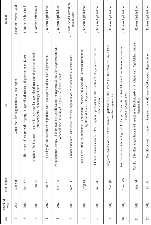
No.Published yearFirst authorTitleJournal 12000Seo CHSenial Macular Degeneration: A case reportJ Korean Oriental Med 22010Park KHThe results of Nation-wide registry of age-related macular degeneration in KoreaJ Korean Ophthalmol 32011Cho YJIntravitreal Ranibizumab therapy for neovascular age-related macular degeneration with a predominantly hemorrhagic lesionJ Korean Ophthalmol 42012Han SYQuaility of life assessment in patients with wet age-related macular degeneration. J Korean Ophthalmol 52012Cho DYPhotodynamic therapy of choroidal neovascularization in age-related macular degeneration with Verteporfin:An analysis of 10 years of clinical results J Korean Ophthalmol 62013Kim CGFactors associated with senile macular degeneration in elders within communitiesJ Korean Acad Community Health Nurs 72013Jang HJLong-Term Effect of Intravitreal Ranibizumab Injection on Choroidal Neovascularization in Age-Related Macular Degeneration J Korean Ophthalmol 82013Kim MSClinical manifestation of retinal pigment epithelial tear after treatment of age-related macular degeneration J Korean Ophthalmol 92013Park JHLong-term observation of retinal pigment epithelial tear after anti-VEGF treatment for age-related macular degeneration J Korean Ophthalmol 102013Choae WSRisk Factors for Retinal Pigment Epithelium Tears after Anti-VEGF Agent Injection in Age-Related Macular Degeneration J Korean Ophthalmol 112013Kim JMMacular Hole after Single Intravitreal Injection of Ranibizumab in a Patient with Age-Related Macular Degeneration J Korean Ophthalmol 122013Kil HKThe efficacy of Vaccinium Uliginosum for early age-related macular degeneration J Korean Ophthalmol
Table 2. Study of Age-related Macular Degeneration Searched in KISS and OASIS
정유진 외 2인 : 나이관련 황반변성에 대한 동서의학적 고찰
71 요법에는 라니비주맙, 베바시주맙, 페갑타민 등이 있
다. Ranibizumab(Lucentis)은 혈관내피세포성장인자 를 저해시키는 대표적인 항체로, 임상실험을 통해 삼 출성 나이관련 황반변성 환자의 1/4~1/3 정도에서 시 력이 2년 이상 안정되었다는 보고가 있다26,27). 또한 국내 연구에서 연령관련 황반변성 환자에게 유리체강 내 라니비주맙 주입술 시행 후 추적관찰하여 후향적 으로 조사하였을 때, 중심황반두께 감소28,29), 황반하 출혈의 감소29)가 나타나는 경과를 보였다. 대장암과 유방암 치료를 위해 만들어진 Bevacizumab(Avastin) 도 혈관내피세포성장인자를 억제하는 효과가 있어 나 이관련 황반변성 치료에 응용된다30). 그러나 주사치료 의 부작용은 국내외 논문에서 여러 차례 보고되고 있 다. 황반변성 환자에게 혈관내피성장인자 억제제 주 입술 후 망막색소상피파열32-34), 황반원공 발생35) 등의 부작용이 발생한 증례 보고가 있었다.
최근 쥐 실험 모델에서 anti-VEGF 주사의 장기치 료는 광수용체나 망막지지세포의 죽음을 초래한다는 연구 보고에서 주사치료법의 한계점이 보고되고 있으
며36,37), 건성 황반변성은 현재까지 뚜렷한 치료법이
없기 때문에, 최근 유전자 치료나 세포 이식 치료가 주목을 받고 있다. 세포 이식 치료법에는 ①세포 현탁 액에 직접 주입하는 방법과 ②RPE 단일층에 외과적 이식을 하는 방법 2종류가 있다31). 최근에는 망막에서 혈액공급을 통해 내부/외부 망막 장벽 역할을 하는 뮐 러아교세포의 연구가 활발히 진행되면서 뮐러아교줄 기세포를 이용한 세포이식치료도 연구되고 있다38). 건성 나이관련 황반변성은 현재까지 뚜렷한 치료법 이 없기 때문에 보존적 치료와 관리가 중요하다. 금 연, 금주, 적당한 신체활동, 비만 환자의 체중감량, 저 시력 재활훈련, 식이 보충제 섭취 등이 있다39). 건성 황반변성에 도움이 된다고 알려진 식이보충제로는 항 산화제, Zinc, Copper, Crocetin, Curcumin, 비타민 등이 있다. 보체(complement)의 과도한 활동으로 망 막색소상피세포에 축적물이 쌓이게 되면 나이관련 황 반변성이 발달하게 되는데, 최근 연구에서 3개월 가량
의 아연의 섭취로 전신 보체 활성화가 감소되고 AMD의 진행을 느리게 하였다는 보고가 있다40). 길 등41)의 연구에서는 초기 황반변성 환자에게 들쭉 추 출물을 복용하여 위약 대조군 비교를 하였을 때, 유의 하게 망막색소상피부터 광수용체의 두께가 얇아지는 것을 방지하고, lipofusin에 의한 산화효과를 억제하 며, 광수용체 위축 예방하는데 효과가 있어 황반변성 의 진행을 억제하는데 도움을 줄 것으로 보고하였다.
2. 나이관련 황반변성에 대한 한의학적 이해 황반변성을 한의학적으로 접근한 논문은 1례에 불 가하다. 서 등5)의 논문에서 연령관련 황반변성증이 있으며, 疲勞, 頭痛, 頭重, 眩暈, 上熱感, 飛蚊症, 心 悸, 不眠症이 있는 환자를 太陰人 燥熱證으로 진단하 여 2개월 가량 淸心蓮子湯加味를 처방하고 上三黃, 下 三皇, 上白, 立白을 자침하여 시력호전 및 임상증상이 호전된 1례를 보고한 바 있지만, 황반변성이 肝腎兩虛 와 관련된다는 기존 한의학적 이해와는 다른 관점에 서 황반변성을 치료한 논문이었다는 점에서 더욱 더 황반변성의 임상 및 기초에 대한 연구가 진행되어야 할 것으로 사료된다.
古代의 醫家들은 眼部를 外에서 內로 “胞瞼, 兩眥, 白睛, 黑睛, 瞳神”의 5개 부분으로 파악하고, 안구의 운동이 바퀴처럼 회전운동을 한다는 의미로 輪字를 써서 각각 “肉輪, 血輪, 氣輪, 風輪, 水輪”으로 구분하 면서 총칭하여 五輪이라고 하였다. 五輪學說의 이론 적 기초는 <靈樞·大惑論>의 “五臟六腑之精氣, 皆上 注于目而爲之精. 精之窠爲眼, 骨之精爲瞳子, 筋之精爲 黑眼, 血之精爲絡, 其窠氣之精爲白眼, 肌肉之精爲約 束, 裹結筋骨血氣之精而與脈幷爲系, 上屬于腦, 後出于 項中”이라 하여 후대의 의가들이 이 이론을 기초로 하 여 장기적인 임상을 통해 五輪學說을 창립하였다43). 망막은 瞳神 즉, 五輪 중 水輪에 해당하며, 水輪은 腎臟에 속하게 된다. 瞳神疾患은 腎과 膀胱의 기능 이 상과 관련된 것을 제외하고는 전신의 장부기능 실조 와 밀접한 관련이 있다고 하였다. 腎精이 충만하면 瞳
한방안이비인후피부과학회지 제28권 제3호(2015년 8월)
72
神이 淸榮하고 眼光이 光彩를 띠게 된다43).
또한, 한의학적으로 황반변성의 증상은 眼昏, 眼花 證 에 속하며 視瞻昏渺, 靑盲에 해당하는 질환이다5). 眼昏은 눈의 외견상의 변화를 초래하지 않으면서 자 각적으로 시력의 저하나 시각의 장애를 유발하는 증 상으로 內障질환에서 흔히 보이는 증상이고, 眼昏은 시력이 떨어짐으로 인해 자각적으로 시야가 맑지 않 음을 말한다3). <素問·臟氣法時論>에서는 “肝이 虛하 면 眼昏이 생긴다.”고 하였고, <靈樞>에서는 “氣가 虛 하면 눈이 밝지 못하다.”고 하였으며, <難經·20難>
에서는 “陰氣가 부족한 환자는 눈이 어둡게된다.”라고 하였으며, <入門·雜病分類·風類·眼>에서는 눈이 어두운 것은 열이 심하기 때문이라고 하였다42). 노인 에 있어 발생하는 眼昏은 대개 血氣가 衰弱하여 肝葉 이 薄하고 膽汁이 감소되어 神膏, 眞精이 제 기능을 하지 못해 발생한다43).
靑盲은 서서히 진행되어서 나중에는 黑白이나 명암 까지 감별하지 못하여 昏盲이 되는 內障질환이며, 망 막질환에서 당뇨병성망막증, 고혈압성망막증, 노인성 황반변성증이 있다. 대부분 虛症에서 肝腎虧損, 心脾 虛損 등으로 精血이 虛少되어서 또는 脾腎陽虛로 精 微가 화생되지 않아 눈을 溫養하지 못하여 神光이 소 실되어 발생된다. 情志抑鬱로 肝氣不舒하여서도 발생 될 수 있다44).
즉, 五臟虛損, 津液損傷, 血虛, 久病 등으로 陰虛하 게 되어 目을 營養하는 작용을 상실하면, 目의 시력기 능에 장애를 주게 되어 眼昏, 眼花, 眼盲의 증상이 생 기게 된다3). 이는 현대 의학에서 황반변성의 조적학 적 marker인 드루젠(Drusen)이 쌓이는 원인이 시세 포외절의 교대 증가, 망막색소상피의 기능이상, 유해 산소, 부르크막의 노화, 면역 저하 등이 있다9)고 한 것과 肝腎不足, 心脾陽虛로 精血이 虧損되어 氣血不濡 養, 目絡壅滯하여 視瞻昏渺가 발생한다42)는 것이 연관 된다. 또한, 최근 서양의학 연구에서 황반변성의 원인 을 유전자 이상으로 보고, 유전자 치료가 각광받고 있 는데, 이 또한 腎의 先天之精과 연관시켜 생각해 볼
수 있다.
건성 황반변성은 현재까지 알려진 치료법이 없으 며, 주사치료 및 세포이식 치료 역시 다양한 부작용이 발표되고 있어, 나이관련 황반변성의 한의학적 접근 을 통한 이해와 치료가 연구 되어야 할 필요성이 있 으며, 널리 응용될 수 있을 것으로 사료된다.
나이관련 황반변성은 老衰와 관련된 만성 眼病이 다. 眼昏, 眼花, 眼盲의 치료는 肝腎不足, 氣血兩虧의 補肝腎治療法이 위주가 된다. 肝腎兩虛와 脾氣虛弱이 그 근본원인이며, 국부 병리는 血熱妄行과 痰瘀水濕 과 관련이 있다. 滋補肝腎과 健脾益氣가 치료의 근본 이 되며, 凉血止血, 化痰祛濕, 活血散瘀가 치료의 標 가 된다. 肝腎虧虛할 경우에는 四物五子丸加減, 脾虛 氣弱에는 補中益氣湯加減, 陽虛火旺에는 知栢地黃湯 合生蒲黃湯加減을 활용할 수 있다43). 침치료는 각 혈 위의 효능과 주치에 따라 精明, 攢竹, 魚腰, 絲竹空, 四白, 瞳子髎 등을 활용하여 자침할 수 있다.
Ⅳ. 결 론
본 연구는 해외 및 국내에 발표된 나이관련 황반변 성에 대한 논문을 정리, 분석하여 나이관련 황반변성 에 대한 동·서양의학의 이해를 도모한 결과 다음과 같은 결과를 얻었다.
1. 나이관련 황반변성은 노화에 의해 황반의 세포와 혈관의 기능이 떨어져, 망막 아래에 노폐물들이 쌓 이게 되면서 망막색소상피세포가 위축되거나 신생 혈관이 생기는 변화를 일으켜, 중심시력의 손상을 유도하는 질환이다. 치료의 서양의학적 접근은 크 게 광역학치료, 주사치료, 병합치료가 있으며 최근 에는 유전자치료, 세포이식, 보존적 치료 등이 주 목받고 있다.
2. 한의학에서는 나이관련 황반변성을 노화의 연장선 으로 보아 肝腎虧損, 脾腎陽虛, 精血虛損의 범주에
정유진 외 2인 : 나이관련 황반변성에 대한 동서의학적 고찰
73 서 이해하였으며, 이에 관한 연구는 아직 미진하
여, 향후 황반변성 치료와 한의학적 이해에 대한 보다 체계적인 연구가 필요하다고 사료된다.
감사의 글
이 논문은 2015학년도 동의대학교 교내 연구비에 의해 연구되었음(2015AA038).
References
1. 3 Blindness diseases: diabetic retinopathy, macular degeneration, glaucoma. Health Josun. 2010;11:60.
2. Friedman DS, O’Colmain BJ, Munoz B, Tomany SC, McCarty C, de Jong PT, et al.
Prevalence of age-related macular degeneration in the United States. Arch Ophthalmol 2004;122:564-72.
3. Lee KH, Noh SS. The study of the etiological cause of blurred vision and blindness. Daejun Univ Institution of oriental medicine.
4(1);1995:1-25.
4. Chen YH, Bedell M, Zhang K. Age-related macular degeneration: genetic and environ- mental factors of disease. Mol Interv. 2010.
Oct;10(5):271-81.
5. Seo CH, Kwon JN, Kim YK. Senial Macular Degeneration. A case report. J Korean Oriental Med. 2000;21(4):260-3.
6. Park KH, Song SJ, Lee WK, Yoon HS, Koh HJ, Kim CG et al. The results of Nation-wide registry of age-related macular degeneration in Korea. J Korean Ophthalmol Soc. 2010;51(4) 516-23.
8. Bressler NM. Early detection and treatment neovascular age-related macular degeneration.
J Am Board Fam Pract. 2002(15):142-52.
9. Gehrs KM, Anderson DH, Johnson LV, Hageman GS. Age-related macular degeneration- emerging pathogenetic and therapeutic concepts. Ann Med. 2006;38:450-71.
10. Smith W. Assink J, Klein R, et al. Risk factors for age-related macular degeneration:
pooled findings from three continetns. J Ophthalmology. 2001;108:697-704.
11. Swaroop A, Chew EY, Rickman CB, Abecasis GR. Unravelling a multifactorial late-onset disease: from genetic susceptibility to disease mechanisnms for age-related macular degeneration. Annu Rev Genomics Hum Genet. 2009;10:19-43.
12. Hirvela H, Luukinen H, Laara E, Sc L, Laatikainen L. Risk factors of age-related maculopathy in a population 70 years of age or older. J Ophthalmology. 1996;103:871-7.
13. Klein R, Klein BE, Moss SE. Relation of smoking to the incidence of age-related maculopathy–The Beaver Dam Eye Study.
1998;147:103-10.
14. Seddon JM, Rosner B, Sperduto RD, Yannuzzi L, Haller JA, Blair NP, et al.
Dietary fat and risk for advanced age-related macular degeneration. Arch Ophthalmol.
2001;119:1191-9.
15. Montezuma SR, Sobrin L, Seddon JM. Review of genetics in age related macular degeneration. Semin Ophthalmol. 2007;22:
229-40.
16. Klein R, Myers CE, Lee KE, Gangnon RE, Sivakumaran TA, Iyengar SK, et al. Small
한방안이비인후피부과학회지 제28권 제3호(2015년 8월)
74
Drusen and Age-Related Macular Degeneration:The Beaver Dam Eye Study. J Clin Med. 2015 Mar;4(3):424-440.
17. Han SY, Bae JH, Song SJ. Quaility of life assessment in patients with wet age-related macular degeneration. J Korean Ophthalmol Soc. 2012;53(4):528-35.
18. Jung JW, Moon YS. Depression and anxiety in Korean patients with age-related macular degeneration. J Korean Opthalmol Soc.
2012;53(6):792-800.
19. Göbel AP, Fleckenstein M, Schmitz- Valckenberg S, Brinkmann CK, Holz FG.
Imaging geographic atrophy in age-related macular degeneration. Ophthalmologica.
2011;226:182-90.
20. Lindblad AS, Lloyd PC, Clemons TE, Gensler GR, Ferris FL 3rd, Klein ML, et al. Change in area of geographic atrophy in the age-related eye disease study:AREDS report No 26. Arch Ophthalmol. 2009;127:1168-74.
21. Schmitz-Valckenberg S, Fleckenstein M, Göbel AP, Sehmi K, Fitzke FW, Holz FG, et al.
Evaluation of autofluorescence imaging with the scanning laser ophthalmoscope and the fundus camera in age-related geographic atrophy. Am J Ophthalmol. 2008 Aug;146 (2):183-92.
22. Haimovici R, Kramer M. Miller JW, et al.
Localization of lipoprotein-delivered benzo- porphyrin derivatiove in the rabbit eye. Curr Eye Res. 1997;16:83-90.
23. Schmidt-Erfurth U, Flotte TJ, Gragoudas ES, et al. Benzoporphyrin-lipoprotein-mediated photodestruction of intraocular tumors Exp Eye Res. 1996;62:1-10.
24. Miller H, Miller B. Photodynamic theraphy of subretinal neovascularization in the monkey eye. Arch Ophthalmol. 1993;111:855-60.
25. Cho DY, Bae SH, Han JR, Kim HK, Nam WH. Photodynamic therapy of choroidal neovascularization in age-related macular degeneration with Verteporfin:An analysis of 10 years of clinical results. J Korean Ophthalmol Soc. 2012;53(1)59-67.
26. Brown DM, Kaiser PK, Michels M, Soubrane G, Heier JS, Kim RY, et al. Ranibizumab versus verteporfin for neovascular age-related macular degeneration. N Engl J Med. 2006 Oct 5;355(14):1432-44.
27. Rosenfeld PJ, Brown DM, Heier JS, Boyer DS, Kaiser PK, Chung CY, et al.
Ranibizumab for neovascular age-related macular degeneration. N Engl J Med. 2006 Oct 5;355(14):1419-31.
28. Jang HJ, Song SJ, Bae JH. Long-Term Effect of Intravitreal Ranibizumab Injection on Choroidal Neovascularization in Age-Related Macular Degeneration. J Korean Ophthalmol Soc. 2013;54(9):1359-64.
29. Cho YJ, Park SP. Intravitreal Ranibizumab therapy for neovascular age-related macular degeneration with a predominantly hemorrhagic lesion. J Korean Ophthalmol Soc. 2011;52(7):838-45.
30. Avery RL, Pieramici DJ, Rabena MD, Castellarin AA, Nasir MA, Giust MJ.
Intravitreal bevacizumab (Avastin) for neovascular age-related macular degeneration.
Ophthalmology. 2006 Mar;113(3):363-372.
31. David L, Lincoln V, Dennis O. Cellular models and therapies for age-related macular
정유진 외 2인 : 나이관련 황반변성에 대한 동서의학적 고찰
75 degeneration. Dis Model Mech. 2015 May
1;8(5):421-7.
32. Kim MS, Chae JB. Clinical manifestation of retinal pigment epithelial tear after treatment of age-related macular degeneration. J Korean Ophthalmol Soc. 2013;54(10):1540-5.
33. Park JH, Choae WS, Yoon HS. Long-term observation of retinal pigment epithelial tear after anti-VEGF treatment for age-related macular degeneration. J Korean Ophthalmol Soc. 2013;55(9):1340-6.
34. Choae WS, Park JH, Lee WS, Kim SW, Yoon HS. Risk Factors for Retinal Pigment Epithelium Tears after Anti-VEGF Agent Injection in Age-Related Macular Degene- ration. J Korean Ophthalmol Soc. 2013;45 (10:1546-53.
35. Kim JM, Jang JW, Kyung SE, Chang MH.
Macular Hole after Single Intravitreal Injection of Ranibizumab in a Patient with Age-Related Macular Degeneration. J Korean Ophthalmol Soc. 2013;54(7):1130-4.
36. Ford KM, Saint-Geniez M, Walshe TE, D'Amore PA. Expression and role of VEGF-A in the ciliary body. Invest. Ophthamol. Vis.
Sci. 2012;53:7520-7.
37. Saint-Geniez M, Maharaj ASR, Walshe TE, Tucker BA, Sekiyama E, Kurihara T, et al.
Endogenous VEGF is required for visual function: evidence for a survival role on Müuller cells and photoreceptors. PLoS ONE.
2008;3(11):e3554.
38. Kim SY. Retinal phagocytes in age-related macular degeneration. Macrophage (Houst).
2015;2(1).
39. Buschini E, Fea AM, Lavia CA, Nassisi M,
Pignata G, Zola M, Grignolo FM. Recent developments in the management of dry age-related macular degeneration. Clin Ophthalmol. 2015 Apr 1;9:563-74.
40. Smailhodzic D, van Asten F, Blom AM, Mohlin FC, den Hollander AI, van de Ven JP, et al. Zinc supplementation inhibits complement activation in age-related macular degeneration. PLoS One. 2014 Nov 13;9(11).
41. Kil HK, Song YM, Chun KI. The efficacy of Vaccinium Uliginosum for early age-related macular degeneration. J Korean Ophthalmo.
2013;54(8):1255-60.
42. Heo J. Donguibogam. Bubinmunhwasa.
2009:635.
43. Ko WS, Kwon K, Kim KJ, Kim KS, Kim YB, Kim JH, et al. Korean Medicine Ophthalmology. 2015:40-41,62,199.
44. Noh SS. Korean Medicine Ophthalmology, Otolaryngology, dermatology. Seoul Inshuaesa.
2011:339,346.