연령, 건강 상태, 활동 정도 및 병변의 크기와 위치, 동반된 퇴행성 변화 등이 고려되어야 한다.
Steadman 등2)이 무릎에서 처음으로 골수 자극술에 대하여 언급 을 하였다. 이는 연골병변을 제거한 이후 노출된 연골하 골을 송곳 (awl)으로 다발성으로 천공하는 방법이다. 이는 병변부 연골하 판의 재혈관화를 통한 중간엽 줄기세포(mesenchymal stem cell)의 침 윤으로 섬유 연골의 분화를 촉진시켜 주는 술식이다. 골수 자극술은 다른 치료들에 비해 안전하고 간단하며 그 결과에서도 우월성을 보 이고 있어 현재 가장 많이 사용되는 술식이다. 그러나 이러한 장점에 도 불구하고 증거 기반 관점에서 보면 등급 4의 논문 자료들로만 구 성되어 있다는 단점도 가지고 있는 술식이다. 또한 장기 비교 결과가 없다는 단점이 있으며 150 mm2 이하의 병변에 국한되어 쓸 수밖에 없다는 한계점을 가지고 있다.3,4) 이에 이 술식에 관한 장점 및 단점 그리고 치료의 적절성 등에 대해 알아보고자 한다.
서 론
거골의 골연골병변(osteochondral lesion of the talus)은 이전 에 박리성 골연골염(osteochondritis dissecans), 경연골 거골 골절 (transchondral talus fracture), 거골 골연골 골절(osteochondral talus fracture) 등으로 불리기도 했었다. 족관절의 이런 관절 내 골 연골 유리체 병변(loose osteochondral fragment)을 처음 보고한 사람은 Alexander Monro였으며 외상에 의해 발생한 것으로 생각 하였다.1) 골연골병변의 치료는 다양한 인자에 의해 결정되며 환자의
This is an Open Access article distributed under the terms of the Creative Commons Attribution Non-Commercial License (http://creativecommons.org/licenses/
CCCopyright 2020 Korean Foot and Ankle Society. All rights reserved. ⓒ
Osteochondral lesion of the talus (OLT) is a broad term used to describe an injury or abnormality of the talar articular cartilage and adjacent bone. Various terms are used to describe this clinical entity, including osteochondritis dissecans, osteochondral fractures, and osteochondral defects. Several treatment options are available; the choice of treatment is based on the type and size of the defect and the treating clinician’s preference. Arthroscopic microfracture (a bone marrow stimulation technique) is a common and effective surgical strategy in patients with small lesions or in those in whom non-operative treatment has failed. This study had the following aims: 1) to review the historical background, etiology, and classification systems of OLT; 2) to describe a systematic approach to arthroscopic bone marrow stimulation for OLT; and 3) to determine the characteristics that are useful for assessing osteochondral lesions, including age, size, type (chondral, subchondral, cystic), stability, displacement, location, and containment of the lesion.
Key Words: Osteochondral lesion of the talus, Microfracture, Foot and ankle, Cartilage
인제대학교 부산백병원 정형외과, *좋은삼선병원 정형외과Operative Treatment of Osteochondral Lesion of the Talus:
Arthroscopic Bone Marrow Stimulation (Multiple Drilling or Microfracture)
Heui-Chul Gwak, Il-soo Eun*
Department of Orthopedic Surgery, Inje University Busan Paik Hospital,
*Department of Orthopedic Surgery, Good Samsun Hospital, Busan, Korea
Received April 20, 2020 Revised May 25, 2020 Accepted May 26, 2020 Corresponding Author: Il-soo Eun
Department of Orthopedic Surgery, Good Samsun Hospital, 326 Gaya-daero, Sasang-gu, Busan 47007, Korea
Tel: 82-51-322-0900, Fax: 82-51-323-3308, E-mail: dreun7@hanmail.net ORCID: https://orcid.org/0000-0001-5863-9729
Financial support: None.
Conflict of interest: None.
www.jkfas.org
본 론
1. 병인
거골의 골연골병변에 대한 분류는 Berndt와 Harty5)에 의해 처 음 보고되었다. 당시에는 발생 원인이 명확하지 않아 경연골 골절 로 지칭하였으며 연골하 골의 국소적 병변으로 인한 골관절염의 전 구 증상으로 이해되었다. 이후 연골하 골의 허혈성 변화로 인한 연골 의 국소적 결손이 주요 병인으로 알려졌다.6,7) 거골 골연골병변의 가 장 흔한 원인은 외상이며 족근 관절 염좌 이후 자주 발생한다. 1955 년에 Bosien 등8)은 전체 족근 관절 염좌의 6.5%에서 골연골병변이 관찰되었으며 대부분 10대부터 30대에서 발생한다고 보고하였다.
하지만 최근 스포츠 손상이 증가함에 따라 거골의 골연골병변의 유 병률도 크게 증가하고 있으며 발생 연령대도 더 다양해지는 추세이 다.9,10)
2. 분류
거골 골연골병변의 특징은 크게 여섯 가지로 나누어서 분류할 수
있으며 이는 병변의 형태, 병변의 안정성, 전위 여부, 위치, 병변의 크기 등으로 구성되어 있다(Table 1). 각각에 따른 예후에 차이가 있 으므로 이 또한 중요한 분류의 기준으로 반드시 유념해야 할 필요가 있다. 1959년 Berndt와 Harty5)는 단순 방사선 소견에 따라 병변의 정도를 4단계로 분류하였으며 이 분류법은 지금까지도 이용되고 있 다(Table 2, Fig. 1). 하지만 거골의 골연골병변이 단순 방사선 소견 에서 나타나지 않는 경우가 많고 병변의 단계를 정확히 구분하기 어 려우며 영상 검사의 발달로 인해 그 유용성은 감소되었다. 따라서 컴 퓨터 단층촬영(computed tomography, CT), 자기공명영상(mag- netic resonance imaging, MRI) 및 관절경적 소견에 따라 분류할 수 있다(Table 3).
3. 비수술적 치료
만성적 거골 골연골병변이나 만성적 병변에 대한 급성 악화의 초 기 치료는 이들 중 대부분이 영상 촬영에 의해 우연히 발견되기 때문 에 비수술적 치료로 시작하게 된다. 전위를 동반한 급성 골연골 골절 은 비수술적 치료에 대한 금기증이다. 비수술적 치료에는 석고나 미 리 제작된 walking boots, 비스테로이드 항염증제(non-steroidal anti-inflammatory drugs) 약물치료, 부목고정, corticosteroid 주 사, 활동의 제한이 포함된다.
Table 1.
Table 1. Characteristics of Osteochondral Lesion of Talus 1. Type of lesion
A. Chondral (cartilage only)
B. Chondral/subchondral (cartilage and underlying bone) C. Subchondral (intact overlying cartilage)
D. Cystic (>5 nm deep) 2. Stability of lesion
A. Stable B. Unstable
3. Displacement of lesion A. Displaced B. Non displaced 4. Location
A. Medial (anterior, central, or posterior) B. Lateral (anterior, central, or posterior) C. Central (anterior, central, or posterior)
Table 2.
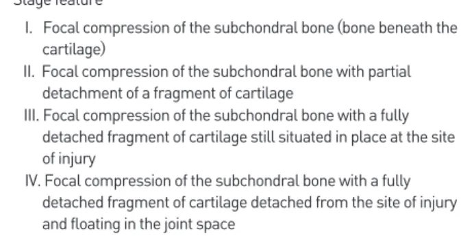
Table 2. Berndt and Harty Classification System Stage feature
I. Focal compression of the subchondral bone (bone beneath the cartilage)
II. Focal compression of the subchondral bone with partial detachment of a fragment of cartilage
III. Focal compression of the subchondral bone with a fully detached fragment of cartilage still situated in place at the site of injury
IV. Focal compression of the subchondral bone with a fully detached fragment of cartilage detached from the site of injury and floating in the joint space
Figure 1.
Figure 1. Berndt and Harty classification system. Data from the article of Berndt et al. (J Bone Joint Surg Am. 1959;41:988-1020).
5)Normal Stage I Stage II Stage III Stage IV
4. 수술적 치료
골수 자극 치료법은 거골의 골연골병변의 1차 치료임에 대부분의 저자들이 동의하고 있다. 이는 기술적으로 쉬우며 비용 면에서 효과 적이고 최소한의 침습적 치료인 동시에 술 후 합병증이 낮으며 술 후 통증도 작다는 이점이 있다.6,7,9,10-12) Zengerink 등13)은 골수 자극 치 료법의 임상적인 성공률을 85%로 보고하였고 이에 따라 보존적 치 료에 실패한 거골 골연골병변에 유용한 치료로 생각된다.
1) 관절경적 다발성 천공
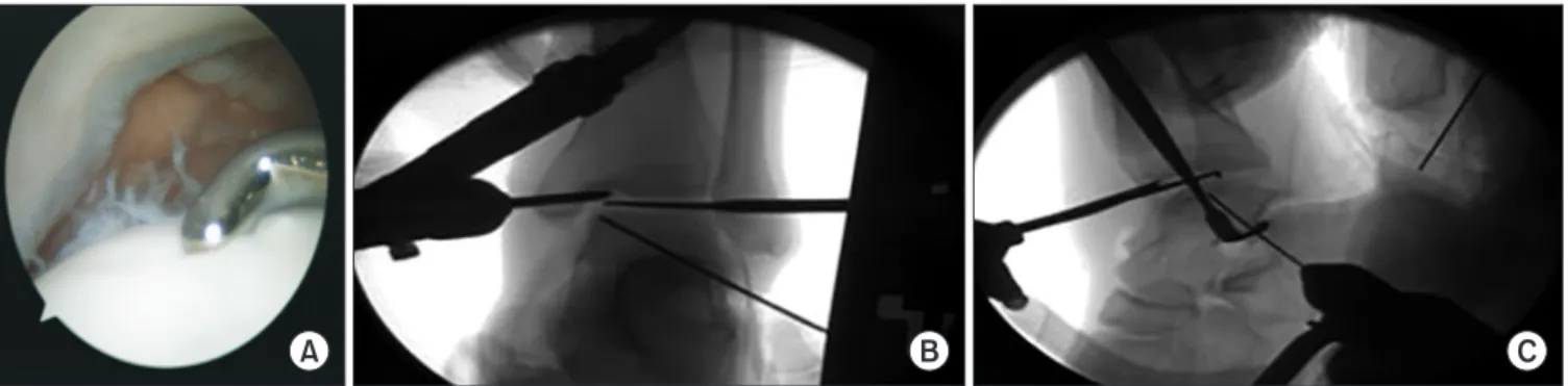
관절경적 역행성 천공술은 연골 손상이 없는 연골하 골 병변의 치 료에 유용한 술식이다.11) 역행성 천공술은 최초 관혈적으로 시행되 었으나 최근 관절경을 이용한 보고가 주를 이루고 있다. Kono 등12) 은 거골의 골연골병변 환자 30명 중 11예를 대상으로 역행성 천공 술을, 19예를 대상으로 경과 천공술(transmalleolar drilling)을 시 행하였고 1년 후 이차 관절경 검사상 역행성 천공술에서의 결과가 더 좋았다고 보고하였다. Anders 등14)은 영상장치하에서 역행성 천
시행 시 병변의 감압과 함께 연골하 골의 괴사 제거 및 연골의 유지 를 위해 연골하 낭종이나 결손 부위에 골이식 등 구조적인 지지가 필 요할 것으로 생각되며, 연골하 낭종에 대한 변연절제술 및 괴사된 골 조직을 제거하였을 때, 공간에 대한 자가 해면골 이식을 시행하는 것 이 바람직한 치료일 것으로 생각된다(Fig. 2). 이와는 달리 관절경을 통한 직접적인 천공술에 대해서도 다양한 연구가 보고되었다. 특히 Ferkel 등15)은 장기 추시를 통한 결과를 보기 위해 거골 골연골병변 환자 50명을 대상으로 관절경적 치료를 시행하였으며 71개월의 장 기 추시 결과에서 임상적으로 우수한 결과를 보고하였다.
2) 관절경적 미세 골절술
골수 자극법은 다발성 천공이나 미세 골절술이 있고 1차적 치료 로 고려된다. 이 술기는 관절경을 통해 시행되며 거골 골연골병변을 안정적인 연골 경계가 될 때까지 제거하는 데 초점이 맞춰져 있다.
Steadman 등2)이 무릎에서 처음으로 골수 자극술에 대하여 언급을 하였다. 이는 연골병변을 제거한 이후 노출된 연골하 골을 송곳으로
nondisplaced fragment
III. Completely detached fragment without displacement
III. Nondisplaced lesion with radiolucency
3. Detached but undisplaced fragment
D. Flap present or bone exposed
IV. Detached and displaced
fragment IV. Displaced fragment 4. Detached and displaced
fragment E. Loose, nondisplaced fragment
V. OLT with subchondral cyst 5. Subchondral cyst formation F. Displaced fragment MRI: magnetic resonance imaging.
Figure 2.
Figure 2. (A-C) Retrograde drilling.
A B C
www.jkfas.org
진시켜 주는 술식이다. 그러나 아쉽게도 증거 기반에 의한 등급 1에해당하는 보고는 아직까지 없는 것이 현실이다. 환자들의 다양한 케 이스를 바탕으로 한 보고들이 주를 이루고 있으며 이에 대해서 아주 다양한 결과들이 보고되고 있다. Lee 등16)은 35예의 환자에서 관절 경하 미세 골절술을 시행하였으며 89%에서 양호한 결과를 보고하 였고 예후 인자로 증상의 지속기간이 오래될수록 결과에 영향을 주 었다고 보고하였다. 장기 예후에 대한 정확한 예측은 불가능하지만 Ferkel 등15)은 평균 71개월의 추시 관찰에서 72%에서의 양호한 결 과를 보고하였고 van Bergen 등17)은 평균 12년(8~20년)의 추시 관 찰에서 78%에서의 양호한 결과를 보고하였다. Polat 등18)이 82명의 환자에서 시행한 평균 121개월 추시 연구에서는 71.6%에서 양호한 결과를 보였고 4기 관절염으로의 악화 소견은 없었으며 32.9%에서 한 단계 더 진행한 관절염이 관찰되었으므로 거골 골연골병변의 좋 은 치료법이라고 주장하였다. 또한 거골 골연골병변에 대한 치료에 있어서 임상 결과에 영향을 미치는 예후 인자들이 있는데 이를 정리 하면 다음과 같다.
3) 예후에 영향을 미치는 인자 (1) 나이(age)
거골 골연골병변의 관절경적 치료 후 나이와 임상학적 결과 사이 의 연관성에 대한 많은 연구가 발표되었다. Cuttica 등19)은 130명의 환자에 대해 후향적으로 평가하여 나이는 임상적 결과에 영향을 미 친다고 하였으며 33세까지 나이가 증가할수록 위험도가 증가한다고 발표하였다. Deol 등20)은 20세 이하의 환자에서 좋은 예후를 보인 다고 발표했다. 또한 이전 연구에서 슬관절에서 미세 골절술을 동반 한 관절경적 치료 후 나이가 임상적 결과와 중요한 차이를 보인다고 증명하였다.21) 반면 다른 연구에서는 서로 다른 나이의 그룹 사이에 서 연관성은 없었으며 환자의 수가 적기 때문에 통계적 유의성을 제 시하는 데 증거가 부족하다고 하였다.22) 또한 Choi 등23)은 170명의 환자를 6개의 그룹으로 나누어 비교를 진행한 연구 결과에서 나이에 따른 임상적 결과나 환자의 만족도에는 차이가 없음을 보고하였다.
이에 대해서는 조금 더 연구가 필요할 것으로 생각된다.
(2) 결손 크기(defect size)
관절경적 치료 이후 악화될 예측인자는 연골 결손의 크기뿐 아 니라 동반된 병소의 심각성(severity)이다.24) 많은 이전의 조사자들 은 결손 크기와 임상학적 결과 사이의 연관성에 대해 언급해 왔다.
Christensen 등25)은 18구의 사체(fresh cadaver)에서 압력 감지 촬 영을 이용하여 발목관절의 특징에 대해 연구하였다. 그 결과 병소의 크기가 발목에서 접촉 stress를 변화시킨다고 하였으며 통계적으로 7.5 mm×15.0 mm보다 큰 병소일 경우에 유의하다고 제시하였다.
그들은 이 결과가 병소의 크기가 연골 결손 환자에서 예후 인자로 제 시될 수 있다고 주장하였다. Choi 등26)은 150 mm2 크기를 기준으 로 결과에 있어서 차이를 보였다고 하였으며 이 크기를 MRI에서 미 리 측정하여 치료 계획을 세우는 것을 추천하였다. Giannini 등27)은 150 mm2 이하의 병소는 관절경적 치료가 필요한 반면, 150 mm2 이상의 병소를 지니거나 이전 수술에서 실패한 경우에는 자가골연골 이식술(osteochondral autograft transplantation) 혹은 자가연골 세포 이식술(autologus chondrocyte implantation)의 치료가 반 드시 필요하다고 하였다.
(3) 관절하 낭종(subchondral cyst)
새로운 진단적 도구들의 개발과 함께 분류 체계는 CT와 MRI를 사용하여 새롭게 만들어졌다. Anderson 등,28) Hepple 등29)은 거골 연골하 낭종을 stage IIA와 V를 포함한 새로운 분류 체계를 고안하 였고 이러한 낭종성 병변들의 병리적인 이론을 기술하였다. 골과 연 골의 결손부를 통하여 윤활막이나 윤활액이 침범한다는 것으로 이 이론에 따르면 연골하 판을 침범한 연골 골절은 윤활액의 강한 유입 을 통한 압력 형성으로 낭종 형성에 기인한다고 한다. 또 다른 이론 은 골수강내 결합 조직의 점액성 퇴행에 대한 것인데 아마도 국소적 허혈이나 무혈성 괴사가 선행되었을 것이다. Ogilvie-Harris와 Sar- rosa30)는 연골하 낭종의 유무가 예후에 영향을 줄 수 있다고 하였다.
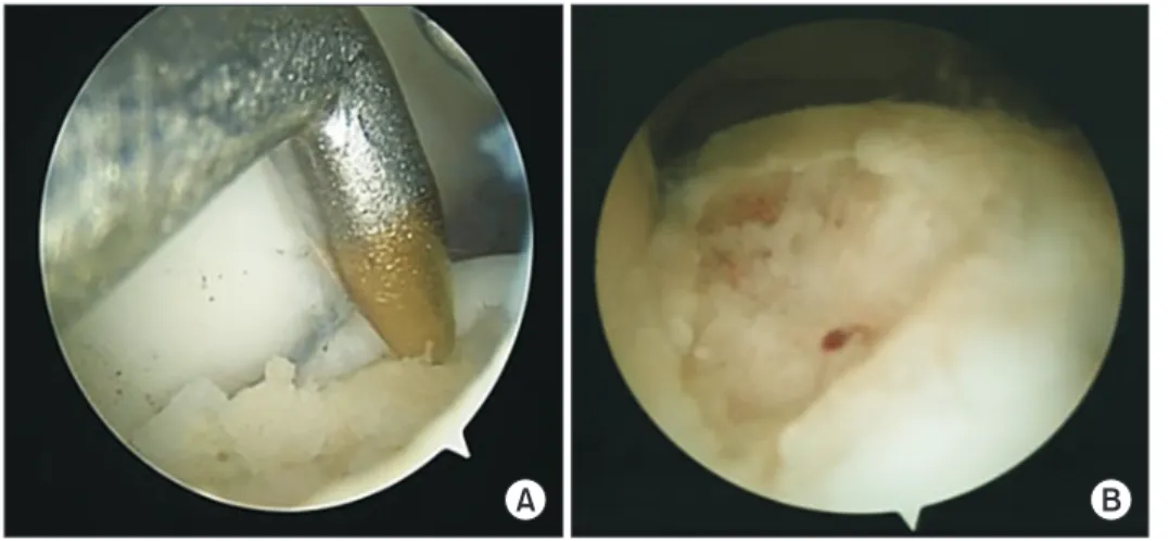
Figure 3.
Figure 3. (A) Arthroscopic photographs showing that microfractures were performed of the osteochondral lesion of the talus. (B) Adequate bleeding occurred from the micro- fracture holes.
A B
공술과 자가골 이식술을 시행하였다. 평균 32개월(2~8년) 추시하였 으며 두 군 모두에서 수술 후 임상적 결과는 호전되었고 두 군 간의 차이는 없었으나 골이식술을 시행한 군에서 운동으로의 복귀가 훨씬 더 늦었음을 보고하였다. 또한 Hu 등33)은 17명을 대상으로 10 mm 이상의 낭종에 대해 자가 장골 이식을 시행하였고 임상적 결과의 호 전을 보고하였으며 이 중 13명에 있어서는 2차 관절경 검사에서 연 골의 회복이 양호함을 보고하였다. 큰 낭종이 있을 경우 효과적인 치 료법이 될 것으로 생각된다.
(4) 위치(location)
거골 골연골병변의 위치에 대한 상대적인 예후 인자로서의 중요 성에 대해서는 논란이 많다. 거골 천장부에서의 안정형(contain- ment) 정도에 의한 거골 골연골병변의 구분의 유용성과 타당성에 대해서는 지속적으로 논란이 되고 있으나 Cuttica 등19)은 안정형 결 손(contained defect)과 불안정 결손(uncontained defect)에 따른 임상적 결과는 명백히 차이가 있다고 보고하였다. 연골 결손부의 불 안정하고 큰 결손부에서 기능적으로 낮은 결과를 보인다고 하였다.
Schimmer 등34)은 내측 거골 골연골병변을 가진 환자들에서 외측 병변을 가진 환자와 비교 시 더 좋은 결과를 보인다고 제시한 바 있 다. Saxena와 Eakin32)은 전외측 병변을 가진 환자들이 일상 생활로 의 복귀 시간이 짧고 AOFAS 점수가 더 높았다 보고하고 있다.
(5) 동반 병변(combined lesion)
Lee 등35)은 420예의 환자에 대한 골수 자극 치료법을 시행하였 는데 이 중 만성 족관절 불안정성이 동반된 74예의 환자와 단독 병 변을 가진 148예의 환자를 비교하였을 때 불안정성이 동반된 환자 에게서 연골 결손부위가 더 컸으며 외측부 연골병변이 더 자주 관찰 되었고, 경골의 내과 부분에 연골병변이 더 자주 관찰되었으며, 수 술 결과도 불안정성이 동반된 군에서 안 좋은 것으로 보고하였다. 또 한 Becher 등36)은 골연골병변에서 연골병변보다 더 호전이 되는 것 으로 보고하였고 자연 치유력도 더 좋다고 하였다. 반면에 Park과 Lee37)는 104예의 환자를 대상으로 두 군 간에 임상적인 차이가 없음 을 보고하였다.
거골의 골연골병변에서의 동통의 원인에 대해서는 여러 주장이
극 치료를 시행할 때 연골하 골과 연골하 판의 중요성에 대한 연구가 있다. 연골하 골과 연골하 판이 연골의 재생과 항상성(homeostasis) 에 중요한 역할을 하며 문제 발생 시 연골 조직의 질과 수명을 단축 시킬 수 있다.39,40) Shimozono 등41)은 42명의 환자에서 관절경적 미세 천공술을 시행한 후 MRI를 이용하여 관찰을 시행하였다. 미세 천공술 후 4~6년까지 연골하 판의 상태는 회복되지 않았고 2년 추 시보다 4~6년 추시에서 연골하 골과 연골하 낭의 상태는 더 악화되 었다. 또한 연골하 골의 상태는 Foot and Ankle Outcome Score (FAOS)와 상관관계를 보여 연골하 골과 연골하 판의 상태가 임상적 결과에 영향을 미칠 수 있다고 하였다. Reilingh 등42)은 58명의 환자 에서 관절경적 골수 자극 치료를 시행하여 술 후 2주, 1년째에 CT를 비교해서 평가하였다. 술 후 2주째에는 병변의 크기는 증가하였으나 1년째에는 크기가 감소하였다고 보고하였다. 단지 14명에서 연골병 변이 완전히 회복되었으나 그 크기와 임상적 결과와의 연관성은 없 었다고 하였다. 또한 second-look 관절경술은 연골 손상이 어느 정 도 복구되어 있는지를 확인할 수 있는 방법으로 Lee 등43)은 미세 골 절술 후 12개월에 시행한 second-look 관절경 소견상 약 40%에서 불완전한 연골 재생 상태를 보였으나 대부분의 환자에서 양호한 임 상 결과를 보였다고 보고하였다.
(6) 부가 치료(additional treatment)
골수 자극술 시행 후 생물학적 제제의 사용이 시도되고 있다. 골 수 흡인 농축술(bone marrow aspirate concentrate, BMAC)에는 성장 인자(growth factor)와 사이토카인(cytokines)이 있어 연골세 포의 분화를 유도하는 능력이 있으며 장골 능(iliac crest), 근위 또는 원위 경골, 종골에서 골수를 추출한 이후 원심 분리하여 추출한다.44) Fortier 등45)은 12예의 말을 대상으로 골수 자극술 단독군과 골수 자 극술 후 BMAC을 시행한 군 간의 비교 연구를 시행하였고 BMAC을 시행한 군에서 미세 천공술 단독군보다 급성의 전층(full thickness) 연골 결손에서의 치유가 우수하다고 보고하였다.
(7) 골수 자극술 치료 실패 이후의 치료
관절경적 치료 실패 이후 재수술 시 골수 자극술을 재시행하여 임상적 호전을 보인 결과는 여러 연구에서 보고되었다.46.47) 그러나
www.jkfas.org
인다고 하였다. 그러므로 골수 자극술을 다시 시행함에 있어서는 신중을 기해야 한다.
결 론
증상이 있는 거골 골연골병변의 치료는 어렵고 한계가 있다. 그 이 유는 발목관절에의 접근이 제한되어 있으며 관절 연골의 재생력이 낮기 때문이다. 보존적 치료에 호전이 없는 경우에는 수술적 가료를 시행하여야 하며 1차 치료 시 150 mm2 이하의 병변에서는 골수 자 극술만으로도 만족할 만한 결과를 얻을 수 있다. 이 치료가 실패할 경우 다른 대안 수술법에 대해서도 인지하고 있어야 할 것이다. 또한 술자는 실패 시의 원인과 함께 거골 골연골병변의 관절경적 치료 결 과에 영향을 줄 수 있는 다른 여러 인자들에 대해서도 이해해야 한 다. 위험 인자들의 존재는 술자로 하여금 새로운 치료 방법을 모색하 고 환자 상담 방법을 변화하는 데에 도움을 줄 것이다. 이 부분에 대 해서는 앞으로 더 많은 연구가 필요하며 지속적인 관심을 가져야 할 것이다.
ORCID
Heui-Chul Gwak, https://orcid.org/0000-0003-1062-0580
REFERENCES