• 검색 결과가 없습니다.

Inhibitory effects of fenbendazole, an anthelmintics, on lipopolysaccharide-activated mouse bone marrow cells

N/A
N/A
Protected

Academic year: 2021

Share "Inhibitory effects of fenbendazole, an anthelmintics, on lipopolysaccharide-activated mouse bone marrow cells"

Copied!
7
0
0

로드 중.... (전체 텍스트 보기)

전체 글

(1)

2021 The Korean Society of Veterinary Science.

This is an open-access article distributed under the terms of the Creative Commons Attribution Non-Commercial license (http://creativecommons.

org/licenses/by-nc/4.0/), which permits unrestrict- ed non-commercial use, distribution, and repro- duction in any medium, provided the original work is properly cited.

서론

Fenbendazole ( FBZ )은 수의에서 유용하게 쓰이는 구충제 중 하나이다[ 1 ]. FBZ 은 실 험용 설치류, 반려동물, 산업동물에서 회충, 십이지장충, 편충 등 내부 기생충 감염의 예방 및 치료제로 허가 받아 사용되고 있다[ 2 , 3 ]. 구충 작용기전으로는 기생충의 세포 분열 과정 중 미세소관의 형성을 방해하여 효과를 나타내는 것으로 알려져 있다[ 4 , 5 ].

최근 FBZ 이 항암효과가 있다는 보고[ 6 ]와 일부 임상사례를 시작으로 커뮤니티, 인터 넷을 통해 FBZ 을 임의로 복용하는 사례가 증가하였다. FBZ 은 인간 암세포의 미세소관 에 친화성을 나타내고 micromolar 농도에서 항암효과를 보였으며, 새로운 항암기전으 로 GLUT transporter hexokinase 의 발현 억제로 glucose uptake 를 효과적으로 억제

Fenbendazole의 억제 효과

박서로1, 주홍구1,2*

1

제주대학교 수의과대학 수의약리학실

2

제주대학교 수의과학연구소

Inhibitory effects of fenbendazole, an anthelmintics, on lipopolysaccharide- activated mouse bone marrow cells

Seo-Ro Park 1 , Hong-Gu Joo 1,2*

1

Laboratory of Veterinary Pharmacology, College of Veterinary Medicine, Jeju National University, Jeju 63243, Korea

2

Veterinary Medical Research Institute, Jeju National University, Jeju 63243, Korea pISSN 2466-1384 · eISSN 2466-1392

Korean J Vet Res 2021;61(3):e22 https://doi.org/10.14405/kjvr.2021.61.e22

*Corresponding author:

Hong-Gu Joo

Laboratory of Veterinary Pharmacology, College of Veterinary Medicine, Jeju National University, 102 Jejudaehak-ro, Jeju 63243, Korea

Tel: +82-64-754-3379 Fax: +82-64-756-3354 E-mail:[email protected] ORCID:

https://orcid.org/0000-0002-1505-8761 Conflict of interest:

The authors declare no conflict of interest.

Received: June 7, 2021 Accepted: July 22, 2021

Original Article

Fenbendazole (FBZ) is a commonly used anthelmintics in veterinary medicine that has recently been found to have anticancer effects in humans. On the other hand, few studies have examined the anti-inflammatory effects of FBZ, and its mechanism is unknown. In this study, mouse bone marrow cells (BMs) were treated with lipopoly- saccharide (LPS), a representative inflammation-inducing substance, to generate a sit- uation similar to osteomyelitis in vitro. The effect of FBZ on inflammatory BMs was examined by measuring the metabolic activity, surface marker expression, cell nuclear morphology, and mitochondrial membrane potential (MMP) of BMs. FBZ decreased the metabolic activity and MMP of LPS-treated BMs. Annexin V-fluorescein isothiocy- anate/propidium iodide staining and Hoechst 33342 staining showed that FBZ re- duced the number of viable cells and induced the cell death of inflammatory BMs. In addition, FBZ reduced the proportion of granulocytes more than B lymphocytes in LPS-treated BMs. Overall, FBZ induces cell death by destabilizing the MMP of LPS-in- duced inflammatory BMs. In addition to anthelmintic and anticancer agent, FBZ can play a role as an anti-inflammatory agent.

Keywords: fenbendazole; bone marrow cells; inflammation; lipopolysaccharides; os-

teomyelitis

(2)

하는 사실이 알려졌다[ 6 ].

골수는 혈액세포와 면역세포가 유래하는 기관으로 면역체계에서 매우 중요한 역할을 한다. 우리는 FBZ 이 항암제로서의 가능성이 대 두되고 있는 가운데 골수세포에 대한 항염증 효과 또한 있는지 의문 이 들었다. 하지만 앞선 연구에서는 조류에서 선충 감염으로 인해 발생한 골수염에 FBZ 을 투여하여 치료한 사례만이 존재하였다[ 7 ].

이와 같이, FBZ 의 골수염에 대한 효과에 관한 연구는 많이 부족하 다. 따라서 본 연구에서는 in vitro 에서 골수염과 유사한 상황을 만들 기 위해 대표적인 염증 유발물질인 lipopolysaccharide ( LPS )를 골 수세포에 처리하여 FBZ 의 항염증 효과 유무와 작용기전을 알아보 았다. 이를 위해 FBZ 을 농도별로 처리한 후 골수세포의 대사활성 도, 표면 마커 발현, 세포의 핵 형태, 미토콘드리아 막전위( mito- chondrial membrane potential ) 등을 측정하여 골수세포에서 FBZ 의 항염증 효과에 대해 연구하였다.

재료 및 방법

실험동물과 시약

실험동물은 OrientBio ( Korea )에서 구입하여 제주대학교 실험동 물센터에서 유지하였다. 동물실험에서 8 - 12 주령 사이의 C57BL / 6 마우스가 사용되었고, 제주대학교 동물실험윤리위원회의 승인을 받 아 시행되었다(승인번호, 2018 - 0011 ). FBZ LPS ( Escherichia coli O55 )는 Sigma 사( USA )에서 구입하였으며, FBZ dimethyl sulf- oxide , LPS 는 인산완충액에 녹인 후 사용하였다.

골수세포의 분리와 물질 처리

본 실험실에서 확립된 방법으로 골수세포를 분리하였다[ 8 ]. CO

2

gas 로 마우스를 안락사 시킨 뒤 대퇴골과 경골을 적출하여 골수조직 을 채취하였다. 이 조직을 ammonium chloride - potassium lysis buffer 로 처리하여 적혈구를 제거한 후 70 μm cell strainer 에 걸러 single cell 을 획득하였다. 이를 계수하여 96 - 또는 6 - well culture plates 에 배양하였다. FBZ LPS 를 처리한 후 37°C , 5 % CO

2

의 조 건에서 배양하였고 분석에 이용하였다.

골수세포의 대사활성도 측정

골수세포를 1 × 10

6

cells / mL 의 농도로 96 - well culture plate 넣은 후 FBZ LPS ( 1 μg / mL )를 농도별로 처리한 후 배양하였다.

배양이 끝난 후 골수세포에 3 -( 4 , 5 - dimethylthiazol - 2 - yl )- 2 , 5 - di- phenyltetrazoliumbromide ( MTT , Sigma ) 용액을 0 . 5 mg / mL 도로 넣고 4 시간 동안 처리하였다[ 9 ]. 살아있는 세포에 의해 생긴 crystal violet 을 녹이기 위해 10 % sodium dodecyl sulfate 용액을 well 100 μL 씩 넣어 2 시간 동안 반응시켰다. 그 후 microplate reader ( Molecular Devices 사, USA )를 이용하여 흡광도( 570 nm ) 를 측정하였다.

유세포 분석

마우스 골수세포를 6 - well culture plate 1 × 10

6

cells / mL 농도로 배양하고 FBZ LPS 를 농도별로 처리하였다. 3 일간 배양 후 미토콘드리아 막전위를 측정하기 위해 rhodamine 123 용액을 10 μg / mL 의 농도로 암실조건에서 30 분간 염색하였다. 골수세포의 세포사( cell death ) 측정을 위해 annexin Ⅴ- fluorescein isothiocya- nate ( FITC )와 propidium iodide ( PI ) 용액으로 염색하였다[ 10 ]. 또 한, 과립구와 B 림프구의 비율을 측정하기 위해 allophycocyanin - la- beled anti - Gr - 1 항체와 biotin - labeled anti - B220 항체, FITC - avi- din 을 사용하였다. 유세포 분석은 CytoFLEX CytExpert soft- ware ( Beckman Coulter , USA )를 이용해 분석하였다.
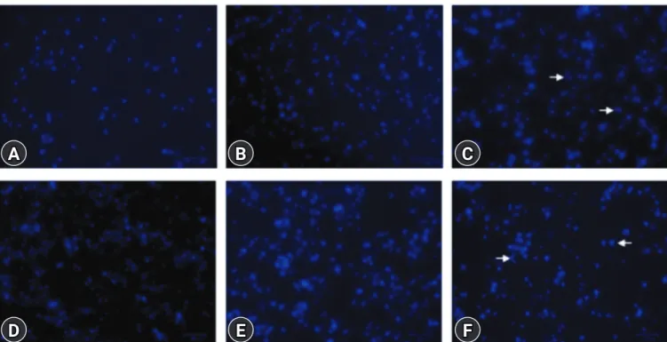
Hoechst 33342

염색을 이용한 골수세포 핵 관찰

골수세포의 세포사를 확인하기 위해 핵 모양을 관찰하였다. 세포 의 핵을 염색하기 위해 Hoechst 33342 용액을 2 . 5 μg / mL 농도로 처리하여 37°C 에서 10 분간 염색하였다[ 11 ]. 염색된 세포는 형광현 미경( ZOE Fluorescent Cell Imager ; BIO - RAD , USA )을 이용해 관찰하였고 사진 촬영을 했다.

통계분석

각 실험은 2 - 3 회 반복하여 시행되었다. Fig . 1 은 평균 ± 표준편 차로 나타냈다. One way analysis of variance 분석 후에 Tur- key - Kramer multiple comparison test ( GraphPad Prism ; Graph- Pad Software , USA )로 유의성을 확인하였다. 통계처리 후 p - value

OD at 57 0 nm

0 0.125 0.25 FBZ (μM)

0.5

##

###

** *** ***

1 2

1.0 0.8 0.6 0.4 0.2 0

FBZ FBZ + LPS

Fig. 1. Fenbendazole (FBZ) decreases the metabolic activity of lipopolysaccharide (LPS)-treated and untreated bone marrow cells (BMs). BMs (1 × 10

6

cells/mL) were incubated in 96-well culture plates and treated with FBZ in the absence or presence of 1 µg/

mL LPS. The concentration range of FBZ was 0 to 2 µM and treat-

ed by 2-fold dilution stepwise. MTT assay was performed at 3 days

after the drug treatment. The data are presented as the mean ±

standard deviation. ***, ### indicate p < 0.001 and **, ## indicate

p < 0.01, compared to the BMs treated without LPS or LPS alone

(FBZ 0 µM), respectively. OD, optical density.

(3)

가 0 . 05 미만인 경우 유의한 것으로 판단하였다.

결과

FBZ

이 골수세포의 대사활성도에 미치는 영향

FBZ 단독과 FBZ + LPS ( 1 μg / mL ) 처리군에서 골수세포의 대사 활성도를 알아보기 위해 MTT assay 를 수행하였다( Fig . 1 ). MTT assay 결과 FBZ 단독에 비해 FBZ + LPS 처리군은 모든 농도( 0 - 1 µM )에서 보다 높은 대사활성도를 나타내었으며, FBZ 의 농도가 증 가함에 따라 대사활성도가 감소됨을 확인하였다. 특히, FBZ 단독의 0 . 5 - 2 µM 농도에서 유의하게 대사활성도가 감소하였고, FBZ + LPS 처리군에서는 1 - 2 µM 농도에서 유의한 감소를 보였다. 이러한 결과는 FBZ LPS 와 같은 염증 물질에 의해 자극된 골수세포의 활

동을 억제할 수 있음을 나타낸다.

FBZ

에 의한 골수세포의 미토콘드리아 막전위 변화

FBZ 이 골수세포에 미치는 영향을 조사하기 위해 FBZ LPS 처리한 골수세포의 미토콘드리아 막전위를 측정하였다. 이를 위해 rhodamine 123 용액을 사용하여 골수세포를 염색한 후 유세포 분 석을 하였다( Fig . 2 ). LPS 는 골수세포의 미토콘드리아 막전위를 높 이는 반면, FBZ LPS 로 증가된 미토콘드리아 막전위를 크게 감 소시켰다. 특히, FBZ 0 . 25 µM 농도와 FBZ 1 µM 농도의 fluores- cence intensity 평균값의 차이는 5 , 034 로, FBZ 0 µM 농도와 FBZ 0 . 25 µM 농도의 평균값의 차이인 1 , 083 보다 크다. 이 결과는 0 . 25 - 1 µM 사이 농도의 FBZ LPS 에 의해 자극된 골수세포의 미토콘드 리아 이중막의 구조를 더욱 불안정하게 할 수 있음을 나타낸다.

Rhodamine 123 A

D

B

E

C

F

Fig. 2. Fenbendazole (FBZ) reduces the mitochondrial membrane potential of both lipopolysaccharide (LPS)-treated and untreated bone marrow cells (BMs). Mouse BMs in 6-well culture plates were treated with or without 1 µg/mL LPS for 2 days, followed by an FBZ treat- ment. The treated BMs were stained with Rhodamine 123 solution. The number in histograms indicates the mean fluorescence intensi- ties. (A) FBZ 0 µM, (B) FBZ 0.25 µM, (C) FBZ 1 µM, (D) FBZ 0 µM + LPS 1 µg/mL, (E) FBZ 0.25 µM + LPS 1 µg/mL, and (F) FBZ 1 µM + LPS 1 µg/mL.

Tube 1 all event

Count

200

100

0

150

100

50

0

100

50

0

150

100

50

0 200

100

0 150

100

50

0

Count Count Count Count Count

FITC-A 10555

25384

9574

24301

8547

19267 10

2

10

2

10

2

10

2

10

2

10

2

10

3

10

3

10

3

10

3

10

3

10

3

10

4

10

4

10

4

10

4

10

4

10

4

10

5

10

5

10

5

10

5

10

5

10

5

10

6

10

6

10

6

10

6

10

6

10

6

FITC-A

Tube 4 all event

Tube 2 all event

FITC-A

FITC-A Tube 5 all event

Tube 3 all event

FITC-A

FITC-A

Tube 6 all event

(4)

FBZ

에 의한 골수세포의 세포사 증가

유세포 분석은 골수세포의 세포사를 확인하기 위해 annexin V - FITC / PI 염색 후 수행되었다( Fig . 3 ). FBZ 단독 또는 FBZ + LPS 처리된 세포의 viable cells ( annexin V -/ PI -)의 비율이 유사하 게 감소하였다. 이 감소는 특히 Fig . 2 와 같이 FBZ 1 µM 농도에서 현저하게 감소함을 보였다. Viable cells FBZ 단독일 때 FBZ 0 - 0 . 25 µM 2 . 3 %, FBZ 0 . 25 - 1 µM 13 . 8 % 감소하였다. FBZ + LPS 처리군에서도 FBZ 0 - 0 . 25 µM 3 . 4 %, FBZ 0 . 25 - 1 µM 9 . 3 % 감소하였다. 죽은 세포 중 necrotic cells ( annexin V -/ PI +)의 수는 FBZ 0 - 1 µM 농도에서 5 %에서 6 %로 증가한 FBZ 단독에 비 해 FBZ + LPS 처리군에서 더욱 현저한 변화를 보였다. 그러나 ear- ly apoptosis cells ( annexin V +/ PI -)에서는 FBZ 단독은 6 %에서 12 %로 증가한 반면, FBZ + LPS 처리군에서는 10 %로 일정하였다.

이러한 결과는 FBZ LPS 처리 여부와 관계없이 골수세포에서 세 포사를 유도할 수 있음을 보여준다.

골수세포 내의 과립구와

B

림프구의 구성에

FBZ

이 미치는 영향

마우스 골수세포의 주요 구성 세포 중에는 과립구와 B 림프구가 포함되어 있다. 두 가지 세포 구성에 FBZ 이 미치는 영향을 조사하 기 위해, FBZ LPS 로 처리된 골수세포를 Gr - 1 또는 B220 특이적 인 항체로 염색한 후 유세포 분석을 실시하였다( Fig . 4 ). Dot plot Gr - 1 + 구역은 호중구를 포함한 과립구이다. FBZ 0 µM 농도에서 FBZ 단독은 27 %, FBZ + LPS 처리군은 25 %인 것으로 보아 LPS 과립구의 비율을 크게 증가시키지 않았다. 반면, FBZ 0 - 1 µM 사이 의 변화를 보면 FBZ 단독에서는 27 %에서 15 %로 감소하였고 FBZ + LPS 처리군에서는 25 %에서 19 %로 감소하였다. 골수세포에 LPS

A

D

B

E

C

F Annexin V

PI

Fig. 3. Fenbendazole (FBZ) induces the apoptosis and necrosis of lipopolysaccharide (LPS)-treated and untreated bone marrow cells (BMs).

The cells were cultured in 6-well culture plates and treated with FBZ (0, 0.25, 1 µM) and LPS (1 µg/mL). Annexin V-FITC/PI staining was performed after 2 days of treatment. The cells in the quadrants indicate necrosis (upper left, UL), late apoptosis (upper right, UR), viable (lower left, LL), and early apoptosis (lower right, LR) cells, respectively. The number in quadrants indicates the percentage of cells. (A) FBZ 0 µM, (B) FBZ 0.25 µM, (C) FBZ 1 µM, (D) FBZ 0 µM + LPS 1 µg/mL, (E) FBZ 0.25 µM + LPS 1 µg/mL, and (F) FBZ 1 µM + LPS 1 µg/mL. PI, propidium iodide.

10

2

10

2

10

2

10

2

10

2

10

2

10

3

10

3

10

3

10

3

10

3

10

3

10

4

10

4

10

4

10

4

10

4

10

4

10

5

10

5

10

5

10

5

10

5

10

5

10

6

10

6

10

6

10

6

10

6

10

6

10

2

10

3

10

4

10

5

10

6

FITC-A FITC-A FITC-A

FITC-A FITC-A

FITC-A

PE-A PE-A PE-A PE-A PE-A PE-A

10

2

10

2

10

2

10

2

10

2

10

3

10

3

10

3

10

3

10

3

10

4

10

4

10

4

10

4

10

4

10

5

10

5

10

5

10

5

10

5

10

6

10

6

10

6

10

6

10

6

Tube 1 all event

Tube 4 all event

Tube 2 all event

Tube 5 all event

Tube 3 all event

Tube 6 all event

5% 5% 6%

10%

7%

5%

16% 18% 25%

23%

17%

15%

6% 7% 12%

10%

10%

10%

(5)

Gr-1 A

D

B

E

C

F

Fig. 4. Fenbendazole (FBZ) significantly decreases granulocytes than B cells in bone marrow cells (BMs). BMs (1 × 10

6

cells/mL) were seeded in 6-well culture plates and treated with FBZ and lipopolysaccharide (LPS). The cells were stained with anti-B220 or Gr-1 specif- ic antibody. The cells were analyzed using flow cytometry. The upper squares in dot plots indicate the population of Gr-1 positive cells, granulocytes. The lower squares in dot plots indicate the population of B220 positive cells, B cells. Irrespective of the presence of LPS, the proportion of granulocytes decreased, but that of B cells increase. (A) FBZ 0 µM, (B) FBZ 0.25 µM, (C) FBZ 1 µM, (D) FBZ 0 µM + LPS 1 µg/mL, (E) FBZ 0.25 µM + LPS 1 µg/mL, and (F) FBZ 1 µM + LPS 1 µg/mL.

처리와 관계없이 과립구의 비율은 현저하게 감소시키는 사실을 알 수 있다. Dot plot B220 + 구역은 B 림프구이다. B 림프구의 비율 은 LPS 에 의해 증가하지 않았고, 28 %에서 20 %로 오히려 감소한 모 습을 보였다. 반면, LPS 유무와 관계없이 FBZ 의 농도가 증가함에 따라 B 림프구의 비율은 증가하였다.

FBZ

에 의한 골수세포의 핵 형태 변화

골수세포를 Hoechst 33342 용액으로 염색한 후, 형광현미경을 사용하여 FBZ 에 의한 골수세포의 핵 형태 변화를 관찰하였다 ( Fig . 5 ). FBZ 단독 처리군에서는 1 µM 농도에서 눈에 띄게 분절된 핵을 가진 세포의 수가 증가하였다. LPS 처리로 골수세포의 수가 현 저하게 증가되었고 FBZ 1 µM 농도에서 분절된 핵을 가진 세포의 수가 증가하였다. 핵이 분절된 세포가 증가하는 것은 세포사의 증가 를 의미하므로 FBZ 1 µM LPS 유무와 관계없이 골수세포의 세포

사를 증가시키는 사실을 확인했다.

고찰

FBZ 은 수의에서 광범위하게 사용되는 항기생충제제 중 하나이며 [ 1 - 3 ], 최근 종양세포를 이용한 연구에서 항암효과를 갖는다는 사실 이 밝혀졌다[ 6 ]. 하지만 FBZ 의 항염증 효과에 대한 연구는 매우 미 흡하며 그 작용기전도 알려져 있지 않다. 본 연구에서는 FBZ 의 항 염증 효과를 알아보기 위해 골수세포에 대표적인 염증성 유도물질 중 하나인 LPS FBZ 과 함께 복합 처리한 후 분석하였다.

FBZ 이 세포활성도와 미토콘드리아 막전위를 감소시켜 골수세포 의 활성을 억제하였다. 또한, annexin V - FITC / PI 염색과 Hoechst 33342 염색을 통해 FBZ 이 염증성 골수세포의 세포사를 유도하는 사실을 확인하였다. 이러한 결과는 LPS 처리로 만들어진 염증성 골

27% 21% 15%

19%

24%

25%

28% 29% 34%

28%

21%

20%

B220

10

2

10

3

10

4

10

5

10

6

10

2

10

3

10

4

10

5

10

6

FITC-A

APC-A

Tube 1 all event

10

2

10

3

10

4

10

5

10

6

10

2

10

3

10

4

10

5

10

6

FITC-A

APC-A

Tube 2 all event

10

2

10

3

10

4

10

5

10

6

10

2

10

3

10

4

10

5

10

6

FITC-A

APC-A

Tube 3 all event

10

2

10

3

10

4

10

5

10

6

10

2

10

3

10

4

10

5

10

6

FITC-A

APC-A

Tube 6 all event

10

2

10

3

10

4

10

5

10

6

10

2

10

3

10

4

10

5

10

6

FITC-A

APC-A

Tube 5 all event

10

2

10

3

10

4

10

5

10

6

10

2

10

3

10

4

10

5

10

6

FITC-A

APC-A

Tube 4 all event

(6)

Fig. 5. Fenbendazole (FBZ) increased the number of bone marrow cells (BMs) with fragmented nuclei. The cells were set up and treated with FBZ and lipopolysaccharide (LPS), as in Fig. 2. After 2 days, Hoechst 33342 staining was performed. The BMs treated with FBZ 1 µM contain many cells with fragmented nuclei (arrows). (A) FBZ 0 µM, (B) FBZ 0.25 µM, (C) FBZ 1 µM, (D) FBZ 0 µM + LPS 1 µg/mL, (E) FBZ 0.25 µM + LPS 1 µg/mL, and (F) FBZ 1 µM + LPS 1 µg/mL.

A

D

B

E

C

F

수세포에서 FBZ 이 세포 수와 미토콘드리아 기능을 모두 감소시킨 다는 것을 보여준다. 과립구와 B 림프구의 구성 비율 변화를 분석한 결과 FBZ LPS 가 처리된 골수세포에서 B 림프구보다 과립구의 비 율을 더 감소시켰다.

FBZ 이 과립구의 발현을 선택적으로 억제하는 정확한 기전은 밝 혀지지 않았지만, 이를 설명할 수 있는 선행연구들이 있다. 천식 동 물모델에서 FBZ 이 알레르기성 기도염증과 Th2 사이토카인의 생산 을 감소시켰다[ 12 ]. FBZ 은 기생충 감염증 치료에 다양하게 이용된 다[ 13 ]. 일반적으로 기생충 감염 시 호산구를 포함한 과립구가 증가 되고[ 14 ], FBZ 이 기생충 치료에 이용되므로, FBZ 이 과립구의 숫자 또는 기능에 영향을 미칠 수 있음을 유추할 수 있다. 또한, squirrel monkey 를 이용한 앞선 연구에서 총 백혈구, 호중구, 단핵구 및 호 산구 수 비교는 FBZ 을 처리하지 않은 대조군에 비해 FBZ 을 처리한 군에서 현저히 감소한 모습을 보였다[ 15 ]. 이를 종합하면 FBZ 은 골 수세포 중에서도 과립구를 특히 감소시킨다고 생각된다. 반면, FBZ 의 농도가 증가함에 따라 전체 생존 세포 수와 과립구는 줄어드는 데, 림프구의 절대값은 크게 변하지 않아 상대적인 비율로 림프구가 증가한다고 추측할 수 있다. 다른 연구에서도 절대 림프구 수는 FBZ 치료에 영향을 거의 받지 않았다[ 15 ]. 하지만 FBZ 은 마우스의 면역체계에 상당한 영향을 미치는 것으로 보아[ 16 ], B 림프구의 기능 이 감소될 가능성은 있다.

다른 연구에서는 설치류에서 FBZ 에 의한 골수 억제가 발생하지

않음을 보고하였다[ 17 ]. 하지만 본 연구에서는 골수세포에 대한 FBZ 의 직접적인 효과를 측정하여 실험조건이 다르다. 또한 LPS 의해 과도한 염증 반응이 발생한 골수세포를 이용하여 FBZ 의 항염 증 효과에 대한 연구를 수행했으며, FBZ 을 항염증제로 사용할 수 있는 가능성을 확인했다.

이러한 결과를 종합하면, LPS 처리로 인해 염증성 상태가 된 골수 세포에서 FBZ 은 미토콘드리아 막전위를 불안정하게 만들어 세포사 를 유도시켜 항염증 효과를 일으키는 것으로 추정된다. 향후 미토 콘드리아와 FBZ 의 관련성, 다른 항염증제와의 상승 효과, 다양한 염증이 유도된 동물질환 모델에서의 약효 실험이 추가적으로 필요 하다.

ORCID

Seo-Ro Park, https://orcid.org/0000-0002-6012-4668 Hong-Gu Joo, https://orcid.org/0000-0002-1505-8761

References

1. Campbell WC. Benzimidazoles: veterinary uses. Parasitol To- day 1990;6:130–133.

2. Pritchett KR, Johnston NA. A review of treatments for the

(7)

eradication of pinworm infections from laboratory rodent colonies. Contemp Top Lab Anim Sci 2002;41:36–46.

3. Petersen MB, Friis C. Pharmacokinetics of fenbendazole fol- lowing intravenous and oral administration to pigs. Am J Vet Res 2000;61:573–576.

4. Dawson PJ, Gutteridge WE, Gull K. A comparison of the in- teraction of anthelmintic benzimidazoles with tubulin isolated from mammalian tissue and the parasitic nematode Ascaridia galli. Biochem Pharmacol 1984;33:1069–1074.

5. Friedman PA, Platzer EG. Interaction of anthelmintic ben- zimidazoles and benzimidazole derivatives with bovine brain tubulin. Biochim Biophys Acta 1978;544:605–614.

6. Dogra N, Kumar A, Mukhopadhyay T. Fenbendazole acts as a moderate microtubule destabilizing agent and causes cancer cell death by modulating multiple cellular pathways. Sci Rep 2018;8:11926.

7. Hawkins MG, Couto S, Tell LA, Joseph V, Lowenstine LJ.

Atypical parasitic migration and necrotizing sacral myelitis due to Serratospiculoides amaculata in a prairie falcon (Falco mexicanus). Avian Dis 2001;45:276–283.

8. Kim HJ, Kim MH, Byon YY, Park JW, Jee Y, Joo HG. Radio- protective effects of an acidic polysaccharide of Panax ginseng on bone marrow cells. J Vet Sci 2007;8:39–44.

9. Kim SY, Joo HG. Evaluation of adjuvant effects of fucoidan for improving vaccine efficacy. J Vet Sci 2015;16:145–150.

10. Moon SY, Joo HG. Anti-inflammatory effects of 4,4'-diamino- diphenyl sulfone (dapsone) in lipopolysaccharide-treated spleen cells: selective inhibition of inflammation-related cyto-

kines. Korean J Vet Res 2015;55:199–204.

11. Jang JY, Moon SY, Joo HG. Differential effects of fucoidans with low and high molecular weight on the viability and func- tion of spleen cells. Food Chem Toxicol 2014;68:234–238.

12. Cai Y, Zhou J, Webb DC. Treatment of mice with fenbenda- zole attenuates allergic airways inflammation and Th2 cyto- kine production in a model of asthma. Immunol Cell Biol 2009;87:623–629.

13. Prichard RK, Ranjan S. Anthelmintics. Vet Parasitol 1993;

46:113–120.

14. Rajamanickam A, Munisankar S, Bhootra Y, Dolla CK, Nut- man TB, Babu S. Elevated systemic levels of eosinophil, neu- trophil, and mast cell granular proteins in Strongyloides ster- coralis infection that diminish following treatment. Front Im- munol 2018;9:207.

15. Nehete PN, Wilkerson G, Nehete BP, Chitta S, Ruiz JC, Scholt- zova H, Williams LE, Abee CR, Vanchiere JA. Cellular im- mune responses in peripheral blood lymphocytes of Giardia infected squirrel monkey (Saimiri boliviensis boliviensis) treated with Fenbendazole. PLoS One 2018;13:e0198497.

16. Landin AM, Frasca D, Zaias J, Van der Put E, Riley RL, Alt- man NH, Blomberg BB. Effects of fenbendazole on the mu- rine humoral immune system. J Am Assoc Lab Anim Sci 2009;48:251–257.

17. Villar D, Cray C, Zaias J, Altman NH. Biologic effects of fen-

bendazole in rats and mice: a review. J Am Assoc Lab Anim

Sci 2007;46:8–15.

수치

Fig. 1. Fenbendazole (FBZ) decreases the metabolic activity of  lipopolysaccharide (LPS)-treated and untreated bone marrow cells  (BMs)
Fig. 2. Fenbendazole (FBZ) reduces the mitochondrial membrane potential of both lipopolysaccharide (LPS)-treated and untreated bone  marrow cells (BMs)
Fig. 3. Fenbendazole (FBZ) induces the apoptosis and necrosis of lipopolysaccharide (LPS)-treated and untreated bone marrow cells (BMs)
Fig. 4. Fenbendazole (FBZ) significantly decreases granulocytes than B cells in bone marrow cells (BMs)
+2

참조

관련 문서

A and E, In control group, a small amount of new bone was observed at the margin of bone defect (40×); B and F, In experimental group 1, a large amount of new bone was formed

Inhibitory effects of classified methanol extracts of Smilax china L on the COX-2 and iNOS expression of human colorectal cancer cell lines... Inhibitory

Effects of pulse frequency of low-level laser thrapy (LLLT)on bone nodule formation in rat calvarial cells.. Low-level laser therapy stimulats

Histologic evaluation of early human bone response to different implant surfaces2. Histologic evaluation of human bone integration on machined and

Consequently, the present study was designed to investigate the effect of adding 60 mg of raloxifene hydrochloride daily to women treated with GnRH-a for severe endometriosis

Approved clinical use of bone marrow stem cells for myocardial infarction treatment... Cardiac

Biochemical markers of bone turnover can be classified according to the process that underlie in markers of bone formation, products of the osteoblast

Fig 4 : Inhibitory effects of classified methanol extracts of Acalypha australis the COX-2 protein expression of the human oral cavity carcinoma KB cells....