• 검색 결과가 없습니다.

Very Early Onset of Amiodarone-Induced Pulmonary Toxicity Wonho Lee, MD

N/A
N/A
Protected

Academic year: 2022

Share "Very Early Onset of Amiodarone-Induced Pulmonary Toxicity Wonho Lee, MD"

Copied!
3
0
0

로드 중.... (전체 텍스트 보기)

전체 글

(1)

699 Copyright © 2013 The Korean Society of Cardiology

Korean Circulation Journal

Introduction

Amiodarone is a benzofuran derivative antiarrhythmic drug that has been commonly used to treat atrial fibrillation as well as supra- ventricular and ventricular tachycardia. Amiodarone-induced pul- monary toxicity (APT) is one of the various side effects of amioda- rone therapy. Because the clinical manifestation of APT resembles pulmonary infection, heart failure, pulmonary thromboembolism and restrictive pulmonary disease,

1)

diagnosis of APT is difficult and therefore is frequently delayed without strong clinical suspicion.

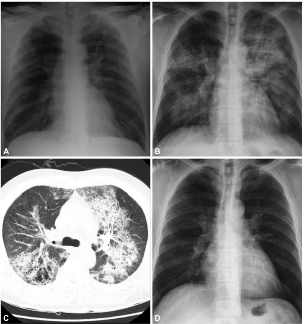
APT has been described mostly in patients who have received a lar- ge dose of amiodarone over prolonged periods of time after cardiac surgery or pulmonary angiography. We report here a case of APT after a short course of therapy for post-myocardial infarction ven- tricular tachycardia. This study will be helpful for increasing aw- areness of a very early onset of APT.

Case Report

http://dx.doi.org/10.4070/kcj.2013.43.10.699 Print ISSN 1738-5520 • On-line ISSN 1738-5555

Very Early Onset of Amiodarone-Induced Pulmonary Toxicity

Wonho Lee, MD 1 , Dong Rueol Ryu, MD 1 , Seon-Sook Han, PhD 1 , Sook-Won Ryu, MD 2 , Byung Ryul Cho, MD 1 , Hyucki Kwon, MD 1 , and Bo Ra Kim, MD 1

1

Departments of Internal Medicine and

2

Laboratory Medicine, Kangwon National University Hospital, Chuncheon, Korea

Amiodarone is a widely used antiarrhythmic agent. Among its various adverse effects, amiodarone-induced pulmonary toxicity (APT) is the most life threatening complication, which has been described mostly in patients who have been in treatment with high accumulative doses for a long duration of time. However, amiodarone therapy in short-term duration induced APT was rarely reported. We describe a case of a 54-year-old man who is presented with symptoms of APT after a few days of therapy for post-myocardial infarction ventricular ta- chycardia. For early diagnosis and successful treatment, awareness and high suspicion of this rare type of early onset APT is crucial in pa- tients with amiodarone therapy. (Korean Circ J 2013;43:699-701)

KEY WORDS: Amiodarone; Drug toxicity; Myocardial infarctions; Arrhythmias, cardiac.

Received: April 15, 2013 Revision Received: May 9, 2013 Accepted: May 29, 2013

Correspondence: Dong Rueol Ryu, MD, Department of Internal Medicine, Division of Cardiology, Kangwon National University Hospital, 156 Baeng- nyeong-ro, Chuncheon 200-722, Korea

Tel: 82-33-258-9439, Fax: 82-33-258-2432 E-mail: [email protected]

• The authors have no financial conflicts of interest.

This is an Open Access article distributed under the terms of the Creative Commons Attribution Non-Commercial License (http://creativecommons.

org/licenses/by-nc/3.0) which permits unrestricted non-commercial use, distribution, and reproduction in any medium, provided the original work is properly cited.

Case

A 54-year-old man was admitted to the hospital with chest pain and ST elevation myocardial infarction. He had no medical history, except for a 60 pack-year smoking history and that he was current smoker. A cardiac catheterization showed total occlusion with Th- rombolysis in Myocardial Infarction 0 flow at the proximal left ante- rior descending artery. He had percutaneous coronary intervention with stenting at the proximal left anterior descending coronary ar- tery. On the night of the catheterization, ventricular tachycardia de- veloped. He was electrically cardioverted, which resulted in sinus rhythm, and amiodarone therapy was started at an initial loading dose of 150 mg intravenously (i.v.), followed by a daily dose of 730 mg i.v. for 2 days. A transthoracic echocardiogram demonstrated anterior and anteroseptal hypokinesis of the left ventricle with an ejection fraction of 35%. After 2 days of amiodarone therapy - a total of 1635 mg of amiodarone - low-grade fever developed with mild dy- spnea. Pulse oximetry was 88% in oxygen saturation on room air.

Compared with the initial chest X-ray finding, a follow-up chest ra- diography demonstrated bilateral diffuse infiltration (Fig. 1A and B).

Blood tests showed a leukocyte count of 15800/mm

3

and a serum

creatinine value of 1.2 mg/dL. Pneumonia was suspected and intra-

venous therapy with piperacillin, tazobactam and levofloxacin was

started. The results of blood and sputum culture were negative. Gr-

am’s stain and a smear for tuberculosis organisms were also nega-

tive, as were serologic tests for Legionella and Chlamydia trachoma-

tis. Mycoplasma pneumoniae polymerase chain reaction tests were

also negative. Values for antinuclear antibody, double-stranded de-

(2)

700 Amiodarone-Induced Pulmonary Toxicity Occurred within a Few Days

http://dx.doi.org/10.4070/kcj.2013.43.10.699 www.e-kcj.org

oxyribonucleic acid antibodies, rheumatoid factor, as well as C’3 and C’4 were all within normal limits. Pulmonary edema was also suspected and intravenous diuretics therapy (Lasix 100 mg/day for 3 days) was started; however, no interval change was noted on chest radiography after 3 kg of weight loss. A high resolution computed tomography scan of the chest showed bilateral diffuse consolida- tion, ground glass opacity and underlying emphysema (Fig. 1C). Ow- ing to a high suspicion of amiodarone pulmonary toxicity (APT), amiodarone was discontinued after a total dose of 4035 mg over more than 8 days. Steroid therapy was not started due to mild re- spiratory symptoms. Pulmonary function tests showed mild obst- ructive impairment and carbon monoxide diffusing capacity (DLCO) was checked as 16.1 L (79%). As the clinical status was stabilized,

bronchoscopy was performed. Bronchoalveolar lavage revealed a cell count of 200 cells/μL with prominent eosinophilia (8%) and foa- my macrophages (Fig. 2). No bacteria or fungi were identified via microscopic examination or culture of the lavage fluid. Ten days after amiodarone was discontinued, the patient was discharged with mild dyspnea. Three months later, APT appeared to be reversible with the resolution of chest radiography findings (Fig. 1D) and recovery of symptoms.

Discussion

Amiodarone is a highly effective antiarrhythmic that is used to treat atrial fibrillation due to its high conversion rate, rapid onset of A

C

B

D

Fig. 1. A: the initial chest X-ray study shows no active lung lesion. B: the follow-up chest X-ray study showed bilateral alveolar and interstitial infiltration.

C: the high resolution computed tomography scan of the lung shows bilateral patchy infiltrations with ground glass opacity. D: three months after dis-

charge, the chest X-ray study shows resolution of bilateral infiltration.

(3)

701 Wonho Lee, et al.

http://dx.doi.org/10.4070/kcj.2013.43.10.699 www.e-kcj.org

action and minimal myocardial depression. However, amiodarone therapy has been associated with various side effects, including pul- monary toxicity, which may occur in up to 5-10% of treated pa- tients.

1)

APT can present as several manifestations: chronic intersti- tial pneumonitis, organizing pneumonia, acute respiratory distress syndrome and solitary pulmonary mass.

2)

The mechanisms of ami- odarone-induced pulmonary injury are incompletely understood. Two major hypotheses include direct cytotoxicity and a hypersensitivity reaction.

3)

Risk factors for APT include a daily dose greater than 400 mg/day, a duration of therapy exceeding two months, increased patient age, preexisting lung disease, undergoing operation and pulmonary an- giography.

4)

In the present case, the patient usually had no respira- tory symptoms; however, a pulmonary function test and high resolu- tion chest computed tomography showed a possibility of chronic lung disease. Additionally, cardiac catheterization was performed.

Other cases of APT occurring within a week after cardiac catheteriz- ation exist.

5)6)

Thus, these could be the risk factors for APT. However, in our case, APT occurred only 2 days after 1635 mg of amiodarone.

To our knowledge, this represents the most rapidly developed APT.

Amiodarone-induced pulmonary toxicity is a diagnosis of exclu- sion. Due to its non-specific nature and its ability to mimic other disease processes, such as congestive heart failure, acute pulmo- nary respiratory syndrome and pneumonia, the diagnosis of APT may be delayed or missed. The presence of foamy macrophages is suggestive of APT, but is not diagnostic. Increased lung attenuation on computed tomography, increased gallium uptake and abnormal Fig. 2. Bronchoalveolar lavage reveals foamy macrophages (Wright-Giem- sa stain, magnification ×1000).

pulmonary function tests are nonspecific. A reduction in DLCO of 15% is a strong indicator for the diagnosis of pulmonary toxicity due to amiodarone, with a sensitivity of 68-100% and a specificity of 69-95%.

7)

Lung biopsy may be helpful in excluding alternative etiologies and may be helpful in identifying the pathologic type of disease (e.g., chronic interstitial pneumonitis); yet, this procedure is also nonspecific for amiodarone-induced disease. In the present case, initial treatment included antibiotics and diuretics because pneumonia and pulmonary edema were suspected. Despite these treatments, symptoms and a chest X-ray were not improved until amiodarone was discontinued. Additionally, foamy macrophages were present in the bronchoalveolar lavage. Thus, our diagnosis was convincing.

Discontinuation of amiodarone is the primary therapy for amio- darone toxicity. Additionally, for patients with symptomatic pul- monary toxicity, systemic steroids may be valuable.

7)

Due to the long elimination half-life (approximately 45 days) of amiodarone, pul- monary toxicity may initially progress despite the drug discontinu- ation and may recur upon glucocorticoid withdrawal. In the pres- ent case, steroid therapy was not carried out due to mild respiratory symptoms.

Very early onset of APT appears to be unusual without surgery.

The present case shows that APT can occur at anytime during ther- apy. As usage of amiodarone increases, one must be aware of a pos- sible APT, even after a short course of amiodarone therapy.

References

1. Dusman RE, Stanton MS, Miles WM, et al. Clinical features of amioda- rone-induced pulmonary toxicity. Circulation 1990;82:51-9.

2. Martin WJ 2nd, Rosenow EC 3rd. Amiodarone pulmonary toxicity. Rec- ognition and pathogenesis (Part I). Chest 1988;93:1067-75.

3. Martin WJ 2nd, Rosenow EC 3rd. Amiodarone pulmonary toxicity. Rec- ognition and pathogenesis (Part 2). Chest 1988;93:1242-8.

4. Ernawati DK, Stafford L, Hughes JD. Amiodarone-induced pulmonary toxicity. Br J Clin Pharmacol 2008;66:82-7.

5. Jang WJ, Chon HR, Jung JS, et al. Amiodarone-induced pulmonary tox- icity within a short period of the initiation of amiodarone therapy: a case report. Korean J Crit Care Med 2011;26:117-21.

6. Tanawuttiwat T, Harindhanavudhi T, Hanif S, Sahloul MZ. Amiodarone- induced alveolar haemorrhage: a rare complication of a common medication. Heart Lung Circ 2010;19:435-7.

7. Papiris SA, Triantafillidou C, Kolilekas L, Markoulaki D, Manali ED. Amio- darone: review of pulmonary effects and toxicity. Drug Saf 2010;33:

539-58.

수치

Fig. 1. A: the initial chest X-ray study shows no active lung lesion. B: the follow-up chest X-ray study showed bilateral alveolar and interstitial infiltration

참조

관련 문서

The definitive fixation using Steinmann pin & 4.0, 6.5 cannulated screws was performed(A-D), Postoperative X-ray (A : ankle lateral view, B : Calcaneal axial view, C

XAFS: X-ray absorption fine structure XES: X-ray emission spectroscopy XRF: X-ray fluorescence.. Use of x-rays; a probe based

Electron probe micro-analyzer image of Zr-4%Si binary alloys; (a).. X-ray diffraction profiles of as-cast Zr-xSi binary alloys, which shows the dual phases.. Magnetic

Excitation Detection X-ray photoelectron spectroscopy (XPS) Photons(X-ray) Electrons UV photoelectron spectroscopy (UPS) Photons (UV) Electrons

The structure and film optical properties were investigated by X-ray diffraction(XRD), the particle size and thickness were investigated by scanning

Single crystal X-ray structure determination clearly revealed structural authenticity of each compound and alluded electronic alteration via investigation on the

the intersection of several such symmetry elements.. International Tables for X-ray Crystallography.. International Tables for X-ray Crystallography.. International Tables

radiography, X-ray generation, and the simulation of weapons effects, such as nuclear electromagnetic pulse (EMP), to packaged pulsed power for directed energy weapons