© 2013 The Korean Ophthalmological Society
This is an Open Access article distributed under the terms of the Creative Commons Attribution Non-Commercial License (http://creativecommons.org/licenses /by-nc/3.0/) which permits unrestricted non-commercial use, distribution, and reproduction in any medium, provided the original work is properly cited.
Original Article
Long-term Evaluation of Endothelial Cell Changes in Fuchs Corneal Dystrophy: The Influence of Phacoemulsification and
Penetrating Keratoplasty
Yong Woo Kim1,2, Mee Kum Kim1,2, Won Ryang Wee1,2
1Department of Ophthalmology, Seoul National University Hospital, Seoul National University College of Medicine, Seoul, Korea
2Seoul Artificial Eye Center, Seoul National University Hospital Biomedical Research Institute, Seoul, Korea
Purpose: To evaluate the natural course of the long-term endothelial cell changes in Fuchs corneal dystrophy (FCD) patients and investigate the effects of phacoemulsification on the annual rate of change in endothelial indices in FCD patients.
Methods: Thirty-four patients diagnosed with FCD at Seoul National University Hospital from 1994 to 2010 were retrospectively reviewed. Sixteen patients who had been followed up for more than 1 year were selected and classified into 3 groups: group A, patients with no ocular surgery; group B, patients who had undergone phacoemulsification only; and group C, patients who had undergone penetrating keratoplasty with cataract surgery. Endothelial cell density, polymegethism, pleomorphism, and pachymetry were measured and the ex- ponential rates of endothelial cell and pachymetry change were analyzed.
Results: A non-linear mixed model of non-operated FCD patients showed that only pachymetric data tended to increase with statistical significance (p = 0.001) with a mean follow-up period of 4.15 years. Using an exponen- tial regression analysis fitting curve, the mean rates of annual endothelial cell loss were 0.82%/yr, 20.39%/yr, and 29.27%/yr in groups A, B, and C respectively, and statistical significance was seen only in group C (p <
0.05).
Conclusions: Retrospective long-term follow-up data showed that changes in endothelial density did not sig- nificantly decrease over at least 4 years in middle-aged FCD patients. The changes in pachymetric corneal thickness appeared to increase over the same period. Considering that no exponential changes were aggra- vated after performing cataract surgery alone, cataract surgery would be a preferable option in FCD patients compared to an approach of “wait-and-do” penetrating keratoplasty combined with cataract surgery.
Key Words: Endothelial cell density, Fuchs corneal dystrophy, Pachymetry, Phacoemulsification
Fuchs corneal dystrophy (FCD) is a bilateral, slowly pro- gressive disease causing focal excrescences of Descemet’s
membrane, or so-called guttata, which commonly appears during the fifth to seventh decades of life [1-3]. FCD occurs predominantly among women and is inherited in an auto- somal dominant fashion with incomplete penetrance [4,5].
The exact pathogenic mechanism of progressive endotheli- al cell loss has not yet been determined. Currently, evi- dence is emerging that decreased expression of anti-apop- totic genes or aquaporin genes in the cornea may be
Received: October 9, 2012 Accepted: February 8, 2013
Corresponding Author: Mee Kum Kim, MD. Department of Ophthal- mology, Seoul National University College of Medicine, #103 Dae- hank-ro, Jongno-gu, Seoul 110-799, Korea. Tel: 82-2-2072-2665, Fax: 82- 2-741-3187, E-mail: [email protected]
involved in the pathogenesis of FCD [6-9]. Secondary stro- mal edema due to progressive endothelial cell loss often leads to loss of vision.
With regard to time-dependent changes, endothelial cell density normally decreases with age by 0.6% per year in the normal population [10,11]. Some diseases, including an- gle closure glaucoma, uveitis, and pseudoexfoliation syn- drome are involved in more progressive endothelial cell loss than usually occurs [12,13]. In addition, intraocular surgery is also a critical factor involved in progressive en- dothelial cell loss, considering that the rate of endothelial cell loss increases to 2.5% per year with cataract surgery and 4.2% per year with penetrating keratoplasty [10,14,15].
It is unclear how fast endothelial loss or dysfunction pro- gresses during the long-term natural course of FCD. This is very concerning when cataract surgery is required.
Seitzman et al. [16] suggested that a preoperative pachym- etry measurement of less than 640 µm indicated tolerable endothelial loss, resulting in the promotion of visual reha- bilitation after cataract surgery in FCD patients. Afshari et al. [17] reported that cataract extraction advanced the time of penetrating keratoplasty by a mean of 3.2 years in a 30- year observational study in FCD patients. The effect of cataract surgery on the natural course of endothelial cell changes in FCD patients should be determined in a quanti- tative manner. This information will help to determine which approach would be better for FCD patients who need cataract surgery.
Thus, for the first time, we evaluated the natural course of long-term endothelial cell changes in FCD patients. We also investigated the effect of phacoemulsification on the annual rate of change in endothelial indices in FCD pa- tients.
Materials and Methods
We retrospectively reviewed the medical records of 34 patients who were diagnosed with FCD at Seoul National University Hospital from 1994 to 2010. FCD was diag- nosed when the patients showed central guttata in both eyes on examination by a slit-lamp and specular micro- scope, without any history of ocular inflammation. Among these patients, we selected 16 patients who had been fol- lowed up for more than 1 year and classified them into 3 groups: group A, patients with no ocular surgery (16 eyes
in 8 patients); group B, patients who had undergone phacoemulsification only (7 eyes in 4 patients); and group C, patients who had undergone penetrating keratoplasty with simultaneous extracapsular cataract extraction (2 eyes in 2 patients) or phacoemulsification afterwards (2 eyes in 2 patients). The follow-up period ranged from 12.3 months to 109.9 months (group A, 18.2 to 109.9 months; group B, 12.3 to 45.8 months; and group C, 41.7 to 85.1 months). Ev- ery patient in group C had a diagnosis of bullous keratopa- thy due to FCD. Age- and sex-matched non-FCD patients who underwent phacoemulsification and penetrating kera- toplasty were also included to compare exponential chang- es in endothelial cell count (ECC) between non-FCD and FCD patients (8 eyes in 7 patients; 6 with viral keratitis, 1 with band keratopathy, and 1 with bullous keratopathy).
Endothelial cell density, polymegethism (coefficient of variance, CV), pleomorphism (hexagonality), and pachym- etry were measured by experienced ophthalmic techni- cians using a specular microscope (Noncon ROBO CA;
Konan Medical, Hyogo, Japan) and ultrasonic pachymeter (Pocket II; Quantel Medical, Clermont-Ferrand, France).
We investigated the annual change in FCDs using a non-linear mixed model with the SAS ver. 9.1 (SAS Insti- tute, Cary, NC, USA). We evaluated the exponential rate of endothelial cell and pachymetry change in each group by exponential curve fitting regression analysis using the SPSS ver. 17.0 (SPSS Inc., Chicago, IL, USA). The Krus- kal-Wallis method was used for comparing demographic findings between groups. A non-linear mixed model was used in group A, which included a sufficient number of patients to show statistical significance.
Results
There were 3 men and 13 women among the 16 patients.
Ages ranged from 22 to 82 years (62.32 ± 13.10 years, mean
± standard deviation), and the mean follow-up period was 3.88 ± 2.25 years. The mean endothelial cell density and pachymetry measurements of the 16 patients at initial ex- amination were 1,413.91 ± 735.42/mm2 and 576.56 ± 74.04 mm, respectively. There were no statistical differences in age, sex, initial endothelial cell density, and initial pachym- etry measurements among the 3 groups (Table 1).
First, we investigated the natural course of FCD among patients who had not undergone surgery. Non-linear mixed
models for time-dependent changes in these patients showed that only pachymetric data tended to increase with statistical significance (p = 0.001), with a mean follow-up of 4.15 years (Table 2). The changes in ECC, CV, and hex- agonality were not significant during these periods using the aforementioned model. This suggests that functional deterioration progresses over a period of 4 years without noticeable morphologic changes in endothelial cells.
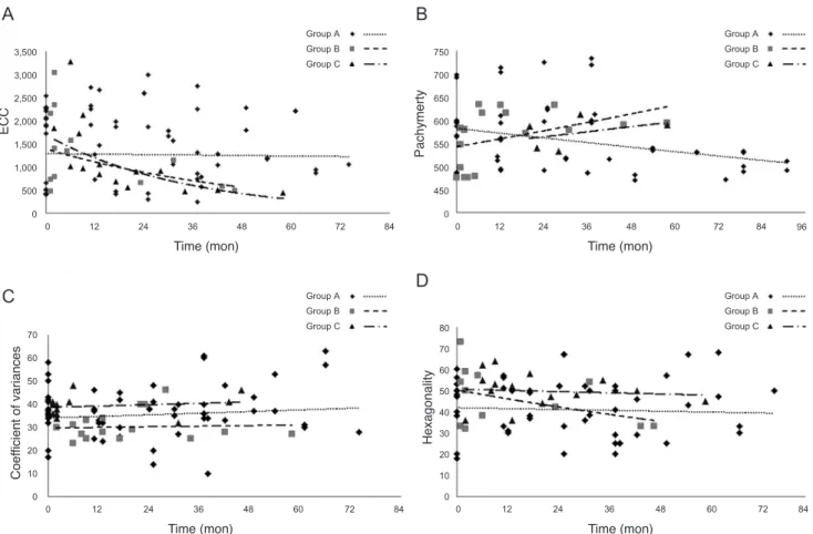
Next, we evaluated whether long-term endothelial changes were worse when FCD patients underwent cata- ract surgery only or penetrating keratoplasty with cataract surgery. Regression analysis showed that the mean rates of annual endothelial cell loss were 0.82%/yr, 20.39%/yr, and 29.27%/yr in groups A, B, and C, respectively, and the rate
in group C showed statistical significance (p < 0.05) (Fig.
1A). Thus, only the patients who underwent penetrating keratoplasty and cataract surgery showed exponential en- dothelial cell loss over time. The mean rates of pachymet- ric change were -1.81%/yr, +1.21%/yr, and +1.89%/yr in groups A, B, and C, respectively, showing no statistical significance (Fig. 1B). The mean rates of change in CV were +1.94%/yr, +1.21%/yr, and +0.48%/yr in groups A, B, and C, respectively, showing no statistical significance (Fig.
1C). The mean rates of hexagonality change were -1.04%/
yr, -8.06%/yr, and -1.19%/yr in groups A, B, and C, respec- tively, showing no statistical significance (Fig. 1D). There was no significant change in the logarithm of the mini- mum angle of resolution (logMAR) visual acuity of group Table 1. Patient demographics in groups A, B, and C
Group A*
(16 eyes) Group B†
(7 eyes) Group C‡
(4 eyes) p-values§
Sex (male : female) 1 : 7 0 : 4 2 : 2 0.081
Mean age (yr)
Range 56.38
22-71 68.5
55-82 67.14
48-79 0.233
Mean follow-up period (yr)
Range 4.15
1.5-9.2 1.99
1.02-3.81 5.23
3.5-7.1 0.003
Mean initial logMAR 0.112 0.642 0.429 0.001
Mean initial ECC (mm2) 1528.33 1456.42 857.00 0.411 Mean initial pachymetry (mm) 566.91 642.50 576.85 0.376
Mean final logMAR 0.185 0.235 1.494 0.032
logMAR = logarithm of the minimum angle of resolution; ECC = endothelial cell count.
*Patients with no ocular surgery; †Patients who had undergone phacoemulsification only; ‡Patients who had undergone penetrating kera- toplasty with cataract surgery; §Kruskall-Wallis method.
Table 2. Annual rate (r) using a nonlinear mixed model (y = e-rt) showed significant change in pachymetry over time in non-operated Fuchs dystrophy patients
Variable Parameter Estimate Standard error p-value*
ECC Rate 0.0423 0.0231 0.0713
ECCinitial 1,628.29 163.28 <0.0001
CV Rate -0.0006 0.0183 0.975
CVinitial 35.6886 2.1734 <0.0001
Hexagonality Rate 0.0150 0.0218 0.4946
HEXAinitial 45.5137 2.9396 <0.0001
Pachymetry Rate -0.0083 0.0024 0.001
PACHYinitial 559.73 12.7416 <0.0001
ECC = endothelial cell count; ECCinitial = estimated initial ECC from nonlinear mixed model; CV = coefficient of variance; CVinitial = estimated initial CV from nonlinear mixed model; HEXAinitial = estimated initial hexagonality from nonlinear mixed model; PACHYinitial
= estimated initial pachymetry from nonlinear mixed model.
*Nonlinear mixed model.
A during the follow up period (p = 0.100, Wilcoxon rank- sum test). However, there was significant improvement of logMAR visual acuity in group B after surgery (from 0.642 to 0.235, p = 0.043, Wilcoxon rank-sum test). The logMAR visual acuity in group C showed a high tendency to deteriorate after surgery, but did not show any statistical significance (from 0.429 to 1.494, p = 0.068, Wilcoxon rank-sum test). Taken together, morphologic changes and functional deterioration were not aggravated in an expo- nential manner in either non-operated FCD patients or those with cataract surgery, but in those with penetrating keratoplasty combined with cataract surgery.
Finally, we checked the exponential changes in endothe- lium parameters in age and sex-matched non-FCD patients (mean age non-FCD patients, 59.0 ± 6.3 vs. FCD patients, 68.5 ± 5.5, p = 0.298; sex distribution, p = 0.428) who un- derwent penetrating keratoplasty and cataract surgery to
indirectly compare these changes with those in FCD pa- tients (Fig. 2). The mean rate of endothelial cell loss was 19.43% per year (with statistical significance) in non-FCD patients and 29.27% per year in FCD patients. Thus, endo- thelial density was exponentially reduced in both FCD and non-FCD patients who underwent penetrating keratoplasty and cataract surgery. The mean rates of pachymetric change were +0.13% per year with no significance in non- FCD patients and +1.89% per year in FCD patients. The mean rates of CVs change were +1.21% per year without any significance in non-FCD patients and +0.48% per year in FCD patients. The mean rates of hexagonality change were -0.48% per year without any significance in non-FCD patients and -1.19% per year in FCD patients. This implies that morphologic changes and functional deterioration were not exponentially worse in either group. No patients experienced corneal stromal edema during the study period.
Fig. 1. Exponential curve fitting model. (A) The mean rates of endothelial cell loss were 0.82% per year, 20.39% per year, and 29.27% per year in groups A, B, and C respectively. However, only the rate in group C was statistically significant (p < 0.05). (B) The mean rates of pa- chymetric change were -1.81% per year, +1.21% per year, and +1.89% per year in groups A, B, and C respectively, but showed no statistical significance. (C) The mean rates of coefficient of variances change were +1.94% per year, +1.21% per year, and +0.48% per year in groups A, B, and C respectively, but showed no statistical significance. (D) The mean rates of hexagonality change were -1.04% per year, -8.06% per year, and -1.19% per year in groups A, B, and C respectively, but showed no statistical significance. ECC = endothelial cell count.
3,500 3,000 2,500 2,000
ECC
Time (mon)
1,500 1,000 500 0
0 12 24 36 48 60 72 84
750 700 650 600
Pachymerty
Time (mon)
550 500 450 0
0 12 24 36 48 60 72 84 96
0 12 24 36 48 60 72 84
70 60 50 40
Coefficient of variances
Time (mon)
30 20 10 0
0 12 24 36 48 60 72 84
70 80
60 50 40
Hexagonality
Time (mon)
30 20 10 0 3,500
3,000 2,500 2,000
ECC
Time (mon)
1,500 1,000 500 0
0 12 24 36 48 60 72 84
750 700 650 600
Pachymerty
Time (mon)
550 500 450 0
0 12 24 36 48 60 72 84 96
0 12 24 36 48 60 72 84
70 60 50 40
Coefficient of variances
Time (mon)
30 20 10 0
0 12 24 36 48 60 72 84
70 80
60 50 40
Hexagonality
Time (mon)
30 20 10 0
A
C
B
D
Group A Group B Group C
Group A Group B Group C
Group A Group B Group C Group A
Group B Group C
Discussion
Our retrospective long-term follow-up data showed that endothelial density did not significantly decrease over the course of four years in middle-aged FCD patients, while changes in pachymetric corneal thickness appeared to in- crease during the same period. This suggested that endo- thelial function progressively deteriorated during this peri- od, although the density and morphologic changes were not severely advanced. This outcome supports Seitzman’s suggestion that the pachymetric index should be given more importance when determining whether cataract sur- gery should be performed in FCD patients [16].
In terms of the semi-quantitative view using exponential curve fitting regression analysis, the mean endothelial cell density loss rate was 1.4-times higher (0.82%/yr) in mid- dle-aged FCD patients (mean age, 62 years) compared to that in the normal population (0.5%/yr) during a mean fol-
low-up of 4.15 years [10]. In addition, the natural course of changes in endothelial density and morphology in FCD pa- tients did not follow an exponential curve; this can provide relief for providers and patients during the long-term fol- low-up of patients in their fifties.
Another interesting finding was that cataract surgery did not significantly worsen the natural course of FCD pa- tients, although the annual decrease in ECC increased. In addition, visual acuity significantly improved an average of 2 years after the phacoemulsification. This may be ex- plained from the clinical impression that the majority of Korean patients had guttata confined to the central cornea, while those of Caucasians are distributed around the entire cornea. Relatively intact peripheral corneal endothelial cells may have facilitated endurance of the mechanical stress of phacoemulsification. Meanwhile, FCD patients who underwent both penetrating keratoplasty and cataract surgery experienced exponential decreases in endothelial Fig. 2. Exponential curve fitting regression model in non-Fuchs patients who underwent penetrating keratoplasty and cataract surgery. (A) The mean rate of endothelial cell loss was 19.43% per year with statistical significance, while the loss rate was 29.27% per year in group C (Fuchs corneal dystrophy patients who underwent keratoplasty and cataract surgery simultaneously). (B) The mean rate of pachymet- ric change was +0.13% per year, and in contrast, +1.89% per year in group C. (C) The mean rate of coefficient of variances change was +1.21% per year, and +0.48% per year in group C. (D) The mean rate of hexagonality change was -0.48% per year and -1.19% per year in group C. ECC = endothelial cell count.
3,500 3,000 2,500 2,000
ECC
Time (mon)
1,500 1,000 500 0
0 12 24 36 48 60 72
600 650 700 750
550 500
Pachymerty 450
Time (mon)
400 350
300 0 12 24 36 48 60 72
0 12 24 36 48 60 72
60 50 40
Coefficient of variances
Time (mon)
30 20 10
00 12 24 36 48 60 72
70 80 90
60 50 40
Hexagonality
Time (mon)
30 20 10 0 3,500
3,000 2,500 2,000
ECC
Time (mon)
1,500 1,000 500 0
0 12 24 36 48 60 72
600 650 700 750
550 500
Pachymerty 450
Time (mon)
400 350
300 0 12 24 36 48 60 72
0 12 24 36 48 60 72
60 50 40
Coefficient of variances
Time (mon)
30 20 10
00 12 24 36 48 60 72
70 80 90
60 50 40
Hexagonality
Time (mon)
30 20 10 0
A
C
B
D
Fuchs Non-Fuchs
Fuchs Non-Fuchs
Fuchs Non-Fuchs
Fuchs Non-Fuchs
density similar to those in non-FCD patients. Although we could not directly compare the loss rate between the groups due to small numbers, the annual decrease rates in endothelial density were higher in FCD patients than in non-FCD patients.
Whenever cataracts that disturb vision develop in FCD patients with moderate-to-low ECC densities, ophthalmol- ogists have to decide whether cataract surgery should be immediately performed or postponed until keratoplasty is required. The better option for visual rehabilitation in FCD patients has long been controversial; it is unclear whether cataract surgery alone or cataract surgery combined with keratoplasty should be performed [18-20]. Our data indi- cated how fast the natural course of ECC changes progress in FCD patients with the intervention of cataract surgery.
Compared with combined cataract surgery and keratoplas- ty, cataract surgery alone appeared to have some benefit in providing less astigmatic visual quality; nevertheless, cata- ract surgery alone exacerbated ECC loss. In addition, con- sidering that no exponential changes were aggravated after performing cataract surgery alone, cataract surgery would be a preferable option in FCD patients compared to an ap- proach of “wait-and-do” penetrating keratoplasty com- bined with cataract surgery.
The present study has two major limitations, namely the small population size and the wide variance in follow-up periods among the groups. Although these limitations weaken the statistical power of the present study, we be- lieve it may be worthy of notice considering that it is a first report of long-term quantitative observation of endothelial cell change in FCD patients. Further prospective well-con- trolled studies with larger patient groups are needed.
Conflict of Interest
No potential conflict of interest relevant to this article was reported.
Acknowledgements
We sincerely appreciate the Medical Research Collabo- rating Center in Seoul National University Hospital for the aid of statistical analysis in this study.
References
1. Borboli S, Colby K. Mechanisms of disease: Fuchs’ endo- thelial dystrophy. Ophthalmol Clin North Am 2002;15:17-25.
2. Adamis AP, Filatov V, Tripathi BJ, Tripathi RC. Fuchs’ endothe- lial dystrophy of the cornea. Surv Ophthalmol 1993;38:149-68.
3. Lietman T, Lee J, Costanza S. Those excrescences on De- scemet’s membrane. Br J Ophthalmol 2003;87:515-6.
4. Cross HE, Maumenee AE, Cantolino SJ. Inheritance of Fuchs’ endothelial dystrophy. Arch Ophthalmol 1971;85:268-72.
5. Magovern M, Beauchamp GR, McTigue JW, et al. Inheri- tance of Fuchs’ combined dystrophy. Ophthalmology 1979;
86:1897-923.
6. Li QJ, Ashraf MF, Shen DF, et al. The role of apoptosis in the pathogenesis of Fuchs endothelial dystrophy of the cor- nea. Arch Ophthalmol 2001;119:1597-604.
7. Gottsch JD, Bowers AL, Margulies EH, et al. Serial analy- sis of gene expression in the corneal endothelium of Fuchs’
dystrophy. Invest Ophthalmol Vis Sci 2003;44:594-9.
8. Macnamara E, Sams GW, Smith K, et al. Aquaporin-1 ex- pression is decreased in human and mouse corneal endo- thelial dysfunction. Mol Vis 2004;10:51-6.
9. Szentmary N, Szende B, Suveges I. Epithelial cell, kerato- cyte, and endothelial cell apoptosis in Fuchs’ dystrophy and in pseudophakic bullous keratopathy. Eur J Ophthalmol 2005;15:17-22.
10. Bourne WM, Nelson LR, Hodge DO. Central corneal en- dothelial cell changes over a ten-year period. Invest Oph- thalmol Vis Sci 1997;38:779-82.
11. Roszkowska AM, Colosi P, D’Angelo P, Ferreri G. Age-re- lated modifications of the corneal endothelium in adults.
Int Ophthalmol 2004;25:163-6.
12. Inoue K, Okugawa K, Oshika T, Amano S. Morphological study of corneal endothelium and corneal thickness in pseu- doexfoliation syndrome. Jpn J Ophthalmol 2003;47:235-9.
13. Sihota R, Lakshmaiah NC, Titiyal JS, et al. Corneal endo- thelial status in the subtypes of primary angle closure glau- coma. Clin Experiment Ophthalmol 2003;31:492-5.
14. Patel SV, Hodge DO, Bourne WM. Corneal endothelium and postoperative outcomes 15 years after penetrating ker- atoplasty. Trans Am Ophthalmol Soc 2004;102:57-65.
15. Ing JJ, Ing HH, Nelson LR, et al. Ten-year postoperative results of penetrating keratoplasty. Ophthalmology 1998;105:1855-65.
16. Seitzman GD, Gottsch JD, Stark WJ. Cataract surgery in patients with Fuchs’ corneal dystrophy: expanding recom- mendations for cataract surgery without simultaneous ker-
17. Afshari NA, Pittard AB, Siddiqui A, Klintworth GK. Clin- ical study of Fuchs corneal endothelial dystrophy leading to penetrating keratoplasty: a 30-year experience. Arch Ophthalmol 2006;124:777-80.
18. Seitzman GD. Cataract surgery in Fuchs’ dystrophy. Curr Opin Ophthalmol 2005;16:241-5.
19. Eghrari AO, Daoud YJ, Gottsch JD. Cataract surgery in Fuchs corneal dystrophy. Curr Opin Ophthalmol 2010;21:15-9.
20. Traish AS, Colby KA. Approaching cataract surgery in pa- tients with fuchs’ endothelial dystrophy. Int Ophthalmol Clin 2010;50:1-11.