Veterinary Science
*Corresponding author
Tel: +1-607-253-3675; Fax: +1-607-253-3083 E-mail: [email protected]
A biosensor assay for the detection of Mycobacterium avium subsp.
paratuberculosis in fecal samples
Vijayarani Kumanan
1, Sam R. Nugen
2, Antje J. Baeumner
2, Yung-Fu Chang
1,*
1
Animal Health Diagnostic Center, Department of Population Medicine and Diagnostic Sciences, College of Veterinary Medicine, and
2Department of Biological and Environmental Engineering, Cornell University, Ithaca, NY, USA
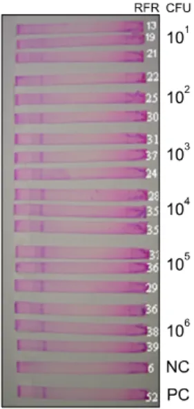
A simple, membrane-strip-based lateral-flow (LF) biosensor assay and a high-throughput microtiter plate assay have been combined with a reverse transcriptase polymerase chain reaction (RT-PCR) for the detection of a small number (ten) of viable Mycobacterium (M.) avium subsp. paratuberculosis (MAP) cells in fecal samples. The assays are based on the identification of the RNA of the IS900 element of MAP. For the assay, RNA was extracted from fecal samples spiked with a known quantity of (10
1to 10
6) MAP cells and amplified using RT-PCR and identified by the LF biosensor and the microtiter plate assay. While the LF biosensor assay requires only 30 min of assay time, the overall process took 10 h for the detection of 10 viable cells.
The assays are based on an oligonucleotide sandwich hybridization assay format and use either a membrane flow through system with an immobilized DNA probe that hybridizes with the target sequence or a microtiter plate well. Signal amplification is provided when the target sequence hybridizes to a second DNA probe that has been coupled to liposomes encapsulating the dye, sulforhodamine B. The dye in the liposomes provides a signal that can be read visually, quantified with a hand-held reflectometer, or with a fluorescence reader. Specificity analysis of the assays revealed no cross reactivity with other mycobacteria, such as M. avium complex, M. ulcerans, M. marium, M. kansasii,
M. abscessus, M. asiaticum, M. phlei, M. fortuitum, M.scrofulaceum, M. intracellulare, M. smegmatis, and M. bovis.
The overall assay for the detection of live MAP organisms is comparatively less expensive and quick, especially in comparison to standard MAP detection using a culture method requiring 6-8 weeks of incubation time, and is significantly less expensive than real-time PCR.
Keywords: feces, lateral flow biosensor assay, liposomes, Mycobacterium avium subsp. paratuberculosis, RT-PCR
Introduction
Mycobacterium avium subsp. paratuberculosis (MAP) is the causative agent of Johne’s disease (JD), a chronic intestinal granulamatous infection affecting domestic and wild ruminants [7,11,15,32]. Although cattle are usually infected early in life, clinical signs do not develop until 2-4 years of age, which makes early diagnosis of this infection a difficult task. JD is considered to be an economically important disease and accounts for an annual loss of $220 million to the US dairy industry [25]. The proposed, but poorly defined association of MAP with Crohn’s disease in human beings, is also of concern [13,18,23,24]. The in vivo diagnosis of MAP infections is quite challenging and difficult in the pre-clinical stages since the majority of infected animals do not show symptoms of the disease.
Although the isolation and identification of MAP is the most definitive test for diagnosis, it is time-consuming and labor-intensive, requiring 8-12 weeks. Contamination is an added problem when MAP is cultured from fecal samples.
Although, PCR for IS900 sequences is of diagnostic value, at times PCR leads to false positive amplification due to the presence of environmental bacteria with similar sequences [10]. Novel sequences recently identified in the genome of MAP appear specific and may also be used in nucleic acid- based diagnostic tests [6,16]. Real time PCR-based assays, which involve high equipment costs and trained personnel, can be used only under well-established laboratory conditions and serological tests may lack sensitivity [8].
Most diagnostic laboratories continue to use traditional culture methods; few laboratories use molecular methods along with culture methods [14,21,26,30,31]. Development of bioanalytical systems, such as biosensors coupled with a reverse transcriptase PCR to achieve low limits of detection, will be useful in the rapid and accurate detection of MAP.
Biosensors based on nucleic acid hybridization and
liposome signal amplification have been shown to be very
useful in developing rapid, inexpensive, and easy-to-
handle systems for the detection and quantification of RNA molecules [1,2,4,5]. A biosensor is a lateral flow assay that provides visual or reflectance data within about 20 min of overall assay time [3]. A biosensor uses a membrane flow- through system with an immobilized DNA probe that hybridizes with the target. Signal amplification is provided when the target sequence hybridizes to a second DNA probe coupled to liposomes encapsulating the dye, sulforhodamine B (SRB). The amount of liposomes captured in the detection zone can be either read visually or quantified with a hand- held reflectometer.
For MAP diagnosis, the IS900 gene, with 15-20 copies [27], has been routinely used in PCR-based detection systems. However, in the past, IS900 primers have also amplified IS900-like PCR products, probably from environmental mycobacteria, and resulting in false positive results [10]. Despite this possibility, IS900 gene amplification should still serve as a good indicator when coupled to a high-specificity hybridization reaction, as proposed here. Apart from IS900, other novel sequences, such as ISMav2 [26] and ISMap02 [20,27], could also be potential candidates in PCR-based assays. In the current study, the development of a rapid biosensor assay for the detection of live MAP organisms employing IS900 gene sequences is described. This is the first time the biosensor assay for MAP has been demonstrated.
Materials and Methods Bacterial strain and growth
Mycobacterium avium subsp. paratuberculosis-66115-98, a clinical isolate available from the Department of Population Medicine and Diagnostic Sciences at Cornell University, was grown in 7H9 medium, supplemented with 10% oleic acid-albumin-dextrose-catalase (Becton, Dickinson and Company, USA) and Mycobactin J (Allied Monitor, USA). The cultures were grown at 37
oC for 8 weeks and used in this study.
RNA extraction
MAP cultures were centrifuged at 12,000 rpm for 10 min.
One ml of Trizol was added to the pellet, and the mixture was passed through the syringe and needle (22 gauge) several times. The mixture was kept at room temperature for 5 min.
Two hundred μl of chloroform was added and mixed vigorously for 15 sec and incubated at room temperature for 3 min. The mixture was spun in a microcentrifuge at 12,000 rpm for 15 min at 4
oC. The supernatant was transferred to a fresh microcentrifuge tube and an equal volume of 70%
alcohol was added at room temperature. The mixture was transferred to the minispin column of a RNeasy kit (Qiagen, USA) and RNA was isolated following the manufacturer’s protocol. The isolated RNA samples were treated with 10 U/ μl of RNase-free DNase I (Qiagen, USA) at 37
oC for 10
min, followed by heat inactivation at 95
oC for 5 min, and then chilled on ice.
Estimation of cell quantity by optical density
MAP organisms were quantified by measuring the optical density at 550 nm as described earlier [17]. An optical density of 0.25 at 550 nm was equivalent to approximately 10
8organisms per ml.
Quantitation of cell number
The organisms were harvested by centrifugation, diluted in phosphate buffered saline (PBS; NaCl, 0.8%; KCl, 0.02%; Na
2HPO
4, 0.115%; and KH
2PO
4, 0.02% [pH 7.2]) containing 0.05% Tween-80, loaded on the platform of an improved Neubauer haemocytometer chamber, and visually counted.
Preparation of spiked fecal samples
Fecal samples were collected from healthy animals for initial standardization. Ten-fold serial dilutions of viable MAP organisms were prepared from a stock suspension of 10
8organisms. Aliquots of each bacterial dilution (900 μl) were added to 100 mg of feces to yield bacterial numbers between 10
1and 10
6. For samples from infected animals, 25-50 gm of fecal samples were collected from 8 calves challenged with 10
7MAP cells/animal in milk replacer for 7 consecutive days. One hundred mg of fecal samples collected 2, 4, 6, 8, and 10 days after challenge were used for RNA isolation.
RNA extraction from spiked fecal samples
RNA was extracted from spiked fecal samples containing 10
1to 10
6organisms using Trizol (Invitrogen, USA) and the extracted RNA was resuspended in 10 μl RNase-free water. The isolated RNA samples were treated with 10 U/ μl of RNase-free DNase I (Qiagen, USA) at 37
oC for 10 min, followed by heat inactivation at 95
oC for 5 min, and then chilled on ice.
Reverse-Transcriptase PCR
RNA isolated from spiked fecal samples was amplified using a one-step RT-PCR kit (Qiagen, USA). IS900 primers were used for amplification. The RT-PCR products were electrophoresed and checked on a 1% agarose gel containing 5 μg of ethidium bromide. The amplified products were used in the biosensor assay.
Preparation of membranes
Polyethersulfone membranes (Pall, USA) were cut into
4.5 mm × 7.5 cm strips. Streptavidin was diluted in 0.4 M
NaHCO
3/Na
2CO
3buffer (pH 9.0) containing 5% methanol
in a final concentration of 20 pmol/ μl. Streptavidin was
spotted on the membrane strips using a Camag Linomat IV
TLC sample applicator (Camag Scientific, USA) and
Function Sequence 5’-3’ Length Location in IS900 Forward primer
Reverse primer Capture probe Reporter probe
ACCGTGCGCCCGGGAATATA GGAGTTGATTGCGGCGGTGA TTGGCCGATGGAGGCGAGGT*
GATCGACCTCAACGCCGG
†20 nt 20 nt 20 nt 18 nt
482-501 358-377 383-402 412-429
*The capture probe is biotinylated at the 5’ end. †The reporter probe had a 20 base oligonucleotide tag (gggggtgggggtgggggtgg) at the 3’ end.