Hepatoid Adenocarcinoma of the Stomach Presenting as a Huge Abdominal Mass
6
0
0
전체 글
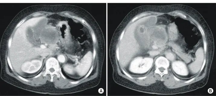
(2) 154. 대한외과학회지:제 66 권 제 2 호 2004. ꠏꠏꠏꠏꠏꠏꠏꠏꠏꠏꠏꠏꠏꠏꠏꠏꠏꠏꠏꠏꠏꠏꠏꠏꠏꠏꠏꠏꠏꠏꠏꠏꠏꠏꠏꠏꠏꠏꠏꠏꠏꠏꠏꠏꠏꠏꠏꠏꠏꠏꠏꠏꠏꠏꠏꠏꠏꠏꠏꠏꠏꠏꠏꠏꠏꠏꠏꠏꠏꠏꠏꠏꠏꠏꠏꠏꠏꠏꠏꠏꠏꠏꠏꠏꠏꠏꠏꠏꠏꠏꠏꠏꠏꠏꠏꠏꠏꠏꠏꠏꠏꠏꠏꠏꠏꠏꠏꠏꠏꠏꠏꠏꠏꠏꠏ. A. B. Fig. 1. Abdominal computed tomography (CT) scan, showing about 10 cm sized large lobulated mass in the upper abdomen, attached from the liver, gall bladder (A) and portal vein to pancreas (B).. B. A. C. 특이소견은 없었다. 혈액 검사에서 B형 간염항원 양성이었 고 AFP가 528 ng/ml 로 증가된 소견을 보였다. 초음파를 시 행하여 담낭과 위전정부, 십이지장 그리고 췌장두부 사이 에 장경 10 cm 이상의 다분엽상을 보이는 종양을 발견하였 다. 복부 전산화단층촬영에서 췌장의 두부와 위의 원위부 그리고 좌측 간 하부 공간에 걸쳐 장경 10 cm 이상의 분엽 을 보이는 조영이 증강된 종양이 있었다(Fig. 1A, B). 이 종. Fig. 2. Upper gastrointestinal radiological study, showing a mass with ulceration of Borrmann type II at the antrum on the greater curvature, suggesting stomach cancer (arrow) (A). Gastroduodenal fibroscopy reveals a 4×3 cm ulcerative lesion at the antrum (B) with extrinsic compression at the lesser curvature of the stomach, the duodenal bulb (arrow) (C).. 양은 십이지장 2번째 부분을 누르는 소견을 보였다. 위장관 조영술에서는 위의 전정부에 3 cm가 약간 넘는 궤양성 종 괴가 관찰되어 Borrmann II형, 위암이 의심되었다(Fig. 2A). 위내시경을 시행하였으며 대만부의 전정부에 중심부에 궤 양성 변화를 동반한 4×3 cm 크기의 병변을 발견하였고 소 만부 및 십이지장 구부가 압박받는 소견을 보였다(Fig. 2B, C). 내시경 조직검사에서 분화가 나쁜(poorly differentiation).
(3) 남소현 외:복강 내 거대 종양형태로 발생한 간세포암종 분화성 위선암. 155. ꠏꠏꠏꠏꠏꠏꠏꠏꠏꠏꠏꠏꠏꠏꠏꠏꠏꠏꠏꠏꠏꠏꠏꠏꠏꠏꠏꠏꠏꠏꠏꠏꠏꠏꠏꠏꠏꠏꠏꠏꠏꠏꠏꠏꠏꠏꠏꠏꠏꠏꠏꠏꠏꠏꠏꠏꠏꠏꠏꠏꠏꠏꠏꠏꠏꠏꠏꠏꠏꠏꠏꠏꠏꠏꠏꠏꠏꠏꠏꠏꠏꠏꠏꠏꠏꠏꠏꠏꠏꠏꠏꠏꠏꠏꠏꠏꠏꠏꠏꠏꠏꠏꠏꠏꠏꠏꠏꠏꠏꠏꠏꠏꠏꠏꠏ. 암종으로 면역조직화학염색에서 cytokeratin 양성, cytokeratin 7과 AFP 양성, 그러나 LCA, cytokeratin 20, vimentin에 는 음성을 보였다. 혈관 조영술을 시행하였고 문맥의 내강 이 경도로 좁아진 소견이 있어 종양에 의한 압박을 의심하 였다(Fig. 3). 이상의 검사소견을 종합해 수술 전 진단으로 위암 및 거대 림프절 전이를 가장 의심하였고 악성 위장관. 기질성종양 혹은 위 및 간 동시암 등을 의심하여 수술을 시행하였다. 수술소견에서 간문에서 십이지장 뒤쪽, 췌장두부의 뒤쪽, 위 소만부의 뒤쪽까지 차지하는 커다란 종양이 주변조직과 유착되어 있었으며 고유 간동맥이 좌우간동맥으로 분리되 는 부위까지 포함되어 있어 위아전절제술, 담낭․간 부분. P V. Fig. 3. Portovenography, showing a mild luminal narrowing of the main portal vein (arrow).. Fig. 4. Macroscopic appearance of the resection specimen, showing a gastric tumor at the antrum along the greater curvature, and large venous invasion in the lesser omentum (P = primary gastric carcinoma; V = venous invasion).. H. V A. B. C. Fig. 5. (A) The tumor was composed of cords of large polygonal cells with abundant eosinophilic cytoplasm arranged in trabecular pattern, hepatocellular carcinoma - like cells (H&E, ×100), (B) Microphotography of venous invasion, hepatoid tumor cells predominantly located in the vein (V = vein; H = intravenous hepatoid adenocarcinoma) (H&E, × 40), (C) Immunohistochemical staining for alphafetoprotein (AFP) shows focally strong positive reaction (arrow)(×100)..
(4) 156. 대한외과학회지:제 66 권 제 2 호 2004. ꠏꠏꠏꠏꠏꠏꠏꠏꠏꠏꠏꠏꠏꠏꠏꠏꠏꠏꠏꠏꠏꠏꠏꠏꠏꠏꠏꠏꠏꠏꠏꠏꠏꠏꠏꠏꠏꠏꠏꠏꠏꠏꠏꠏꠏꠏꠏꠏꠏꠏꠏꠏꠏꠏꠏꠏꠏꠏꠏꠏꠏꠏꠏꠏꠏꠏꠏꠏꠏꠏꠏꠏꠏꠏꠏꠏꠏꠏꠏꠏꠏꠏꠏꠏꠏꠏꠏꠏꠏꠏꠏꠏꠏꠏꠏꠏꠏꠏꠏꠏꠏꠏꠏꠏꠏꠏꠏꠏꠏꠏꠏꠏꠏꠏꠏ. 절제를 동반한 종괴 절제술을 시행하였다. 조직검사에서 위의 대만부에서 기원한 5.0×4.0×1.5 cm 의 간세포암종 분화성 선암종, 진행성 위암, Borrmann III형 을 진단하였다. AFP 염색에서는 부분적으로 강양성을 보였 다. 위의 원발성 종양은 장막하층까지 침범하였으며 정맥 침범이 심하였고 26개의 림프선에서는 종양이 발견되지 않 았다. 또한 11×8 cm의 커다한 종괴는 ‘소망으로 전이된 간 세포암종 분화성 선암종’으로 진단되었다(Fig. 4, 5). 간과는 유착된 소견을 보였으며 절제된 간 및 담낭에서는 종양이 발견되지 않았다. 수술 직후에 시행한 혈중 AFP는 121 ng/ml로 감소된 소견을 보였다. 술 후 환자는 합병증 없이 퇴원하였으며 5-FU, mitomycin으로 항암화학요법을 4차례 시행하였고 혈중 AFP는 1.5 ng/ml으로 감소하였다. 현재 재 발 및 전이의 증거 없이 9개월째 생존하고 있다.. 고. 찰. 간세포암종 분화성 선암종은 간 이외의 장기에서 AFP를 분비하며 조직학적으로 간세포와 유사한 형태 및 기능을 가지는 선암종의 변이라 하여 1985년 Ishikura(1)가 명명하 였다. 이 종양은 위, 식도, 바터 팽대부, 대장, 폐, 담낭, 부신, 신장, 방광, 난소, 자궁, 고환 등에서(3) 발생한 것으로 보고 되었다. AFP를 생성하는 종양은 전체 위암의 1.3∼15%를 차지하고 있으며(4) 이 중 간세포암종 분화성 선암종이 가 장 많다.(16) 다른 장기보다 간세포암종 분화성 선암종은 위에서 가장 많이 발생하고 있는데 그 이유는 정확히 알려 진 바 없다.(3) 다만 간과 위 둘 다 전장에서 기원하는 장기 이며 태생학적으로 위와 간배아(liver bud)가 가까운 위치에 서 발생하는 데서 그 원인을 찾고 있으며(5,8) 또한 첫째로 선암성 종양의 진행과정에서 간세포 표현형(phenotype)을 획득하거나 둘째로는 두 가지 분화능을 가질 것으로 추정 되는 세포가 발암 과정 중 선상피와 간세포 모두의 표현형 으로 분화하는 능력을 갖고 있는 것으로 추측하고 있다.(2) AFP를 생성하는 것은 간세포암의 특징적인 소견이며 간 세포암종 분화성 선암종을 진단하기 위해 혈청 AFP 상승 및 AFP 양성인 암세포의 존재가 필수적이며 이것이 불량한 예후와도 관계가 있다고 여겨져 왔다. 그러나 간세포 이외 에도 난황낭난종, 사람의 수태물(conceptus), 역형성세포(anaplastic cell)에 의해서도 AFP가 분비될 뿐만 아니라(4) AFP 를 분비하지 않는 간세포암종 분화성 선암종이 보고되면서 이 종양의 진단에 대해 새로운 기준이 마련되었다.(2,14) Motoyama는 AFP를 생성하는 위암을 크게 세 종류로 나누 어 보고하고 각각의 예후에 대해 비교한 바 있다.(13) 이는 간세포암종 분화성 선암종, 난황난낭종 유사 종양(yolk sac tumor like tumor), 그리고 태아 위장관 종양형(fetal gastrointestinal type)의 세 가지로 나뉘며 각각의 현미경 소견 및 면역조직화학염색 소견이 다르게 나타났다. 또한 태아 위. 장관 종양형에서 분비되는 AFP는 간세포암종 분화성 선암 종에서 분비되는 AFP와 달라서 다른 범주의 종양임을 시사 한 바 있다. 세 가지 종류의 종양은 각각 다른 예후를 보였 는데 간세포암종 분화성 선암종은 가장 흔하지만 다른 둘 에 비하여 현저히 생존율이 떨어졌다.(16) 조직학적인 특성으로 간세포성 분화세포는 커다란 호산 성 세포질과 둥글거나 난원형의 핵을 함유하고 있으며(2,6) 이 세포들이 판(sheets), 둥우리 형태(rounded nest), 띠(cords) 혹은 주형(trabeculae) 형태로 배열되어 있는 특징을 보인 다.(14) 세포질의 당원함유량에 따라 PAS-양성, diastase 에 저항을 보이는 미립들이 보이며 대부분에서는 세포내 혹은 세포밖 초자양소적(hyaline globule)들을 관찰할 수 있 다.(2,3,5) 때로 담즙 색소가 관찰될 수 있는데 이것은 진단 적 가치를 가진다.(2,17) Kodama 등(4,18)은 간세포암종 분화성 선암종이 면역조 직화학염색에 근거해 두 종류의 조직학적 소견을 보이는 사실을 기술하였다. 한 가지는 수질형(medullary type)으로 커다한 다형성 혹은 다핵거세포가 충실형의 판상 혹은 둥 우리(solid nest or sheets), 주형(trabecular type)으로 배열되는 양상을 보이는 것이며 다른 한 가지는 유두형 혹은 관상형 을 보인다. 이 두 가지는 한 종양 내에서 공존할 수 있으며 (4,8) 각각 다른 면역조직화학염색 특성을 보인다. 간세포성 부위(hepatoid foci)에서는 대개 주형 또는 충실형 배열상을 보였으며(5) AFP 양성, albumin, prealbumin, A1-antitrypsin, A1-antichymotrypsin 양성, CEA 음성 소견을 보인다. 선암종 부위(glandular foci)에서는 대부분 유두-선관형의 배열상이 었고(5) 장상피의 모양(intestinal type)을 보이며 낮은 AFP level, CEA 강양성, AFP 약양성, albumin, prealbumin, A1antitrypsin, A1-antichymotrypsin 음성소견을 보인다.(3,8) 이 두 부위 간의 이행은 점진적으로 나타나며(5) 대부분의 종 양은 비간세포성 분화부위, 즉 선암성 분화부위를 포함하 고 있다.(3) 본 증례에서는 선암성 분화부는 거의 찾아볼 수 없었고 간세포성 분화부위가 대부분을 차지하고 있었으며 종양에서 AFP는 부분적으로 강양성을 보였다. 음성을 보이 는 부위에는 간세포성 분화부가 다수 포함되어 있어서 이 종양을 AFP 양성만으로 진단내릴 수 없다는 앞선 보고들을 뒷받침하고 있다. 이 종양의 예후는 5년 생존율이 11.9%로(14) 비간세포암 종 분화성 선암종의 38.2%에 비해 매우 불량한 것으로 보고 되고 있다. 문헌에서 보고된 간세포암종 분화성 선암종은 수술 시 59예 중 53예에서,(3) 85예 중 70예에서(4) 전이가 보고되었으며(3) 그중 가장 많은 장기는 림프선과 간이었 다.(3,4) 조기위암이라 하더라도 간세포암종 분화성 선암종 은 동시적으로 혹은 향후에(metachronous) 간전이가 35%에 이른다.(19) 또한 Chang의 보고에 의하면 AFP를 분비하지 않는 종양과 비교해 림프선 및 혈관 침범의 양이 비슷한 경우에도 간세포암종 분화성 선암종은 예후가 불량한 것으.
(5) 남소현 외:복강 내 거대 종양형태로 발생한 간세포암종 분화성 위선암. 157. ꠏꠏꠏꠏꠏꠏꠏꠏꠏꠏꠏꠏꠏꠏꠏꠏꠏꠏꠏꠏꠏꠏꠏꠏꠏꠏꠏꠏꠏꠏꠏꠏꠏꠏꠏꠏꠏꠏꠏꠏꠏꠏꠏꠏꠏꠏꠏꠏꠏꠏꠏꠏꠏꠏꠏꠏꠏꠏꠏꠏꠏꠏꠏꠏꠏꠏꠏꠏꠏꠏꠏꠏꠏꠏꠏꠏꠏꠏꠏꠏꠏꠏꠏꠏꠏꠏꠏꠏꠏꠏꠏꠏꠏꠏꠏꠏꠏꠏꠏꠏꠏꠏꠏꠏꠏꠏꠏꠏꠏꠏꠏꠏꠏꠏꠏ. 로 알려져 있다.(20) 불량한 이유로 여러 가지가 제시되고 있다. 그중 하나로 간으로 전이하는 성질이 강한 것을 들고 있다. 1994년 Aizawa의 동물실험에 의해 AFP를 생성하는 세포 중 간세포 성 분화를 보이지 않는 종양에 비해 간세포성분화를 보이 는 종양이 간전이를 훨씬 더 잘한다는 것이 증명되었 다.(15) 다음으로 림프선으로의 과도한 전이를 들 수 있다. 문헌상 보고된 종양의 대부분은 임파선 전이를 가지고 있 었으며(3,4) 심지어 Kang 등의 보고에 의하면 위에서는 전 혀 발견할 수 없었던 간세포분화성 선암종이 장간막의 림 프선에서만 발견되고 혈청 AFP가 상승되었던 증례도 있 다.(12) 셋째는 Ishikura(1)가 제안한 바로 면역조직화학염색 의 특성을 들 수 있다. 이 종양이 AFP, albumin, prealbumin, A1-antitrypsin, A1-antichymotrypsin 등에 양성을 보이는데 이들은 침습력을 항진시킬 수 있으며 면역억제능, 단백분 해효소 억제능을 가지고 있는 것으로 알려져 있다.(4,5,8) 단 백분해효소에 의해 세포독성 T세포가 종양세포를 분해할 수 있는데 이를 방해함으로써 종양의 성장을 돕는 결과는 낳는다. 또한 AFP를 생성하는 종양이 높은 증식력, 약한 세 포소멸능(apoptosis), 풍부한 신혈관생성능을 보이는 특성을 가지고 있으며 항암요법에 반응이 좋지 않은 것으로 연구 보고되었다.(21) 넷째로 혈관 침범력이 강한 것을 들 수 있다. 1997년 Ishikura는 이 종양이 국소 혈관 내에서 과다하게 증 식한다는 사실을 보고하였으며 혈관 내 과다증식으로 인한 두꺼운 종양혈전을 육안적으로 관찰할 수 있었다. 이는 혈관 내에서의 과도한 증식능력이 간세포성간암과 비슷한 성질이 있음을 보여준 것으로써 과다한 혈관 침범을 통해 조직학적 인 공격성을 가짐을 추정해 볼 수 있다.(7) 본 증례에서는 위의 보고와는 달리 림프선, 간의 전이 없 이 오로지 정맥 침범만 심하였다. 간문에서 십이지장 뒤쪽, 췌장두부의 뒤쪽, 위 소만부의 뒤쪽까지 차지하는 거대 종 괴는 위의 원발성 종양의 2배 이상의 크기였으며 조직학적 으로는 소망에 심한 전이를 보이고 있었다. 이는 대만부에 위치한 종양이 정맥 침범을 통해 전이된 것으로 해석할 수 있다. 이상을 종합해 볼 때 간세포암종 분화성 선암종은 혈청 AFP를 상승시킬 수 있는 원발성 위종양으로 확진은 조직학 적 소견, 면역조직화학염색소견에 의해 이루어지며 매우 불량한 예후를 가짐을 알 수 있다. 혈청의 AFP은 예후를 결정하는 결정적 요소는 아니지만 추적관찰에 유용하며 위 장관 종양에서 상승되어 있을 때 간세포암종 분화성 선암 종을 의심해 볼 만한 근거로서 중요하다. 간으로의 전이 성 향이 강하고 혈관 침범을 매우 잘한다는 점, 이 종양이 생성 하는 단백질의 면역 억제능, 그리고 림프선 전이가 빈번한 점 등을 들어 불량한 예후의 원인을 찾을 수 있다.. REFERENCES 1) Ishikura H, Fukasawa Y, Ogasawara K, Natori T, Tsukada Y, Aizawa M. An AFP-producing gastric carcinoma with features of hepatic differentiation. A case report. Cancer 1985 15;56: 840-8. 2) Kishimoto T, Yuichiro Nagai, Kazuki Kato, Ozaki D, Hiroshi Ishikura. Hepatoid adenocarcinoma: a new clinicopathological entity and the hypotheses on carcinogenesis. Medical Electron Microscopy 2000;33:57-63. 3) Roberts CC, Colby TV, Batts KP. Carcinoma of the stomach with hepatocyte differentiation (Hepatoid adenocarcinoma). Mayo Clin Proc 1997;72:1154-60. 4) Inagawa S, Shimazaki J, Hori M, Yoshimi F, Adachi S, Kawamoto T, et al. Hepatoid adenocarcinoma of the stomach. Gastric Cancer 2001;4:43-52. 5) Kang GH, Kim YI. Hepatoid adenocarcinoma of the stomachA pathologic Analysis of 14 cases. Korean J Pathol 1994:28: 620-8. 6) Petrella T, Montagnon J, Roignot P, Van Nieuvanhuyse A, Matagrin C, Michiels-Marzais D, et al. Alphafetoproteinproducing gastric adenocarcinoma. Histopathology 1995;26: 171-5. 7) Ishikura H, Kishimoto T, Andachi H, Kakuta Y, Yoshiki T. Gastrointestinal Hepatoid adenocarcinoma: venous permeation and mimicry of hepatocellular carcinoma, a report of four cases. Histopathology 1997;31:47-54. 8) deLorimier A, Park F, Aranha GV, Reyes C. Hepatoid carcinoma of the stomach. Cancer 1993;71:293-6. 9) Shimoyama S, Nozaki K, Kaminishi M, Motoi N, Murakami T. A rare case of alpha-fetoprotein-producing early gastric cancer. Hepatogastroenterology 2001;48:687-91. 10) Rassidakis GZ, Delladetsima JK, Letsos SP, Polyzos A, Yannopoulos A. Hepatoid adenocarcinoma of the stomach with extensive neuroendocrine differentiation and a coexisting carcinoid tumour. Histopathology 1998;33:186-8. 11) Sugawara Y, Konishi T, Hiraishi M, Sato A, Natomi H, Aoki T, et al. Hepatoid adenocarcinoma of the stomach: a case report. Hepatogastroenterology 1996;43:995-9. 12) Kang GH, Kim YI. Alpha-fetoprotein-producing gastric carcinoma presenting focal hepatoid differentiation in metastatic lymph nodes. Virchows Arch 1998;432:85-7. 13) Motoyama T, Aizawa K, Fujiwara T, Endoh Y, Watanabe H. Coexistence of choriocarcinoma and Hepatoid adenocarcinoma in the stomach. Pathol Int 1994;44:716-21. 14) Nagai E, Ueyama T, Yao T, Tsuneyoshi M. Hepatoid adenocarcinoma of the stomach. A clinicopathologic and immunohistochemical analysis. Cancer 1993;72:1827-35. 15) Aizawa K, Motoyama T, Suzuki S, Tanaka N, Yabusaki H, Tanaka S, et al. Different characteristics of hepatoid and nonhepatoid alpha-fetoprotein producing gastric carcinomas: an.
(6) 158. 대한외과학회지:제 66 권 제 2 호 2004. ꠏꠏꠏꠏꠏꠏꠏꠏꠏꠏꠏꠏꠏꠏꠏꠏꠏꠏꠏꠏꠏꠏꠏꠏꠏꠏꠏꠏꠏꠏꠏꠏꠏꠏꠏꠏꠏꠏꠏꠏꠏꠏꠏꠏꠏꠏꠏꠏꠏꠏꠏꠏꠏꠏꠏꠏꠏꠏꠏꠏꠏꠏꠏꠏꠏꠏꠏꠏꠏꠏꠏꠏꠏꠏꠏꠏꠏꠏꠏꠏꠏꠏꠏꠏꠏꠏꠏꠏꠏꠏꠏꠏꠏꠏꠏꠏꠏꠏꠏꠏꠏꠏꠏꠏꠏꠏꠏꠏꠏꠏꠏꠏꠏꠏꠏ experimental study using xenografted tumors. Int J Cancer 1994;58:430-5. 16) Motoyama T, Aizawa K, Watanabe H, Fukase M, Saito K. alpha-Fetoprotein producing gastric carcinomas: a comparative study of three different subtypes. Acta Pathol Jpn 1993;43: 654-61. 17) Ishikura H, Ishiguro T, Enatsu C, Fujii H, Kakuta Y, Kanda M, et al. Hepatoid adenocarcinoma of the renal pelvis producing alpha-fetoprotein of hepatic type and bile pigment. Cancer 1991;67:3051-6. 18) Kodama T, Kameya T, Hirota T, Shimosato Y, Ohkura H, Mukojima T, et al. Production of alpha-fetoprotein, normal serum proteins, and human chorionic gonadotropin in stomach cancer: histologic and immunohistochemical analyses of 35. cases. Cancer 1981;48:1647-55. 19) Koga S, Kaibara N, Tamura H, Nishidoi H, Kimura O. Cause of late postoperative death in patients with early gastric cancer with special reference to recurrence and the incidence of metachronous primary cancer in other organs. Surgery 1984; 96:511-6. 20) Chang YC, Nagasue N, Abe S, Kohno H, Kumar DD, Nakamura T. Alpha Fetoprotein producing early gastric cancer with liver metastasis: report of three cases. Gut 1991;32: 542-5. 21) Chang YC, Nagasue N, Kohno H, Ohiwa K, Yamanoi A, Nakamura T. Xenotransplantation of alpha-fetoprotein-producing gastric cancers into nude mice. Characteristics and responses to chemotherapy. Cancer 1992;69:872-7..
(7)
수치
관련 문서
The Dutch physicist Pieter Zeeman showed the spectral lines emitted by atoms in a magnetic field split into multiple energy levels... With no magnetic field to align them,
Modern Physics for Scientists and Engineers International Edition,
If both these adjustments are considered, the resulting approach is called a bootstrap-BC a -method (bias- corrected-accelerated). A description of this approach
③ A student who attended Korean course at KNU Korean Language Program and holds TOPIK Level 3 or a student who completed Korean course Level 4 at the KNU Korean Language
Five days later, on 15 January 1975, the Portuguese government signed an agreement with the MPLA, FNLA and UNITA providing for Angola to receive its independence on 11
· 50% exemption from tuition fee Ⅱ for the student with a TOPIK score of level 3 or higher or completion of level 4 or higher class of the Korean language program at the
Since every classical or virtual knot is equivalent to the unknot via a sequence of the extended Reidmeister moves together with the forbidden moves, illustrated in Section 2,
웹 표준을 지원하는 플랫폼에서 큰 수정없이 실행 가능함 패키징을 통해 다양한 기기를 위한 앱을 작성할 수 있음 네이티브 앱과
