INTRODUCTION CASEREPORT C-1RootSchwannomawithAggressiveLateralMassInvasion
4
0
0
전체 글
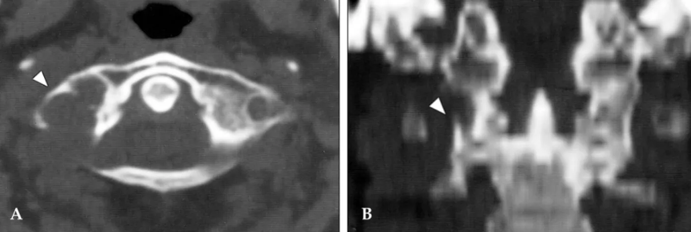
(2) Joo Han Kim, et al.. A. B. Fig. 1. A and B: Pre-operative CT scan with bone setting reveals extensive erosion with marginal sclerosis in the right transverse process and superior articular facet of C-1 (white triangle).. A. B. C. D. Fig. 2. (A, B) Axial T1-weighted MRI scan reveals a multilobulated mass involving the lateral mass of C-1 and enhancing moderately with indefinite tumor margin (white triangle). (C) Coronal T2-weighted MRI scan reveals mass in the right articular facet of C-1 spine (white triangle). (D) After 1 year, MRI scan demonstrates the remnant of the tumor after radiotherapy (white arrow).. according to established protocols. The histopathological diagnosis was a benign schwannoma composed of interwoven bundles of Yonsei Med J Vol. 46, No. 4, 2005. spindle-shaped, fibrillated cells (Fig. 3). An immunohistochemical investigation disclosed a strong reactivity for S-100 protein and vimentin. The.
(3) C1 Root Schwannoma with Lateral Mass Invasion. DISCUSSION. A. B. C Fig. 3. Histological examination reveals a schwannoma composed of interwoven bundles of spindle-shaped, fibrillated cells (hematoxylin and eosin, 200 ×, A) and immunopositivity for vimentin(200×, B) and S-100 (200 ×, C).. patient experienced no neurological deficit or any complication following surgery and she was discharged from the hospital seven days later. Follow-up MRI scans showed good stability of the atlanto-axial joint, but revealed remnants of the tumor which required the patient to undergo radiotherapy (Fig. 2).. Schwannomas represent 29% of spinal tumors, although neurofibromas are encountered less frequently. Schwannomas are typically solitary, circumscribed and encapsulated tumors eccentrically located on proximal nerves or the spinal nerve roots.1 Although schwannomas are relatively common benign tumors in the spinal region, there are only a few reports of C-1 neurinomas including schwannomas.2,4,5 In 1988, Guidetti 5 and Spallone reported three cases of C-1 tumors and Lot and George2 reported four cases of C1 neurinomas (three neurofibromas, one schwannoma) in 1997. Schwannomas are tumors that gradually increase in size, and only occasionally are they accompanied by pressure erosion of the adjacent bone, resulting in a concave deformity of the bony surface or an enlargement of the canal. Therefore, extensive erosion of the bone is usually considered uncommon for benign schwannomas, but common for malignant bone tumors. Schwannomas can involve bone through three possible mechanisms. First, a tumor can arise centrally within the bone (intraosseous schwannoma). Second, an extraosseous tumor can cause secondary erosion of the bone by pressure. Lastly, an extraosseous tumor can also arise within the nutrient canal and grow in a dumbbell-shaped configuration, resulting in the enlargement of the canal.6 In this case, the surgical findings indicated that the lesions were of the extraosseous type and were considered to originate in a C-1 nerve root. C1-root schwannomas can extend through wide spaces behind the lateral mass without extensive invasion because there is no intervertertebral foramen for the C-1 nerve root. Therefore, we speculate that the third mechanism, rather than the second mechanism, is related to the tumor growing in our patient and lead to the extensive expansion into the lateral vertebral mass. In an article on expansile vertebral bone lesions, extradural schwannomas were not included because osteoblastomas, aneurismal bone cysts, and giant-cell tumors are among the tumors usually considered when a patient has an expansile spinal lesion.7 Inoaka et al.6 presented two unusual cases of benign extraosseous neurinomas Yonsei Med J Vol. 46, No. 4, 2005.
(4) Joo Han Kim, et al.. associated with aggressive intravertebral extension in the thoracic and lumbar regions. The case reported here is an extremely rare example of a C-1 schwannoma with invasion of the lateral vertebral mass.. REFERENCES 1. Kalavakonda C, Sekhar LN, Jones RV, Rehaman AB. Intermittent vertebral artery compression caused by C1-root schwannoma: case report. Neurol Res 2000;22: 679-84. 2. Lot G and George B. Cervical neuromas with extradural components: surgical management in a series of 57. Yonsei Med J Vol. 46, No. 4, 2005. patients. Neurosurgery 1997;41:813-20. 3. Barnowsky L, Dalal R. Extradural schwannoma manifested as an expansile vertebral lesion. AJR Am J Roentgenol 1992;159:1352-3. 4. George B, Lot G. Neurinomas of the first two cervical nerve roots: a series of 42 cases. J Neurosurg 1995;82: 917-23. 5. Guidetti B, Spallone A. Benign extramedullary tumors of the foramen magnum. Adv Tech Stand Neurosurg 1988;16:83-120. 6. Inaoka T, Takahashi K, Hanaoka H, Aburano R, Tokusashi Y, Matsuno T, et al. Paravertebral neurinoma associated with aggressive intravertebral extension. Skeletal Radiol 2001;30:286-9. 7. Kumar R, Guinto FC, Jr., Madewell JE, David R, Shirkhoda A. Expansile bone lesions of the vertebra. Radiographics 1988;8:749-69..
(5)
수치

관련 문서
