ISSN 1225-6552, eISSN 2287-7630 https://doi.org/10.7853/kjvs.2018.41.1.57
< Case Report >
Veterinary Service
Available online at http://kjves.org
*Corresponding author: Jeong-Hee Han, Tel. +82-33-250-8657, Fax. +82-33-259-5625, E-mail. [email protected]
말초신경초 종양의 특징을 지닌
개 신경종의 조직병리학적 및 면역조직화학적 진단
이선규ㆍ이재하ㆍ한정희 *
강원대학교 수의과대학 및 동물의학연구소
Canine nervous-tissue tumors with features of peripheral nerve sheath tumor: histopathological and immunohistochemical findings
Sun-Gue Lee, Jae-Ha Lee, Jeong-Hee Han*
College of Veterinary Medicine and Institute of Veterinary Science, Kangwon National University, Chuncheon 24341, Korea (Received 5 March 2018; revised 14 March 2018; accepted 17 March 2018)
Abstract
Canine peripheral nerve sheath tumors (PNSTs) are spindle cell tumors that arise from Schwann cells, perineural cells, fibroblasts or all of them. Based on the morphology and biologic behavior, PNSTs are divided into benign PNST (BPNST) and malignant PNST (MPNST) forms. The aim of this study is to diagnose the two cases of neoplastic tissue samples with features of PNSTs by the histopathology and immunohistochemistry. The study was performed using two specimens from small animal clinic.
The first case, A was a mass, 3∼4 cm in diameter, extruded from vaginal mucosa of 10-year-old spayed female mixed-breed dog. And the second case, B was a subcutaneous mass, 1.5 cm in diameter, which is originated from right hind leg of 9-year-old castrated male mixed-breed dog. Two cases were stained with hematoxylin and eosin (H&E) for histopathological examination. And also im- munohistochemistry (IHC) was performed by the avidin-biotin peroxidase complex (ABC) method with antibodies specific for the following proteins: S-100 protein, smooth muscle actin (SMA) and epidermal growth factor receptor (EGFR). In results, Antoni B schwannoma pattern characterized by pleomorphic, round and fusiform polygonal cells was seen in A. In B, Antoni A pattern, densely packed spindle cells arranged in interlacing bundles was seen in addition to Antoni B pattern. In IHC, cytoplasms of neo- plastic cells were diffusely labeled for S-100 expression in A and B. For SMA, both A and B show negative expression. And for EGFR, A shows negative expression but B shows partially positive ex- pression in areas of Antoni B schwannoma pattern. The histopathologic features of two cases coupled with the S-100 immunoreactivity led to a diagnosis of PNST. For SMA, both A and B show negative expression. The diagnosis of A will be a BPNST with the negative result and B will be a MPNST with the positive result for EGFR.
Key words : PNSTs, Immunohistochemistry, Specific antibodies, Schwannoma pattern
서 론
개 신경종양(Canine nervous-tissue tumor)은 조직병 리학적 특징에 따라 분류하기가 까다롭고 용어도 완
전히 표준화되어 있지 않은 것으로 알려져 있다 (Gaitero 등, 2008; Gisele 등, 2015). 이에 따라 ‘개 연 부조직의 방추형 세포종양’(spindle cell tumor)이라고 도 사용되어 왔다(McColl과 Middleton, 1998). 신경종 양은 신경초종(schwannoma), 신경섬유종(neurofibroma), 혈관주위세포종(hemangiopericytoma)으로 분류된다. 악
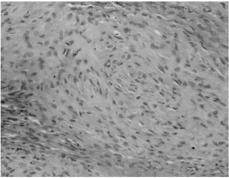
Fig. 1.Histopathological finding of A. Antoni B schwannoma pat- terns characterized by pleomorphic, round, fusiform and polygonal cells are seen. H&E stain. Bar=50 m.
성종양은 병변부가 희고 단단한 결절로 구성된 경우 가 많으며 침습성이 강한 특징이 있다. 이들 말초신 경초 종양(peripheral nerve sheath tumors, PNST)은 림 프계로는 전이되지 않는 특징을 지니며 노령의 개에 서 주로 발생한다(Koestner와 Higgins, 2002).
PNST의 정확한 병인은 알려진 바가 없으나, 외상 을 받았던 부위에 발생하는 경향이 있다. 신경집세포 (Schwann cell)는 손상을 받은 조직이나 세포가 정상 기능을 회복하는데 도움을 주는데, 이 과정에서 종양 으로 발전하는 것으로 추정되지만 규명한 연구결과 는 아직 없다(Sughrue 등, 2008). PNST의 일반적인 임 상증상으로는 원인불명의 앞다리나 뒷다리의 심한 통증, 만성적인 파행, 근육위축, 운동실조, 불완전 단 일마비, 고유수용감각이상, 자가절단, 근긴장저하, 반 사저하 등이 있다(Koestner와 Higgins, 2002). 척수가 종양에 의해 압박을 받는 경우에는 Honer 증후군이 나 마비를 보이기도 한다. 드문 경우지만, 하안검의 미약한 상승이나 동공축소 등을 보이기도 한다(Koestner 와 Higgins, 2002).
PNST의 진단은 면역염색법이 주로 이용되고 있는 데 아직까지는 확실한 진단기준이 되지는 못하며 계 속적으로 적합한 면역염색 marker의 연구가 진행되고 있다(Sirri 등, 2016). S-100단백질은 연부조직 종양이 신경유래인지 아닌지를 감별하는데 사용된다(Abbas 등, 2013). 수의학에서도 S-100 단백질로는 연부조직 종양의 분류가 쉽지 않으나, PNST의 marker로서는 유용하다. SMA (Smooth muscle actin)는 calponin, des- min과 함께 혈관주위세포종을 감별하는데 유용하다 고 알려져 있다(Sawamoto 등, 1999; Sirri 등, 2016).
최근에는 악성 PNST의 marker로서 주목받고 있는 단 백질인 EGFR (epidermal growth factor receptor)은 170 kDa의 RTK (receptor tyrosine kinase) 단백질로서, 사 람 악성 PNST의 80%에서 표현을 보이고 대부분의 마우 스 세포주에서도 표현을 나타냈다. EGFR은 EGFR- STAT3 signaling pathway를 통해 악성종양의 형성을 증진하는 것으로도 알려져 있다(Jianqiang 등, 2014).
본 연구의 목적은 PNST의 특징을 갖는 신경종양의 2예를 조직병리학적 및 면역염색학적 방법을 이용하 여 진단함으로써 소동물 임상에 도움을 주고자 보고 하는 바이다.
재료 및 방법
본 연구는 경기도 고양시 소재 동물병원에서 의뢰 받은 2예를 대상으로 진행하였다. 첫번째 예(증례 1) 는 10년령의 중성화된 잡종 암컷의 질점막으로부터 돌출된 약 3 cm 직경의 종괴였으며, 두번째 예(증례 2)는 9년령의 중성화된 잡종 수컷의 뒷다리에 생긴 약 1.5 cm 직경의 피하 종괴였다. 종괴는 조직학적으 로 검경을 위하여 10% 중성 포르말린에 고정하고 파 라핀으로 포매 과정을 거쳤다. 조직은 4 m로 박절 하여 hematoxylin and eosin (H&E) 염색을 하였다. 면 역염색은, tri-ethoxysilyl propylamin이 처리된 slide glass 에 올려서 진행하였다. ABC법(avidin-biotin peroxidase complex method)을 사용하였으며 발색제로는 3,30 di- aminobenzidine (DAB)를 선택하였다. 또한 sodium cit- rate 용액을 약 90∼100°C로 가열하여 pH 9로 항원 복구과정을 수행하였다. 항체는 각각 S-100 (1:100;
Dako, Denmark), -SMA (1:450; Cell MarqueTM, USA), EGFR (1:100; Santa Cruz Biotechnology, USA) 을 사용하였다.
결 과
병리조직학적 소견으로 증례 1은 Antoni B schwan- noma pattern으로 관찰되었다. 느슨하게 짜여진 다형 태의 둥글고, 방추형의 세포들이 관찰되었으며, 이들 방추형 세포는 점액성 혹은 유리질화된 기질 또는 아
A B
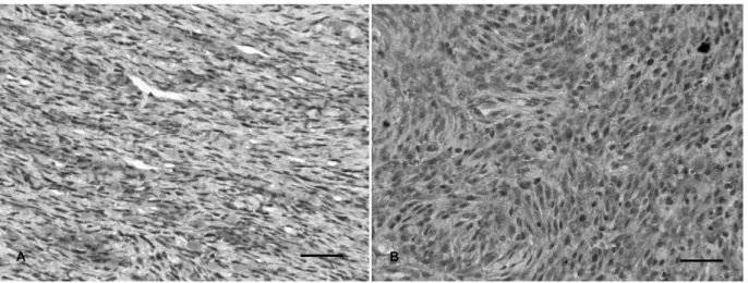
Fig. 3. Expression of S-100. A; Cytoplasms of neoplastic cells were diffusely labeled for S-100 expression. B; Expression of S-100 in areas of Antoni A and B schwannoma patterns. IHC. Bar=50 m.
A B
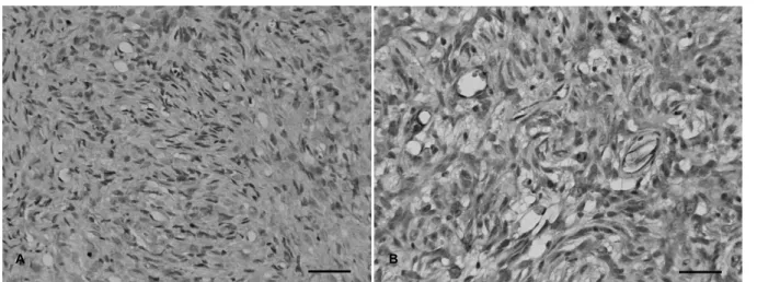
Fig. 4. Expression of -SMA. A and B; Negative expression for SMA. IHC. Bar=50 m.
Fig. 2.Histopathological finding of B. Antoni A patterns, densely packed spindle cells arranged in interlacing bundles are seen. H&E stain. Bar=50 m.
교질 등에 의해 임의적으로 배열되어 있었다(Fig. 1).
증례 2에서는 Antoni A와 B의 schwannoma pattern 이 모두 나타났다. A예처럼 Antoni B pattern을 보이 는 부분이 있는 반면에 빽빽하게 모인 방추형세포가 소용돌이의 다발모양을 형성하며 얽혀있는 양상의 Antoni A pattern을 보이는 부분도 있었다(Fig. 2). A와 B예에서 세포분열상(mitotic figures)은 거의 관찰되지 않았다.
면역염색학적 소견으로 A와 B예의 종양세포들은 세포질부분에서 S-100에 대하여 양성을 보였다(Fig.
3). SMA는 A와 B예의 세포질 및 핵에서 음성으로 나 타났으나 혈관주위만 양성으로 확인되었다(Fig. 4).
EGFR은 A예는 음성을 보였지만 B예에서는 Antoni B schwannoma pattern의 세포들에서 국소적으로 양성을 보였다(Fig. 5).
A B
Fig. 5. Expression of EGFR. A; Negative expression for EGFR. B; Partially positive expression for EGFR especially in areas of Antoni B schwan- noma pattern. IHC. Bar=50 m.
고찰 및 결론
조직병리학적 소견을 통해 증례 1과 2 모두에서 schwannoma pattern이 관찰되었으며 면역염색 소견에 서 S-100 protein에 양성을 나타냈다. 따라서 두 증례 모두 PNST로 진단하였다. S-100 protein은 신경집세 포, 멜라닌세포, 신경아교세포로 구성된 신경릉세포, 연골세포, 근상피세포, 지방세포, 대식세포 등에서 유 래되는 산성(acidic)의 단백질로서 vimentin과 함께 PNST에서 높은 양성율을 나타내는 것으로 알려져 있 기 때문이다(Chijiwa 등, 2004; Franziska 등, 2015; Silvia 등, 2016; Sirri 등, 2016). PNST와 감별진단해야 하는 종양들은 섬유육종(firosarcoma), 혈관주위세포종(he- mangiopericytoma) 그리고 무흑색소성 흑색종(amelanotic melanoma) 등이 있다(Klopfleisch 등, 2013). 섬유육종 은 S-100 protein을 발현하지 않기 때문에 감별진단할 수 있었다. SMA의 결과는, A와 B예에서 발현되지 않 았으며 SMA는 평활근세포의 isoform한 형태로서 혈 관의 평활근세포나 외막세포의 세포질에서 주로 관 찰된다(Omar 등, 1989). SMA와 밀접한 연관이 있는 혈관주위세포종은, 모세혈관의 pericyte에서 유래하는 종양으로 80%가 SMA에 양성발현을(Chijiwa 등, 2004), S-100 protein에 음성발현을 나타낸다(Silvia 등, 2016).
이에 따라 혈관주위세포종과도 감별된다. 사람에서 PNST는 135사례 중 1예에서만 SMA에 양성발현을 보였다(Sirri 등, 2016). EGFR의 결과는, A예는 음성 의 발현을, B예는 Antoni B schwannoma pattern 일부 영역에서 양성 발현을 보였다. EGFR은 세포 증식, 이 주, 부착, 혈관생성에 관여하는 RTK (receptor tyrosine
kinase) 단백질로서, 마우스와 사람의 Schwann cell의 전구세포 또는 악성 PNST 세포에서 발현하는 것으로 알려져 있다(Jianqiang 등, 2014). B예의 Antoni B 영 역에서 EGFR에 양성발현을 보이므로 악성의 PNST 로 진단하였다. A예는 PNST이지만 면역염색과 조직 병리학적 결과를 기초로 하여 양성종양으로 진단하 였다.
PNST의 면역염색의 marker가 확실하게 정립되어 않았으며 확진하는데 일관되게 합치되지 못하는 실 정이다(Sirri 등, 2016). 앞으로 PNST를 확진하는 면역 염색 marker의 지속적인 개발이 필요하다고 사료된다.
사 사
2015년도 강원대학교 대학회계 학술연구조성비로 연구하였음(과제번호-D1000287-01-01).
REFERENCES
Abbas T, Javad J, Fariba K, Ehsan H, Alimohammad B, Mehdi AH , Mohammadmehdi M. 2013. Ulnar malignant pe- ripheral nerve sheath tumour diagnosis in a mixed-breed dog as a model to study human: histologic, immunohis- tochemical and clinicopathologic study. Diagn Pathol 8:
86.
Chijiwa K, Uchida K, Tateyama S. 2004. Immuno- histochemical evaluation of canine peripheral nerve sheath tumors and other soft tissue sarcomas. Vet Pathol 41: 307-318.
Franziska H, Gwendolyna R, Kershaw O, Corinna EJ. 2015.
Malignant peripheral nerve sheath tumor of the third eyelid in a 3-year-old Rhodesian Ridgeback. Clin Case Rep 3: 50-56.
Gaitero L, Anor S, Fondevila D, Pumarola M. 2008. Canine cuta- neous spindle cell tumours with features of peripheral nerve sheath tumours: A histopathological and im- munohistochemical study. J Comp Pathol 139: 16-23.
Gisele S, Daniele M, Fabiana W, Neusa B, Tatiane TN, Gustavo S, Luciana S, David D. 2015. Retrospective canine skin peripheral nerve sheath tumors data with emphasis on histologic, immuno-histochemical and prognostic factors.
Pesq Vet Bras 35: 965-974.
Jianqiang W, Deanna M, Edwin J, David W, Kimberly B, Ami V, Eric B, James R, Timothy P, Anat O, Nancy R. 2014.
EGFR-STAT3 signaling promotes formation of malig- nant peripheral nerve sheath tumors. Oncogene 33:
173-180.
Klopfleisch R, Meyer A, Lenze D, Hummel M, Gruber AD.
2013. Canine cutaneous peripheral nerve sheath tumours versus fibrosarcomas can be differentiated by neuro- ectodermal marker genes in their transcriptome. J Comp Patho 148: 197-205.
Koestner A, Higgins RJ. 2002. Peripheral nerve sheath tumors. pp 731-735 In: Meuten DJ, Tumors in Domestic Animals, 4th ed, Blackwell Publishing, Iowa State Press, Ames,
Iowa, USA.
McColl WM, Middleton DJ. 1998. Cutaneous soft tissue tumours in dogs: classification, differentiation and histogenesis.
Vet Dermatol 9: 43-48.
Omar S, Marie-Francoise P, Marie-Claire P, Giulio G, Patrizia G, Gianni B, Mariella R, Lelio O. 1989. -Smooth muscle actin, a differentiation marker of smooth muscle cells, is present in microfilamentous bundles of pericytes. J Histochem Cytochem 37: 315-321.
Sawamoto O, Yamate J, Kuwamura M, Hagiwara R, Kurisu K.
1999. A canine peripheral nerve sheath tumor including peripheral nerve fibers. J Vet Med Sci 61: 1335-1338.
Sílvia T, Irina A, Alexandra R, Fátima F, Fátima G. 2016.
Molecular heterogeneity of canine cutaneous peripheral nerve sheath tumors: a drawback in the diagnosis refinement. In Vivo 30: 819-828.
Sirri R, Sabattini S, Bettini G, Mandrioli L. 2016. Reclassifica- tion of 21 presumptive canine peripheral nerve sheath tumors (PNST) using a literature-based immuno- histo- chemical panel. Acta Vet Beograd 66: 455-465.
Sughrue ME, Jon L, Barbarol NM. 2008. Pain as a symptom of peripheral nerve sheath tumors: clinical significance and future therapeutic directions. J Brachial Plex Peripher Nerve Inj 3: 6.