INTRODUCTION
Gastric cancer (GC) develops from the lining of the stomach and ranks fourth in incidence and second in mortality among all cancers worldwide.1,2 Since most cases are diagnosed at advanced stages with poor prognosis and limited treatment options, GC remains a major clinical challenge.3,4 Although considerable effort has been directed toward the develop- ment of surgical and chemotherapeutic interventions, as well as increased GC screening rates, the prognosis of GC remains
poor, and the molecular mechanisms of GC progression are not completely understood.5-7
The development of GC is a complex and multifactorial pro- cess involving a number of genetic alterations.8 During this pro- cess, the differential expression of particular genes and mi- croRNA (miRNA) plays a very important role.9 Accumulating evidence suggests that dysregulation of miRNAs is intimately involved in the carcinogenesis, progression, and metastasis of many cancers, including GC and that the alteration of certain miRNAs may be biomarkers of use in detecting early GC.10 As a miRNA, miRNA-381 has been reported to play suppressive or promoting roles in the progression of cancer.11 Liang, et al.12 indicated that the down-regulation of miRNA-381 promotes cell proliferation and invasion in colon cancer. A previous study showed that miRNA-381 inhibits the metastasis of GC by targeting TMEM16A expression.13 Zhang, et al.14 showed that miRNA-381 inhibited migration and invasion in human GC through the down-regulation of SRY-Box 4. Although dif- ferential expression of miRNA-381 in GC has been described in previous study, the detail diagnostic value of miRNA-381 in
Serum microRNA-381: A Potential Marker for Early Diagnosis of Gastric Cancer
Ye Li1, Huihui Sun2, Jie Guan3, Tingting Ji4, and Xinwei Wang5
Departments of 1Gastroenterology and 5General Surgery, The 5th People’s Hospital of Ji’nan, Jinan, Shandong;
2Department of Gastroenterology, Jinan First People’s Hospital, Jinan, Shandong;
3Department of Gastrointestinal Surgery, Shandong Institute of Cancer Prevention and Control, Jinan, Shandong;
4Department of Movement Control Section, Jinan Medical Emergency Center, Jinan, Shandong, China
Purpose: The purpose of this study was to explore the potential early diagnostic value of serum microRNA-381(miRNA-381) in patients with gastric cancer (GC).
Materials and Methods: Patients with advanced gastric cancer (AGC) and early gastric cancer (EGC), as well as healthy individu- als, were enrolled in this study. Expression of miRNA-381 in serum was detected using real-time quantitative PCR. Electrochemi- luminescence analysis was used to investigate the expression of classic tumor markers, including carbohydrate antigen (CA) 199, CA724, and carcinoembryonic antigen. Finally, receiver operating characteristic curve and Kaplan-Meier analysis were used to determine the value of miRNA-381 in clinical diagnosis of GC.
Results: miRNA-381 was differentially expressed among the study groups. AUC analysis showed that the sensitivity and specifici- ty of serum miRNA-381 in the diagnosis of GC were superior to those of other tumor markers. Furthermore, low levels of miR- NA-381 expression were positively correlated with lymph node metastasis and AGC. Finally, Kaplan-Meier survival analysis showed that down-regulation of miRNA-381 was associated with lymph node metastasis and the development of GC.
Conclusion: miRNA381, which was down-regulated in GC, might be a novel early diagnosis marker for patients with GC.
Key Words: Gastric cancer, serum microRNA-381, early diagnosis, tumor markers
pISSN: 0513-5796 · eISSN: 1976-2437
Received: April 24, 2019 Revised: June 5, 2019 Accepted: June 18, 2019
Corresponding author: Xinwei Wang, BM, Department of General Surgery, The 5th People’s Hospital of Ji’nan, No. 24297, Jingshi Road, Jinan, Shandong 250022, China.
Tel: 86-0531-87197138, Fax: 86-0531-87192659, E-mail: [email protected]
•The authors have no potential conflicts of interest to disclose.
© Copyright: Yonsei University College of Medicine 2019
This is an Open Access article distributed under the terms of the Creative Com- mons Attribution Non-Commercial License (https://creativecommons.org/licenses/
by-nc/4.0) which permits unrestricted non-commercial use, distribution, and repro- duction in any medium, provided the original work is properly cited.
Yonsei Med J 2019 Aug;60(8):720-726 https://doi.org/10.3349/ymj.2019.60.8.720
serum in GC has not been fully investigated yet.
In the present study, we aimed to investigate the role of se- rum miRNA-381 in the early diagnosis of GC. We found that the levels of miRNA-381 in advanced gastric cancer (AGC) were significantly lower than those in early gastric cancer (EGC) and that reduced expression of miR-381 was positively correlated with lymph node metastasis and GC progression.
We sought to explore the early diagnostic value of serum miR- NA-381 in GC and to provide a novel strategy for GC therapy.
MATERIALS AND METHODS
Baseline information of patients
Patients with GC confirmed by gastroscopy or surgical pathol- ogy were selected from department of gastroenterology of The 5th People’s Hospital of Ji’nan for May 2016 to May 2018. All GC patients were diagnosed by pathology without any anti- neoplastic treatment. Among them, a total of 80 serum sam- ples were collected from AGC patients (51 males and 29 fe- males; average age of 61.73±10.63 years). Meanwhile, 40 serum samples from EGC patients (26 males and 14 females;
average age of 58.25± 9.67 years) and 40 serum samples from healthy controls (22 males and 18 females; average age of 60.20±10.70 years) were collected among outpatient health examinees of People’s Hospital Affiliated to Inner Mongolia
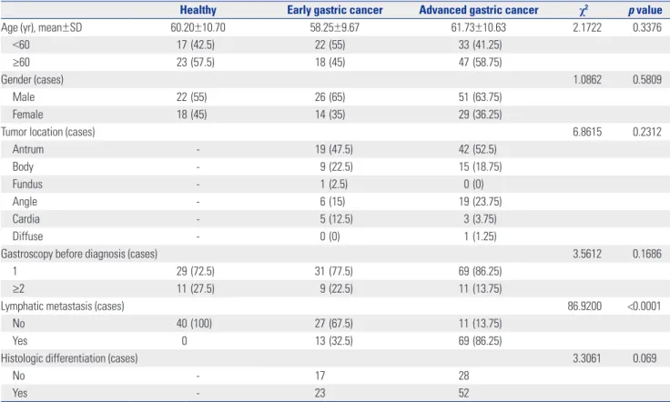
Medical University. The detail baseline information of the GC patients is showed in Table 1. The experimental scheme of this study was approved by the Ethics Committee of The 5th People’s Hospital of Ji’nan (IRB No. 2018066). Patient partici- pation was voluntary, and all patients provided written in- formed consent.
Real-time quantitative PCR analysis
Total RNA was extracted from early morning fasting venous blood using miRcute Serum/Plasma miRNA Isolation Kits (Beijing Tiangen Biochemical Technology Co., Ltd., Beijing, China). Reverse transcription was performed using miRNA RT Reaction Buffer and miRNA RT Enzyme Mix (Beijing Tian- gen Biochemical Technology Co., Ltd.). Real-time quantitative PCR (RT-qPCR) was performed on ABI7500 (Thermo Fisher Scientific, Waltham, MA, USA) using special primers (miR- NA-381, forward: 5'-UAUACAAGGGCAAGCUCUCUGU-3';
reverse: 5'- AGAGAGCUUGCCCUUGUAUAUU- 3'). U6 was used as an internal control (U6, forward: 5'-TCGCCCTTGGCA CAGCA-3'; reverse: 5'-CGAACCATTCAAGTGTTGCT-3'). The PCR program included 95°C for 10 min, 40 cycles of 95°C for 10 s, 60°C for 20 s, and 72°C for 34 s. The relative expression of miRNA-381 was calculated using the 2-ΔΔCt method.15 The RT- qPCR was conducted according to the instructions of miRcute Plus miRNA qPCR Detection Kits (Thermo Fisher Scientific) and repeated three times for each experiment.
Table 1. Baseline Information for Study Participants
Healthy Early gastric cancer Advanced gastric cancer χ2 p value
Age (yr), mean±SD 60.20±10.70 58.25±9.67 61.73±10.63 2.1722 0.3376
<60 17 (42.5) 22 (55) 33 (41.25)
≥60 23 (57.5) 18 (45) 47 (58.75)
Gender (cases) 1.0862 0.5809
Male 22 (55) 26 (65) 51 (63.75)
Female 18 (45) 14 (35) 29 (36.25)
Tumor location (cases) 6.8615 0.2312
Antrum - 19 (47.5) 42 (52.5)
Body - 9 (22.5) 15 (18.75)
Fundus - 1 (2.5) 0 (0)
Angle - 6 (15) 19 (23.75)
Cardia - 5 (12.5) 3 (3.75)
Diffuse - 0 (0) 1 (1.25)
Gastroscopy before diagnosis (cases) 3.5612 0.1686
1 29 (72.5) 31 (77.5) 69 (86.25)
≥2 11 (27.5) 9 (22.5) 11 (13.75)
Lymphatic metastasis (cases) 86.9200 <0.0001
No 40 (100) 27 (67.5) 11 (13.75)
Yes 0 13 (32.5) 69 (86.25)
Histologic differentiation (cases) 3.3061 0.069
No - 17 28
Yes - 23 52
p<0.05 was considered as statistically significant. Variables are presented as a number (percentage) unless otherwise noticed.
Electrochemiluminescence assay
Electrochemiluminescence is chemiluminescence triggered by electrochemical techniques.16 Serum carbohydrate antigen (CA) 724, CA199, and carcinoembryonic antigen (CEA) were measured using the electrochemiluminescence immuno-as- say (Roche cobas e601, Roche Diagnostics GmbH, Mannheim, Germany). The detection thresholds of CA724, CEA, and CA199 were 6.9 U/mL, 6.5 ng/mL, and 27 U/mL, respectively.
Survival analysis
To reveal the prognostic value of factors of interest in patients with GC, survival analysis was performed. Patients with GC were grouped according to clinical information and patholog- ical parameters, including age, sex, stage of GC, expression levels of miRNA-381, lymph node metastasis, and differentia- tion of tumor tissue. Survival rate estimation was performed using the Kaplan-Meier method17 and Cox proportional haz- ards models.18,19
Statistical analysis
Statistical analysis was performed using SPSS, version 17.0 (SPSS Inc., Chicago, IL, USA) and GraphPad Prism 7.0 (GraphPad Softward, San Diego, CA, USA). The chi-square test was used to compare the counting data between groups.
The measurement data are expressed as a mean±SD. The means were compared using Student’s t-test between groups.
One-way ANOVA followed by Tukey’s multiple comparisons test was used for comparison among groups. The area under receiver operating characteristic curves (AUC) was used to analyze the clinical diagnostic value of the detection of miR- NA-381. The survival time of GC patients was tested by the Kaplan-Meier method. Prognosis was analyzed by Cox regres- sion. p<0.05 was regarded as statistically significant.
RESULTS
Optimal GC biomarkers
Normality test results showed that the data of each group con- formed to normal distribution; therefore, the data in each group are expressed as means±SDs. The serum levels of miR- NA-381 in the EGC group were significantly lower than those in the healthy group, while the levels of miRNA-381 in the AGC group were significantly lower than those in the EGC group (all p<0.05) (Fig. 1A). Moreover, the serum levels of CA199 in the EGC group were significantly higher than those in the healthy group, while the serum levels of CA199 in the AGC group were significantly higher than those in the EGC
Fig. 1. The expression of serum miRNA-381, CA199, CA724, and CEA among healthy people, EGC patients, and AGC patients. (A) The expression of miR- NA-381 in three groups. (B) The expression of CA199 in three groups. (C) The expression of CA724 in three groups. (D) The expression of CEA in three groups. One-way ANOVA was used; Tukey’s multiple comparisons test was used to compare the two comparisons after ANOVA analysis. *Compared with healthy group, p<0.05, †compared with EGC group, p<0.05. CA, carbohydrate antigen; CEA, carcinoembryonic antigen; EGC, early gastric cancer;
AGC, advanced gastric cancer.
1.5
1.0
0.5
0.0
40
30
20
10
0
50
40
30
20
10
0
15
10
5
0 EGC
EGC
EGC
EGC Healthy
Healthy
Healthy
Healthy AGC
AGC
AGC
AGC
*†
*†
*†
*†
* *
Relative miR-381 expressionCA724 (U/mL) CA199 (U/mL)CEA (ng/mL)
A
C
B
D
group (all p<0.05) (Fig. 1B). Furthermore, the levels of CA724 and CEA in the AGC group were significantly higher than those in the EGC group and healthy group (all p<0.05) (Fig. 1C and D).
Tumor marker evaluation based on AUC analysis To evaluate the value of miRNA-381 in the diagnosis of GC, receiver operating characteristic curves of serum miRNA-381, CA199, CA724, and CEA were investigated among the study groups. We found that AUC value of miRNA-381 was larger than AUC values for other tumor markers comparing the EGC group versus the healthy group (AUC of miRNA-381: 0.931) (Fig. 2A) and the EGC versus the AGC group (AUC of miR- NA-381: 0.922) (Fig. 2B). The sensitivity and specificity of se- rum miRNA-381 in the diagnosis of GC were better than those for other tumor markers (Table 2).
Prediagnosis evaluation and Kaplan-Meier analysis for miRNA-381 in GC
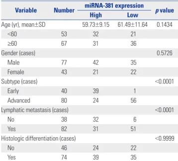
To further reveal the relationship between the expression of miRNA-381 and clinicopathological parameters of GC pa- tients, the mean expression level of miRNA-381 (0.6173) was measured as a node. Meanwhile, a total of 120 GC patients was divided into two groups: high miRNA-381 expression group (n=63) and low miRNA-381 expression group (n=57). In doing so, we found that low expression of miRNA-381 was positively correlated with lymph node metastasis and AGC (p<0.01). Detailed information of miRNA-381 expression and associated clinical characteristics is showed in Table 3.
Kaplan-Meier survival analysis showed that the overall sur- vival rate of GC patients with low expression of miRNA-381 was significantly shorter than that of GC patients with high expression of miRNA-381 (Fig. 3). Cox regression analysis showed that the expression of miRNA-381 [95% confidence
Fig. 2. AUC for miRNA-381, CA199, CA724, and CEA in gastric cancer patients and healthy individual. (A) The AUC values of miRNA-381, CA199, CA724, and CEA in EGC vs. healthy. (B) The AUC values of miRNA-381, CA199, CA724, and CEA in EGC vs. AGC. p<0.05 was considered a significant difference. AUC, area under the receiver operating characteristic curve; CA, carbohydrate antigen; CEA, carcinoembryonic antigen; EGC, early gastric cancer; AGC, ad- vanced gastric cancer.
1.0
0.8
0.6
0.4
0.2
0.0
1.0
0.8
0.6
0.4
0.2
0.0 0.2 0.4 0.6 0.8 1.0 0.0 AUC (C-Early)
0.0 0.2 0.4 0.6 0.8 1.0 AUC (Early-Advanced)
miRNA-381 CA199 CA724 CEA
miRNA-381 CA199 CA724 CEA
1-specificity 1-specificity
Sensitivity Sensitivity
A B
Table 2. Evaluation of Tumor Biomarkers
Tumor markers Cutoff Sensitivity (%)
Specificity (%)
AUC p value
Mean±SD 95% CI
EGC vs. AGC
miR-381 (fold change) 0.6557 83.75 97.5 0.931±0.029 0.875–0.987 <0.0001
CA199 (U/mL) 18.4700 80.00 60.0 0.761±0.047 0.669–0.853 <0.0001
CA724 (U/mL) 9.5200 65.00 87.5 0.843±0.035 0.775–0.911 <0.0001
CEA (ng/mL) 4.1950 68.75 77.5 0.788±0.040 0.709–0.867 <0.0001
EGC vs. healthy
miR-381 (fold change) 0.8687 82.50 92.5 0.922±0.032 0.860–0.984 <0.0001
CA199 (U/mL) 9.1950 72.50 60.0 0.710±0.058 0.596–0.823 0.0012
CA724 (U/mL) 2.2650 65.00 57.5 0.615±0.063 0.491–0.739 0.0766
CEA (ng/mL) 2.3850 70.00 62.5 0.631±0.064 0.506–0.756 0.0438
AUC, area under the receiver operating characteristic curve; CI, confidence interval; CA, carbohydrate antigen; CEA, carcinoembryonic antigen; EGC, early gastric cancer; AGC, advanced gastric cancer.
p<0.05 was considered significantly different.
interval (CI): 1.117–3.217; p=0.009], lymph node metastasis (95% CI: 0.295–0.828; p=0.007), and histologic differentiation (95% CI: 1.416–4.085; p=0.001) were correlated with the prog- nosis of GC (data not shown).
DISCUSSION
GC is one of the leading causes of cancer-related deaths
worldwide with poor diagnosis and few treatment strategies.20 The current study explored the early diagnostic value of se- rum miRNA-381 in patients with GC. Our results showed that miRNA-381 was differentially expressed in not only EGC ver- sus AGC but also EGC versus healthy individuals. Moreover, AUC analysis showed that the sensitivity and specificity of se- rum miRNA-381 in the diagnosis of GC were superior to other tumor markers. Furthermore, low levels of miRNA-381 ex- pression were positively correlated with lymph node metasta- sis and AGC. Finally, Kaplan-Meier survival analysis showed that the deregulation of miRNA-381 was associated with the development of GC.
The down-regulation of miRNA-381 is closed related with the progression of cancer.21 A previous study showed that miRNA-381 is significantly down-regulated in metastatic cells, compared to normal prostatic epithelial cells.22 Liang, et al.12 indicated that the down-regulation of miRNA-381 promotes cell proliferation and invasion in colon cancer. Rothschild, et al.23 showed that miR-381 was significantly down-regulated in human lung adenocarcinomas and that low miR-381 expres- sion levels are correlated with poor prognosis. A clinical in- vestigation on osteosarcoma indicated that osteosarcoma pa- tients with low expression of miRNA-381 experienced a longer survival time after surgical intervention and that miRNA-381 expression promotes cell proliferation and cell invasion abili- ty.24 A previous study showed that the down-regulation of miRNA-148a is associated with lymph node metastasis and poor clinical outcomes and functions as a suppressor of tu- mor metastasis in non-small cell lung cancer.25 Fujino, et al.26 indicated that the down-regulation of miRNA-100 is associat- ed with lymph node metastasis in early colorectal cancer with submucosal invasion. In the current study, the analysis of se- rum miRNA-381expression in GC patients showed that, com- pared with healthy controls, miRNA-381 was down-regulated in both AGC patients and EGC patients. Meanwhile, investi- gation of the relationship between miRNA-381 expression and clinicopathological features revealed that low expression of miRNA-381 was positive correlated with lymph node metas- tasis and AGC.
CEA, CA199, and CA724 are three classic biomarkers of the progression of GC.27 Ucar, et al.28 proved the prognostic values of preoperative CEA, CA199, and CA724 in GC clinical treat- ment. A previous study indicated that CEA and CA199 can be used for postoperative monitoring of recurrence in patients with AGC.29 Meanwhile, another study reported that CA724 (47.7%) showed a higher positivity rate for GC than CEA (25%) and CA 199 (25%).30 Compared with cystic fluid CEA determi- nation, miRNA detection holds great promise as molecular diagnostic tools for assessing cancer risk,31 as miRNAs act as oncogenes or tumor suppressors in a wide variety of human cancers.32 A previous study suggested that the down-regula- tion of serum miRNA-195 is an optimal biomarker in the clini- cal diagnosis of breast cancer, compared to CEA and CA153.33 Table 3. Correlations between Expression of miRNA-381 and Clinico-
pathological Features
Variable Number miRNA-381 expression
p value
High Low
Age (yr), mean±SD 59.73±9.15 61.49±11.64 0.1434
<60 53 32 21
≥60 67 31 36
Gender (cases) 0.5726
Male 77 42 35
Female 43 21 22
Subtype (cases) <0.0001
Early 40 39 1
Advanced 80 24 56
Lymphatic metastasis (cases) <0.0001
No 38 32 6
Yes 82 31 51
Histologic differentiation (cases) <0.9999
No 46 24 22
Yes 74 39 35
p<0.05 was considered significantly different.
Fig. 3. Kaplan-Meier survival analysis for gastric cancer patients based on differential expression of miRNA-381. p<0.05 was considered as a sig- nificant difference.
1.0
0.8
0.6
0.4
0.2
0.0
0 10 20 30 40 50 60 p<0.0001
Survival functions
Low HighLow-censored High-censored
Survival time (month)
Cumulative survival
miRNA-381 expression
Zhang, et al.34 indicated that miRNA-181a functions as an bio- marker in GC by targeting certain tumor suppressor genes.
Moreover, genome-wide miRNA profiles have suggested miR- 378 as a serum biomarker for early detection of GC.35 In the current study, AUC analysis based on potential tumor mark- ers, including miRNA-381, CA199, CA724, and CEA showed that the sensitivity and specificity of serum miRNA-381 in the diagnosis of GC were superior to the other tumor markers.
In conclusion, we suggest that miRNA-381, which is down- regulated in GC, might be a novel early diagnosis marker for patients with GC. Furthermore, we discovered that the down- regulation of miRNA-381 is positively correlated with lymph node metastasis and advanced stage disease in GC.
AUTHOR CONTRIBUTIONS
Conceptualization: Ye Li. Data curation: Ye Li. Formal analysis: Ye Li.
Investigation: Huihui Sun. Methodology: Jie Guan. Project adminis- tration: Jie Guan. Resources: Tingting Ji. Software: Tingting Ji. Super- vision: Ye Li, Huihui Sun. Validation: Xinwei Wang. Visualization:
Xinwei Wang. Writing—original draft: Ye Li, Huihui Sun, Jie Guan, Tingting Ji, Xinwei Wang. Writing—review & editing: Ye Li, Huihui Sun, Jie Guan, Tingting Ji, Xinwei Wang.
ORCID iDs
Ye Li https://orcid.org/0000-0002-9377-5504 Huihui Sun https://orcid.org/0000-0002-6976-0046 Jie Guan https://orcid.org/0000-0002-6189-8825 Tingting Ji https://orcid.org/0000-0002-1736-6856 Xinwei Wang https://orcid.org/0000-0003-1953-8946
REFERENCES
1. Piazuelo MB, Correa P. Gastric cancer: overview. Colomb Med (Cali) 2013;44:192-201.
2. Britto AV. [Stomach cancer: risk factors]. Cad Saude Publica 1997;
13 Suppl 1:7-13.
3. Brenner H, Rothenbacher D, Arndt V. Epidemiology of stomach cancer. Methods Mol Biol 2009;472:467-77.
4. Choi NK, Youn KE, Heo DS, Lee SM, Kim Y, Park BJ. Stomach can- cer incidence, mortality and survival rate in Korean Elderly Phar- macoepidemiologic Cohort (KEPEC) in 1994~1998. Cancer Res Treat 2003;35:383-90.
5. Yoshida K, Yamaguchi K, Okumura N, Osada S, Takahashi T, Tana- ka Y, et al. The roles of surgical oncologists in the new era: mini- mally invasive surgery for early gastric cancer and adjuvant sur- gery for metastatic gastric cancer. Pathobiology 2011;78:343-52.
6. Layke JC, Lopez PP. Gastric cancer: diagnosis and treatment op- tions. Am Fam Physician 2004;69:1133-40.
7. Lee EY, Lee YY, Suh M, Choi E, Mai TTX, Cho H, et al. Socioeco- nomic inequalities in stomach cancer screening in Korea, 2005- 2015: after the introduction of the National Cancer Screening Program. Yonsei Med J 2018;59:923-9.
8. McLean MH, El-Omar EM. Genetics of gastric cancer. Nat Rev Gas- troenterol Hepatol 2014;11:664-74.
9. Zhang T, Liu C, Huang S, Ma Y, Fang J, Chen Y. A downmodulated microRNA profiling in patients with gastric cancer. Gastroenterol
Res Pract 2017;2017:1526981.
10. Ishimoto T, Baba H, Izumi D, Sugihara H, Kurashige J, Iwatsuki M, et al. Current perspectives toward the identification of key players in gastric cancer microRNA dysregulation. Int J Cancer 2016;138:
1337-49.
11. Zhou S, Ye W, Ren J, Shao Q, Qi Y, Liang J, et al. MicroRNA-381 in- creases radiosensitivity in esophageal squamous cell carcinoma.
Am J Cancer Res 2014;5:267-77.
12. Liang Y, Zhao Q, Fan L, Zhang Z, Tan B, Liu Y, et al. Down-regula- tion of MicroRNA-381 promotes cell proliferation and invasion in colon cancer through up-regulation of LRH-1. Biomed Pharma- cother 2015;75:137-41.
13. Cao Q, Liu F, Ji K, Liu N, He Y, Zhang W, et al. MicroRNA-381 in- hibits the metastasis of gastric cancer by targeting TMEM16A ex- pression. J Exp Clin Cancer Res 2017;36:29.
14. Zhang M, Huang S, Long D. MiR-381 inhibits migration and inva- sion in human gastric carcinoma through downregulatedting SOX4. Oncol Lett 2017;14:3760-6.
15. Livak KJ, Schmittgen TD. Analysis of relative gene expression data using real-time quantitative PCR and the 2(-Delta Delta C(T)) Method. Methods 2001;25:402-8.
16. Zhou H, Yang Y, Li C, Yu B, Zhang S. Enhanced iridium complex electrochemiluminescence cytosensing and dynamic evaluation of cell-surface carbohydrate expression. Chemistry 2014;20:
14736-43.
17. Bland JM, Altman DG. Survival probabilities (the Kaplan-Meier method). BMJ 1998;317:1572.
18. Alberti C, Timsit JF, Chevret S. [Survival analysis - the log rank test].
Rev Mal Respir 2005;22(5 Pt 1):829-32.
19. Crichton N. Cox proportional hazards model. J Clin Nurs 2002;11:723.
20. Van Cutsem E, Sagaert X, Topal B, Haustermans K, Prenen H.
Gastric cancer. Lancet 2016;388:2654-64.
21. Wang J, Wu S, Huang T. Expression and role of VEGFA and miR- 381 in portal vein tumor thrombi in patients with hepatocellular carcinoma. Exp Ther Med 2018;15:5450-6.
22. Formosa A, Markert EK, Lena AM, Italiano D, Finazzi-Agro’ E, Levine AJ, et al. MicroRNAs, miR-154, miR-299-5p, miR-376a, miR-376c, miR-377, miR-381, miR-487b, miR-485-3p, miR-495 and miR-654-3p, mapped to the 14q32.31 locus, regulate prolifer- ation, apoptosis, migration and invasion in metastatic prostate cancer cells. Oncogene 2014;33:5173-82.
23. Rothschild SI, Tschan MP, Jaggi R, Fey MF, Gugger M, Gautschi O.
MicroRNA-381 represses ID1 and is deregulated in lung adeno- carcinoma. J Thorac Oncol 2012;7:1069-77.
24. Li Y, Zhao C, Yu Z, Chen J, She X, Li P, et al. Low expression of miR-381 is a favorite prognosis factor and enhances the chemo- sensitivity of osteosarcoma. Oncotarget 2016;7:68585-96.
25. Chen Y, Min L, Zhang X, Hu S, Wang B, Liu W, et al. Decreased miRNA-148a is associated with lymph node metastasis and poor clinical outcomes and functions as a suppressor of tumor metas- tasis in non-small cell lung cancer. Oncol Rep 2013;30:1832-40.
26. Fujino Y, Takeishi S, Nishida K, Okamoto K, Muguruma N, Kimu- ra T, et al. Downregulation of microRNA-100/microRNA-125b is associated with lymph node metastasis in early colorectal cancer with submucosal invasion. Cancer Sci 2017;108:390-7.
27. Shimada H, Noie T, Ohashi M, Oba K, Takahashi Y. Clinical signif- icance of serum tumor markers for gastric cancer: a systematic review of literature by the Task Force of the Japanese Gastric Can- cer Association. Gastric Cancer 2014;17:26-33.
28. Ucar E, Semerci E, Ustun H, Yetim T, Huzmeli C, Gullu M. Prog- nostic value of preoperative CEA, CA 19-9, CA 72-4, and AFP lev- els in gastric cancer. Adv Ther 2008;25:1075-84.
29. Takahashi Y, Takeuchi T, Sakamoto J, Touge T, Mai M, Ohkura H,
et al. The usefulness of CEA and/or CA19-9 in monitoring for re- currence in gastric cancer patients: a prospective clinical study.
Gastric Cancer 2003;6:142-5.
30. Mattar R, Alves de Andrade CR, DiFavero GM, Gama-Rodrigues JJ, Laudanna AA. Preoperative serum levels of CA 72-4, CEA, CA 19-9, and alpha-fetoprotein in patients with gastric cancer. Rev Hosp Clin Fac Med Sao Paulo 2002;57:89-92.
31. Henry JC, Bassi C, Giovinazzo F, Bloomston M. MicroRNA from pancreatic duct aspirate differentiates cystic lesions of the pan- creas. Ann Surg Oncol 2013;20 Suppl 3:S661-6.
32. Slattery ML, Herrick JS, Mullany LE, Samowitz WS, Sevens JR, Sa- koda L, et al. The co-regulatory networks of tumor suppressor
genes, oncogenes, and miRNAs in colorectal cancer. Genes Chro- mosomes Cancer 2017;56:769-87.
33. Zhao FL, Dou YC, Wang XF, Han DC, Lv ZG, Ge SL, et al. Serum microRNA-195 is down-regulated in breast cancer: a potential marker for the diagnosis of breast cancer. Mol Biol Rep 2014;41:
5913-22.
34. Zhang X, Nie Y, Li X, Wu G, Huang Q, Cao J, et al. MicroRNA-181a functions as an oncomir in gastric cancer by targeting the tumour suppressor gene ATM. Pathol Oncol Res 2014;20:381-9.
35. Liu H, Zhu L, Liu B, Yang L, Meng X, Zhang W, et al. Genome- wide microRNA profiles identify miR-378 as a serum biomarker for early detection of gastric cancer. Cancer Lett 2012;316:196-203.