http://e-nrp.org
Fermentation of purple Jerusalem artichoke extract to improve the α -glucosidase inhibitory effect in vitro and ameliorate blood glucose in db/db mice
Zhiqiang Wang
1, Seung Hwan Hwang
1, Sun Youb Lee
3,4and Soon Sung Lim
1,2§1Department of Food Science and Nutrition, Hallym University, 1 Hallymdeahak-gil, Chuncheon, 24252 Korea
2Institute of Natural Medicine, Hallym University, Chuncheon, 24252 Korea
3Natural Resources Commercialization, Chuncheon Bioindustry Foundation, Chuncheon, 24252 Korea
4Frontbio Co. Ltd., Chuncheon, 24252 Korea
BACKGROUND/OBJECTIVES: Jerusalem artichoke has inhibitory activity against α-glucosidase and decreases fasting serum glucose levels, which may be related to its fructan content. The biological activity of fructan can be influenced by the degree of polymerization. Thus, in this study, the inhibitory effects of original and fermented purple Jerusalem artichoke (PJA) on α-glucosidase were compared in vitro. Additionally, the anti-diabetes effect of Lactobacillus plantarum-fermented PJA (LJA) was studied in a non-insulin-dependent diabetes mellitus animal model (C57BIKsJ db/db).
MATERIALS/METHODS: The water extract of PJA was fermented by L. plantarum, and two strains of Bacillus subtilis to compare their anti-α-glucosidase activities in vitro by α-glucosidase assays. The anti-diabetes effect of LJA was studied in a non-insulin-dependent diabetes mellitus animal model (C57BIKsJ db/db) for seven weeks. During the experiment, food intake, body weight, and fasting blood glucose were measured every week. At the end of the treatment period, several diabetic parameters and the intestinal α-glucosidase activity were measured.
RESULTS: The LJA showed the highest α-glucosidase inhibitory activity in vitro. In the in vivo study, it resulted in a significantly lower blood glucose concentration than the control. Serum insulin and HDL cholesterol levels were significantly higher and the concentrations of triglycerides, non-esterified fatty acids, and total cholesterol were significant lower in mice treated with LJA after seven weeks. In addition, the intestinal α-glucosidase activity was partially inhibited.
CONCLUSIONS: These results suggested that LJA regulates blood glucose and has potential use as a dietary supplement.
Nutrition Research and Practice 2016;10(3):282-287; doi:10.4162/nrp.2016.10.3.282; pISSN 1976-1457 eISSN 2005-6168
Keywords: α-Glucosidase, diabetes mellitus, jerusalem artichoke, Lactobacillus plantarum, fructan
INTRODUCTION
5)Diabetes mellitus is a chronic metabolic disease affecting 4%
of the population worldwide [1]. The disease is divided into two main forms, insulin-dependent diabetes mellitus (IDDM) and non-insulin-dependent diabetes mellitus (NIDDM), and approximately 90% of patients are NIDDM with increased concentrations of glucose in the blood [2]. Epidemiological studies and clinical trials strongly support the notion that hyperglycemia is the principal cause of diabetic complications affecting the eyes, kidneys, nerves, and arteries [3]. Thus, effective blood glucose control is the key to preventing or reversing diabetic complications and improving quality of life in NIDDM patients.
α -Glucosidases are a group of key intestinal enzymes involved
in the digestion of carbohydrates [4]. Therapeutic measures for the treatment of NIDDM include the use of α-glycosidase inhibitors, such as acarbose, miglitol, and voglibose, to delay the absorption of carbohydrates from the small intestine and thus lower postprandial blood glucose [5]. However, these drugs have certain gastrointestinal adverse effects, which may limit long-term compliance to therapy [6]. Currently, there is growing interest in plant foods due to their less frequent adverse effects compared with therapeutic agents for the treatment of diabetes mellitus [7,8].
Jerusalem artichoke (Helianthus tuberosus Linne) is native to North America and is in the daisy family; its tubers are available in the produce section of many grocery stores worldwide, including Korea, and are a commercial source of fructan [9].
Several researchers have reported that Jerusalem artichoke
We thank Soo Kyeong Lee for helpful experiments in this study. This research was supported by Basic Science Research Program through the National Research Foundation of Korea (NRF) funded by the Ministry of Education (NRF-2012R1A1A2008842) and Priority Research Centers Program through the National Research Foundation of Korea (NRF) funded by the Ministry of Education, Science and Technology (NRF-2009-0094071), Hallym University Research Fund (HRF-201510-013).
§Corresponding Author: Soon Sung Lim, Tel. 82-33-248-2144, Fax. 82-33-251-0663, Email. [email protected] Received: October 13, 2015, Revised: November 30, 2015, Accepted: December 15, 2015
This is an Open Access article distributed under the terms of the Creative Commons Attribution Non-Commercial License (http://creativecommons.org/licenses/by-nc/3.0/) which permits unrestricted non-commercial use, distribution, and reproduction in any medium, provided the original work is properly cited.
decreases fasting serum glucose levels, and this effect may be related to its fructan [10-12]. Generally, fructan with a degree of polymerization from 2 to greater than 60 is labeled inulin, which is the major form in the Jerusalem artichoke. Fructooli- gosaccharides, which are produced by the partial enzymatic hydrolysis of inulin, are defined by a degree of polymerization of less than 10 [13].
In this study, the α-glucosidase inhibitory effects of purple Jerusalem artichoke (PJA) hydrolyzed by Lactobacillus plantarum, Bacillus subtilis, and Bacillus subtilis (produced by S&D Co.Ltd, Chuncheon, Korea) were investigated and compared in vitro. Furthermore, we studied the effect of hydrolyzed L.
plantarum-fermented PJA (LJA) on blood glucose and α -glucosidase activity in a type 2 diabetic animal model (C57BL/
ksJ, db/db mouse).
MATERIALS AND METHODS Materials
α -Glucosidase, acarbose, aniline, diphenylamine, phosphoric acid, sulfuric acid, bovine serum albumin, sodium azide, p-nitrophenyl-α-d-glucopyranoside, sodium chloride, potassium chloride, edetic acid, sucrose, maltose, and lactose were purchased from Sigma-Aldrich Chemical Co. (St. Louis, MO, USA). Methanol was purchased from Avantor Performance Materials, Inc. (Center Valley, PA, USA). All other chemicals and solvents, unless otherwise specified, were guaranteed reagent grade and purchased from Sigma-Aldrici Chemical Co. (St, Louis, MO, USA).
Preparation of extract samples and fermented samples The PJA was purchased from a local market in Chuncheon, Korea. The specimens were authenticated by Emeritus Prof. H.J.
Chi, Seoul National University, Korea. The dried PJA powder (5 g) was extracted by water (500 ml) with sonication at 45°C for 1 h. The PJA extract was fermented by L. plantarum, B. subtilis, and B. subtilis (produced by S&D Co.Ltd, Chuncheon, Korea) at 37°C for 36 h. After centrifuging the samples at 5,741 × g for 15 min, the supernatants were freeze dried.
Measurement of fructan concentration
The total fructan concentration of LJA was measured accor- ding to the AOAC method 999.03 using the Megazyme Fructan Assay Kit (Megazyme International Ireland Ltd., Wicklow, Ireland). Fructan flour was used as a control. The fructan concentration was calculated as follows:
Fructan (%, w/w) = ΔA × F × V/W × 2.48,
where ΔA is the difference in absorbance between the sample and the blank, V is the extract volume, W is the weight of the extracted sample, F is the conversion factor from absorbance to the weight of fructose.
α -Glucosidase assay
The α-glucosidase assay was performed as previously described, with slight modifications [14]. Briefly, distilled water (500 μl), sample solution (50 μl, 50 mg/ml in distilled water), and p-nitrophenyl-α-d-glucopyranoside solution (150 μl, 2 mM in 0.4 mg/ml phosphate-buffered saline (PBS) containing 0.4
mg/ml bovine serum albumin and 0.04 mg/ml NaN
3, pH 7) were mixed in a tube. Then, 300 μl of enzyme solution (0.5 units/ml in 0.4 mg/ml PBS buffer containing 0.4 mg/ml bovine serum albumin and 0.04 mg/ml NaN
3, pH 7) was added to each mixture, and absorbance was measured at 405 nm using a spectrophotometer (Secoman, Alès, France). The inhibitory activity was calculated as follows:
Inhibition % = (1-(A
1-A
2)/A
3-A
4)) × 100%,
where A
1is the absorbance of the test samples with the enzyme, A
2is the absorbance of the test samples without the enzyme, A
3is the absorbance of the solvent with the enzyme, and A
4is the absorbance of the solvent without the enzyme.
Animals
Six-week-old C57BL/KsJ db/db mice and C57BL/KsJ db/+ mice were purchased from Central Lab, Animal Inc. (Seoul, Korea).
All animals were acclimatized to the laboratory environment for 1 week before the experiment. Mice were allowed free access to drinking water and food under a constant room temperature (22 ± 1°C) and humidity (60 ± 5%) and an automatic 12-h light/12-h dark cycle. The mice were cared for and treated in accordance with the guidelines of the Committee on Care and Use of Laboratory Animal Resources, National Research Council, USA. The mice were randomly divided into 4 groups: lean mice were db/+ mice fed the standard diet only as a control; con were db/db mice fed the standard diet as diabetic control; LJA were db/db mice fed the standard diet with LJA (1.5 g/kg of diet); AB were db/db mice fed the standard diet with acarbose (0.5 g/kg of diet) as a positive control. During the 7-week experiment, food intake, body weight, and fasting blood glucose were measured once every week. All animal experiment procedures were conducted in accordance with the guidelines and approval of the Institutional Animal Care and Use Committees (IACUC) of Hallym University (Hallym-2012-37).
Oral glucose tolerance test
The mice were fasted for 10 h prior to the experiment, and then the soluble starch (1 g/kg of body weight) was orally administered with or without additives. LJA 200 was given LJA 200 mg/kg of body weight; LJA 400 was given LJA 400 mg/kg of body weight; AB 60 was given acarbose 60 mg/kg of body weight. The blood was withdrawn from the tail vein at 0 min, 30 min, and 120 min after glucose administration. Blood glucose levels were determined by the glucose oxidase method.
Blood biomarkers
Blood samples were drawn from an orbit vein. Serum was separated immediately by centrifugation (1,722 × g at 4°C for 15 min) and stored at -70°C. Serum concentrations of trigly- cerides, total cholesterol, and non-esterified fatty acids were determined using Blood Chemistry Kornelab 20XT (Thermo, Vantaa, Finland). Serum HDL-cholesterol levels were measured using a commercial kit (Asan Pharmaceutical Corp., Seoul, Korea). Blood glycated hemoglobin (HbA1c) was measured on an HbA1c Analyzer (HLCr-723GHb G7, Tosoh, Aarhus, Denmark).
Serum insulin and adiponectin were analyzed using a mouse
insulin ELISA (enzyme-linked immunosorbent assay) kit and a
Samples Yield (%) Fructan (%, w/w) α-Glucosidase inhibition (%)*
1 Purple Jerusalem artichoke 77.0 15.94 12.01
2 Purple Jerusalem artichoke hydrolyzed by Lactobacillus plantarum 78.5 13.58 49.34
3 Purple Jerusalem artichoke hydrolyzed by Bacillus subtilis 79.0 15.70 12.45
4 Purple Jerusalem artichoke hydrolyzed by Bacillus subtilis, produced by S&D Co.Ltd, Chuncheon, Korea 73.5 13.54 21.18
* Final concentration was 2.5 mg/ml.
Table 1. Yield, fructan content, and α-glucosidase inhibitory activity of purple Jerusalem artichoke before and after hydrolysis.
Fig. 1. Thin-layer chromatograms (TLC) of purple Jerusalem artichoke before and after fermentation. The TLC elution solvent was acetonitrile and water (7:3, v/v). (A) Visualized by aniline and diphenylamine solution; (B) visualized by sulfuric acid solution.
"1" is purple Jerusalem artichoke; "2" is purple Jerusalem artichoke hydrolyzed by Lactobacillus plantarum; "3" is purple Jerusalem artichokehydrolyzed by Bacillus subtilis;
"4" is purple Jerusalem artichokehydrolyzed by Bacillus subtilis produced by S&D Co.Ltd, Chuncheon, Korea; "5" is glucose standard; "6" is fructose standard.
(A)
(B)
Fig. 2. Oral glucose tolerance tests (A) after 12 h of food deprivation in db/db mice. (B) Area under the blood-glucose concentration curve was measured over 120 min (AUC-120 min). Values represent means ± SE (n = 10). "Lean" represents normal mice fed starch 1 g/kg of body weight; "Con" represents diabetic mice fed starch 1 g/kg of body weight; "LJA 200" represents diabetic mice fed starch 1 g/kg and LJA 200 mg/kg of body weight; "LJA 400" represents diabetic mice fed starch 1 g/kg and LJA 400 mg/kg of body weight; "AB 60" represents diabetic mice fed starch 1 g/kg and acarbose 60 mg/kg of body weight. *P< 0.05 vs. Con; **P< 0.01 vs. Con; ***P< 0.001.
mouse/rat high-molecular-weight adiponectin ELISA kit (both from Shibayagi Co. Ltd., Shibukawa, Japan).
Measurement of intestinal α -glycosidase activity
The small intestine was removed and washed with ice-cold saline. The intestine was cut into three equal segments (proximal, middle, and distal). Each section was homogenized with buffer (0.5 M NaCl, 0.5 M KCl, 5 mM EDTA, pH 7.0) and centrifuged at 8,037 × g for 30 min. The pellet was washed by saline and centrifuged again. The pellet was homogenized in saline and centrifuged at 200 × g for 10 min and the supernatant was used as the enzyme solution. The intestinal glycosidase was measured. Briefly, the enzyme solution (100 μl), disaccharides (100 μl, 2 mM maltose, 40 mM lactose, or 10 mM sucrose), and phosphate PBS (200 μl, 0.1 M, pH 7.0) were incubated at 37°C for 30 min. Then, the protein was denatured by heat at 100°C for 5 min and removed by centrifugation. The glucose concentrations were determined by Trinder’s method [15] and protein concentrations by the Lowry method [16].
Statistical analysis
Data are expressed as mean values ± SD and comparisons among data were carried out using Student’s unpaired t-tests or one-way analyses of variance, as appropriate. Mean values were considered significantly different when P < 0.05.
RESULTS
Fermentation of purple Jerusalem artichoke extract
The PJA extract was fermented by L. plantarum, B. subtilis, and B. subtilis (produced by S&D Co.Ltd, Chuncheon, Korea).
The composition, yield, and fructan concentration of fermented and original samples were analyzed and the results are shown in Fig. 1 and Table 1. The yields of fermented and original samples were similar, but the composition of PJA changed after fermentation, which resulted in the formation of glucose and fructose. Moreover, the compositions of PJA fermented by different microbes were different. L. plantarum and B. subtilis (produced by S&D Co.Ltd, Chuncheon, Korea) had higher PJA fermentation activities than B. subtilis owing to their lower fructan concentrations.
Evaluation of fermented PJA α -glucosidase inhibitory activity in vitro
The α-glucosidase inhibitory activities of fermented and
original PJA were compared in vitro. As shown in Table 1, the
α -glucosidase inhibitory activities of original PJA, LJA, B. subtilis-
fermented PJA, and B. subtilis (produced by S&D Co.Ltd,
Chuncheon, Korea)-fermented PJA were 12.01%, 49.34%, 12.45%,
and 21.18%, respectively. LJA showed the best inhibitory effect
on α-glucosidase and was used for further in vivo study.
Lean Con LJA AB
Initial body weight (g) 24.10 ± 1.24b 33.48 ± 1.21a 33.80 ± 0.99a 32.48 ± 0.87a
Final body weight (g) 32.68 ± 1.76b 47.73 ± 5.15a 49.53 ± 3.37a 47.73 ± 1.93a
Food intake (g/day) 3.19 ± 0.22b 6.98 ± 0.50a 7.34 ± 0.40a 6.97 ± 0.63a
Food efficiency ratio 0.077 ± 0.024b 0.058 ± 0.035a 0.061 ± 0.031a 0.062 ± 0.028a
Insulin (ng/ml) 9.25 ± 1.66ab 7.51 ± 0.57b 9.84 ± 1.65a 12.35 ± 4.08a
HbA1c (%) 3.62 ± 0.08d 10.85 ± 0.35a 9.73 ± 0.17b 8.33 ± 0.34c
Adiponectin (μg/ml) 5.03 ± 0.15a 4.04 ± 0.14b 4.07 ± 0.13b 4.10 ± 0.08b
Plasma factors
Triglyceride (mg/dl) 133.28 ± 14.76c 457.97 ± 45.02a 159.01 ± 16.01c 214.00 ± 12.46b
Non-esterified fatty acids (mEq/l) 0.521 ± 0.031c 1.461 ± 0.076a 1.252 ± 0.179b 1.352 ± 0.103ab
Total cholesterol (mg/dl) 125.24 ± 8.22b 186.57 ± 0.92a 211.03 ± 26.53a 200.31 ± 26.48a
HDL-cholesterol (mg/dl) 66.34 ± 9.44ab 60.30 ± 10.41b 83.75 ± 14.05a 70.72 ± 15.23ab
HTR (%) 52.97 ± 5.21a 32.32 ± 4.25c 39.69 ± 2.93b 35.30 ± 2.75bc
Lean, normal mice; Con, diabetic mice; LJA, diabetic mice fed LJA 1.5 g/kg of diet; AB, diabetic mice fed acarbose 0.5 g/kg of diet. Food efficiency ratio = body weight gain (g)/food intake (g). Values represent mean ± SE (n = 10). Different letters in the same column indicate significant differences, P< 0.05.
Table 2. Metabolic parameters after the administration of LJA in db/db mice.
(A)
(B)
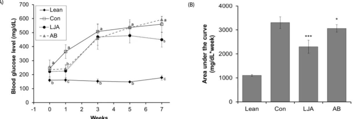
Fig. 3. Effect of Lactobacillus plantarum-hydrolyzed Jerusalem artichoke on fasting blood glucose levels in db/db mice. Values represent means ± SE (n = 10). "Lean" represents normal mice; "Con" represents diabetic mice; "LJA" represents diabetic mice fed LJA (1.5 g/kg of diet); "AB" represents diabetic mice fed acarbose (0.5 g/kg of diet). Different letters in same week indicate significant differences, P< 0.05. *P< 0.05 vs. Con; ***P< 0.001.
Anti-diabetes effect of LJA in vivo Effect of LJA on oral glucose tolerance
Oral glucose tolerance tests were performed to determine the effect of a single oral dose of LJA on glucose tolerance using db/db mice. The results are summarized in Fig. 2. Compared with the lean group, the glucose level of the control group was substantially higher. However, the LJA-treated groups had suppressed blood glucose levels at 30 and 120 min after starch load (Fig. 2A). When the area under the curve was compared between groups, the LJA 200 and LJA 400 groups showed 31.34% and 42.25% reductions, respectively, compared to the control group (Fig. 2B).
Effect of LJA on blood glucose levels
We studied the effects on blood glucose after the admini- stration of LJA at 1, 3, 5, and 7 weeks in db/db mice. The results showed the presence of a high blood glucose level in the control group, but lower levels in the LJA-treated group (Fig.
3A). However, their differences were not significant, except at first week, and the decline in blood glucose levels reached a maximum after 1 week. When the AUC was compared between groups, the LJA groups showed 30.46% reduction than the control group (Fig. 3B).
Effect of LJA on body weight, food intake, and FER We studied the effects in body weight, food intake, and feed efficiency ratio (FER) after LJA administration for 7 weeds in db/db mice. The differences in body weight and food intake between the mice treated with LJA and controls were not significant (Table 2). Changes in body weight and FER did not differ between the groups.
Effect of LJA on intestinal glycosidase activity
The activities of intestinal glycosidases (sucrose, maltase, and lactase) in the small intestine after LJA administration are shown in Fig. 4. LJA showed no significant inhibitory effect on maltase activity. However, it inhibited sucrose activity in the distal segment of the intestine. Moreover, lactase activity in the proximal segment was inhibited significantly by treatment with a low dose of LJA. Small intestinal glycosidase activity can be partially inhibited by treatment with LJA in db/db mice.
Effect of LJA on serum metabolic parameters
To determine the mechanism underlying the effect of LJA,
we studied the effect of LJA on serum metabolic parameters,
including insulin concentration, HbA1c percentage, adiponectin
concentration, and serum lipid concentration (Table 2). The
(A)
(B)
(C)
Fig. 4. Disaccharidase activities in the small intestines of LJA-administered db/db mice. Values represent means ± SE (n = 10). "Lean" represents normal mice; "Con"
represents diabetic mice; "LJA" represents diabetic mice fed LJA (1.5 g/kg of diet); "AB"
represents diabetic mice fed acarbose (0.5 g/kg of diet). Different letters in the same group indicate significant differences, P< 0.05.
serum insulin levels in the LJA-treated mice were increased significantly, by 1.3-fold, compared to the control group.
Furthermore, the HbA1c levels were significantly lower (10.32%
lower) in LJA-treated mice than control mice. However, adiponectin concentrations did not differ between mice treated with LJA and the control group.
Triglyceride levels were 65.27% lower in the LJA group than the control group. The non-esterified fatty acid concentration of LJA-treated mice was also significantly lower than that of the control group. The total cholesterol level in the LJA-treated mice showed no significant differences, but the HDL-cholesterol levels in the LJA group were significantly higher (by 38.89%) than those in the control group. Finally, a significant increase (13.52%) in HDL-total cholesterol ratio (HTR) in the LJA group was observed compared with the control group.
DISCUSSION
Sustained reductions in hyperglycemia are associated with a decreased risk of developing complications in NIDDM patients
[17]. However, it is difficult to reverse the dietary and lifestyle trends of NIDDM patients. Thus, the dietary supplements are used for regulation of blood glucose and the α-glucosidase inhibitors are used as a dietary intervention.
It has been reported that the tubers of Jerusalem artichoke contained sugars, fructans, coumarins and lectins as its main components [18-20]. Jerusalem artichoke showed α-glucosidase inhibitory activity without adverse effects and may increase insulin sensitivity, which may be related to its inulin [13,21].
Also, Jerusalem artichoke decrease fasting serum glucose levels perhaps due to it is rich in fructan and coumarins [13,22].
Jerusalem artichoke had anti-diabetic effects due to high in fructoligossacharides that may decrease insulin resistance by mechanisms that remain unknown [23]. Therefore, the degree of polymerization of fructan is considered as a factor that influences hyperglycemia amelioration and α-glucosidase inhibitory activity.
The aim of this study was to investigate the α-glucosidase inhibitory effect and hyperglycemia amelioration of fermented PJA for NIDDM treatment. Therefore, the α-glucosidase inhibitory activity of the PJA extract and its fermented products were compared in vitro. Our results demonstrated that the inhibitory effect of PJA against α-glucosidase depends not only on the degree of polymerization of fructan, but also on the change in its composition by fermentation with different microbes. LJA showed the highest α-glucosidase inhibition activity and was used to investigate its effects on hyperglycemia using NIDDM animal models.
LJA does not influence body weight, food intake, or FER.
However, we observed that LJA significantly reduces blood glucose and HbA1c levels in db/db mice, indicating that there is a significantly higher rate of glucose disposal after LJA treatment and the effect of LJA on glucose metabolism can last for a long period. There was also a significant increase in the levels of serum insulin after LJA administration to db/db mice. Accordingly, LJA may exert hypoglycemic action in diabetic mice by potentiating the effect of insulin in serum or by increasing either the pancreatic secretion of insulin from existing beta cells or its release from the bound form. Our OGTT data in db/db mice indicated that, after LJA treatment, glucose disposal increased significantly, suggesting an improvement in glucose tolerance via LJA, presumably due to an increase in insulin sensitivity.
The main features of insulin resistance include dyslipidemia,
which is characterized by high triglycerides, high non-esterified
fatty acids, low high-density lipoproteins, and low adiponectin
in the blood. Even though there were no changes in
adiponectin after LJA treatment, there were significant decreases
in triglycerides and non-esterified fatty in the LJA-treated
groups. Moreover, although a slight increase in total cholesterol
was observed after LJA treatment, HDL-cholesterol and HTR
increased significantly. The inclusion of fructan in the diet of
saturated fat-fed rats significantly reduced the triglyceride
content in the blood and liver. Delzenne and Kok [23] suggested
that the triacylglycerol-lowering effect of oligofructose occurs
via a reduction in low-density lipoprotein-triacylglycerol secretion
from the liver as a result of the reduction in the activity of
lipogenic enzymes, and in the case of fatty acid synthase, via
modification of lipogenic gene expression.
Small intestinal α-glucosidase activity was found to be partially inhibited by LJA. In particular, LJA significantly inhibited sucrose in the distal segment and lactase in the whole intestine.
For this reason, carbohydrate digestion can be delayed. Therefore, another possible mechanism underlying the anti-hyperglycemic effect of LJA may be the decreased activity of small intestinal α -glucosidase, which converts carbohydrates into glucose, preventing the rapid increase in postprandial blood glucose levels and slow absorption of carbohydrates in the small intestine.
We observed that the inhibitory effect of PJA on α -glucosidase activity could be improved dramatically by L.
plantarum fermentation. LJA has a significant anti-hyperglycemia effect in db/db mice by increasing the level of insulin, decreasing insulin resistance, and delaying the absorption of carbohydrates. LJA has potential as a food supplement for the treatment of NIDDM.
REFERENCES
1. Chen L, Magliano DJ, Zimmet PZ. The worldwide epidemiology of type 2 diabetes mellitus--present and future perspectives. Nat Rev Endocrinol 2012;8:228-36.
2. Fuller JH, Shipley MJ, Rose G, Jarrett RJ, Keen H. Coronary-heart- disease risk and impaired glucose tolerance. The Whitehall study.
Lancet 1980;1:1373-6.
3. American Diabetes Association. Diagnosis and classification of diabetes mellitus. Diabetes Care 2010;33 Suppl 1:S62-9.
4. Krasikov VV, Karelov DV, Firsov LM. α-Glucosidases. Biochemistry (Mosc) 2001;66:267-81.
5. Scheen AJ. Is there a role for α-glucosidase inhibitors in the prevention of type 2 diabetes mellitus? Drugs 2003;63:933-51.
6. Holman RR, Turner RC. Oral agents and insulin in the treatment of NIDDM. In: Pickup JC, Williams G, editors. Textbook of Diabetes.
Oxford: Blackwell; 1991. p. 467-9.
7. Kim SH, Hyun SH, Choung SY. Anti-diabetic effect of cinnamon extract on blood glucose in db/db mice. J Ethnopharmacol 2006;104:119-23.
8. Rojo LE, Ribnicky D, Logendra S, Poulev A, Rojas-Silva P, Kuhn P, Dorn R, Grace MH, Lila MA, Raskin I. In vitro and in vivo anti-diabetic effects of anthocyanins from Maqui Berry (Aristotelia chilensis).
Food Chem 2012;131:387-96.
9. Chernenko TV, Glushenkova AI, Rakhimov DA. Lipids of Helianthus tuberosus tubers. Chem Nat Compd 2008;44:1-2.
10. Kim HS, Han GD. Hypoglycemic and hepatoprotective effects of Jerusalem artichoke extracts on streptozotocin-induced diabetic rats. Food Sci Biotechnol 2013;22:1121-4.
11. Rumessen JJ, Bodé S, Hamberg O, Gudmand-Høyer E. Fructans of Jerusalem artichokes: intestinal transport, absorption, fermentation, and influence on blood glucose, insulin, and C-peptide responses in healthy subjects. Am J Clin Nutr 1990;52:675-81.
12. Park BS. Effect of oral administration of Jerusalem artichoke inulin on reducing blood lipid and glucose in STZ-induced diabetic rats.
J Anim Vet Adv 2011;10:2501-7.
13. Kaur N, Gupta AK. Applications of inulin and oligofructose in health and nutrition. J Biosci 2002;27:703-14.
14. Mohamed Sham Shihabudeen H, Hansi Priscilla D, Thirumurugan K. Cinnamon extract inhibits α-glucosidase activity and dampens postprandial glucose excursion in diabetic rats. Nutr Metab (Lond) 2011;8:46.
15. Barham D, Trinder P. An improved colour reagent for the determination of blood glucose by the oxidase system. Analyst (Lond) 1972;97:142-5.
16. Lowry OH, Rosebrough NJ, Farr AL, Randall RJ. Protein measurement with the Folin phenol reagent. J Biol Chem 1951;193:265-75.
17. Jung HW, Jung JK, Ramalingam M, Yoon CH, Bae HS, Park YK.
Anti-diabetic effect of Wen-pi-tang-Hab-Wu-ling-san extract in streptozotocin-induced diabetic rats. Indian J Pharmacol 2012;44:
97-102.
18. Baldini M, Danuso F, Turi M, Vannozzi GP. Evaluation of new clones of Jerusalem artichoke (Helianthus tuberosus L.) for inulin and sugar yield from stalks and tubers. Ind Crops Prod 2004;19:25-40.
19. Rakhimov DA, Arifkhodzhaev AO, Mezhlumyan LG, Yuldashev OM, Rozikova UA, Aikhodzhaeva N, Vakil MM. Carbohydrates and proteins from Helianthus tuberosus. Chem Nat Compd 2003;39:312-3.
20. Suseelan KN, Mitra R, Pandey R, Sainis KB, Krishna TG. Purification and characterization of a lectin from wild sunflower (Helianthus tuberosus L.) tubers. Arch Biochem Biophys 2002;407:241-7.
21. Boillot J, Alamowitch C, Berger AM, Luo J, Bruzzo F, Bornet FR, Slama G. Effects of dietary propionate on hepatic glucose production, whole-body glucose utilization, carbohydrate and lipid metabolism in normal rats. Br J Nutr 1995;73:241-51.
22. Luo J, Rizkalla SW, Alamowitch C, Boussairi A, Blayo A, Barry JL, Laffitte A, Guyon F, Bornet FR, Slama G. Chronic consumption of short-chain fructooligosaccharides by healthy subjects decreased basal hepatic glucose production but had no effect on insulin- stimulated glucose metabolism. Am J Clin Nutr 1996;63:939-45.
23. Yang HJ, Kwon DY, Kim MJ, Kang S, Kim DS, Park S. Jerusalem artichoke and chungkookjang additively improve insulin secretion and sensitivity in diabetic rats. Nutr Metab (Lond) 2012;9:112.
24. Delzenne NM, Kok N. Effect of non-digestible fermentable carbohy- drates on hepatic fatty acid metabolism. Biochem Soc Trans 1998;
26:228-30.