INTRODUCTION
Central venous stenosis (CVS) is a common problem in patients with a long standing implanted central venous catheter or a compressing mediastinal mass and this can lead to a critical situation with facial and neck swelling, distended neck veins, headache due to cerebral edema, dyspnea and in severe cases stridor and mental changes (1).
In this situation stenting of the affected venous vessels (superior vena cava [SVC], left brachiocephalic vein [LBV], right brachiocephalic vein [RBV] left/right internal jugular vein [LJV/RJV] and left/right subclavian vein [LSV/RSV]) via the endovascular approach is an effective treatment
Stenting of the Superior Vena Cava and Left
Brachiocephalic Vein with Preserving the Central Venous Catheter in Situ
Peter Isfort, MD
1, 2, Tobias Penzkofer, MD
1, 2, Fabian Goerg, MD
1, Andreas H. Mahnken, MD, MBA, MME
1, 21Department of Diagnostic and Interventional Radiology, University Hospital RWTH Aachen, 52074 Aachen, Germany; 2Department of Applied Medical Engineering, Helmholtz-Institute for Biomedical Engineering, RWTH Aachen, 52074 Aachen, Germany
Stenting of the central veins is well established for treating localized venous stenosis. The techniques regarding catheter preservation for central venous catheters in the superior vena cava have been described. We describe here a method for stent implantation in the superior vena cava and the left brachiocephalic vein, and principally via a single jugular venous puncture, while saving a left sided jugular central venous catheter in a patient suffering from central venous stenosis of the superior vena cava and the left brachiocephalic vein.
Index terms: Central venous stenosis; Venous PTA and stenting; Central venous catheter protection
Received December 11, 2010; accepted after revision March 10, 2011.
Corresponding author: Peter Isfort, MD, Department of Applied Medical Engineering, Helmholtz-Institute for Biomedical Engineering, RWTH Aachen, Pauwelsstrasse 20, 52074 Aachen, Germany.
• Tel: (49241) 8035824/8085462 • Fax: (49241) 8082442
• E-mail: [email protected]
This is an Open Access article distributed under the terms of the Creative Commons Attribution Non-Commercial License (http://creativecommons.org/licenses/by-nc/3.0) which permits unrestricted non-commercial use, distribution, and reproduction in any medium, provided the original work is properly cited.
pISSN 1229-6929 · eISSN 2005-8330 Korean J Radiol 2011;12(5):629-633
option (2). However, in-situ catheter-material such as port- or hemodialysis catheters pose the problem of catheter- fixation to the vessel wall and this is possibly followed by functional deficiency due to catheter compression or catheter defects caused by the expanded stent (3).
Therefore, the catheter material may temporarily be removed before stent implantation. So far this procedure has only been described for the SVC (3-6).
In this case report we describe a complex procedure with an in situ port catheter which was temporarily repositioned for stent implantation in the SVC and LBV.
CASE REPORT
A 54 year-old male patient with a central bronchial carcinoma (UICC stage IIIb) was referred to our clinic with symptoms of CVS, including distended neck veins, plethora of the face and upper extremities and dyspnea, for palliative SVC stenting. He had undergone left-sided subclavian port implantation nine months before for chemotherapy with Cisplatin and Vinorelbin in combination with external beam radiotherapy. Since there was no sufficient response to the first-line therapy, the medication was replaced by second- line therapy with Gemzar and Navelbine. Because of tumor
progression, his CVS symptoms were considered to be due to tumor-related SVC stenosis and the patient was referred to our hospital for SVC stenting. The RJV was punctured with an 18G needle and biplane diagnostic angiography revealed a compression of the SVC at the level of the tumor which was causing a high grade SVC stenosis (Fig. 1A, G).
Thereafter, a 7 Fr-introducer sheath (Pinnacle Introducer sheath, Terumo, Tokyo, Japan) was inserted and additional diagnostic angiography of the LBV was performed: this showed a subtotal stenosis of the LBV in its central two- thirds (Fig. 1B, G). After repositioning the tip of the port-catheter into the RBV using a 15 mm Andra-snare (Angiopro, Speyer, Germany), a self-expandable stent (14 mm diameter; 40 mm length Sinus-Repo-stent, Optimed, Ettlingen, Germany) was placed in the SVC without prior balloon dilatation (Fig. 1C, H). Balloon dilation was performed after placement of the self expandable stent in the SVC using a 10 mm diameter, 40 mm length Fox balloon (Abbott Laboratories, Abbott Park, IL) (Fig. 1C, D).
Thereafter, the LJV was punctured and a 7 Fr-introducer sheath (Terumo, Tokyo, Japan) was introduced (Fig. 1D).
Using the same 15 mm Andra-snare, the port-catheter was repositioned in the distal part of the LBV and it was fixed there by keeping the loop-wire of the snare around the port catheter, while the catheter of the snare was removed (Fig.
1E, I). Then the LBV stenosis was crossed with a guide- wire via the same sheath while the loop wire remained in place and two stents (12 mm diameter/40 mm length, and 12 mm diameter/60 mm length, respectively, Sinusflex- Repo, Optimed, Ettlingen, Germany) were placed via the same introducer sheath in the LBV, extending from the SVC to the LBV after pre-ballooning the stenosis with a 8 mm diameter/40 mm length Fox balloon (Abbott Laboratories) (Fig. 1I). Finally, the snare`s catheter was inserted again and the port catheter was repositioned through the newly implanted catheter into the SVC (Fig. 1F, J). After manual compression the introducer sheaths were removed. The signs of CVS regressed within a few hours and the patient was discharged the same day with markedly short- and midterm symptom relief. The patient died two months later due to tumor progression without any signs of CVS.
DISCUSSION
Central venous stenosis is a common problem in patients with mediastinal tumors and/or central venous catheters with an incidence of 11% to 50% depending on the
catheter type and access route (6-8). Interestingly, catheter associated stenosis occurs more often in patients with a subclavian central venous access (42%) than in patients with an internal jugular venous access (10%). CVS is more often seen with left sided central venous catheters than with right sided central venous catheters, and this is possibly due to a longer and more tortuous course of the left-sided catheters through the LBV (9). The treatment of CVS using percutaneous transluminal angioplasty (PTA) and stenting is a well accepted technique with usually rapid symptom relief in these patients (2, 5, 8). The complications of this procedure, including rupture of the SVC or stent migration, are rare (2). An in situ central venous catheter is usually removed before stenting to prevent catheter fixation between the stent and the vessel wall with the associated risk of catheter dysfunction or catheter loss (3). To avoid catheter removal and the associated strain and risks of a new central venous puncture (e.g. bleeding, pneumothorax and air embolism) for the patient, a technique that allows stenting of the SVC without removal of central venous catheters has been proposed by several authors (5, 6).
In this case report we describe the combined stenting of the SVC and the LBV with an in-situ central-venous catheter in a patient with SVC stenosis due to bronchial cancer, which was treated with external beam radiation therapy and multiple courses of chemotherapy, and a second stenosis in the proximal two-thirds of the LBV in the presence of a central venous port catheter. Due to the coincidence of the stenosis and catheter position in the LBV, we suspected the LBV stenosis as being the result of catheter-related scarring (Fig. 1B, G). To prevent the removal of the central venous catheter, it has to first be dislocated in the RJV before SVC stent implantation (Fig. 1H). Thereafter, it was dislocated in the distal LBV using a snare (Fig. 1E, I). The removal of the snare-catheter while the central venous catheter was held in place with the snare-wire allowed the simultaneous introduction of the stents through the same 7 Fr sheath and this avoided another venous puncture and sheath insertion (Fig. 1F, I). Further, another angiographic control procedure of the central venous catheter could be avoided prior to repositioning because the central venous catheter was constantly fixed with the snare and it was ready to be advanced in its original position. This approach is on the one hand convenient for the experienced interventionalist and it is also convenient for the patient since no new central venous catheter has to be placed. Further, one venous puncture and sheath insertion were avoided by using
A
C
B
D
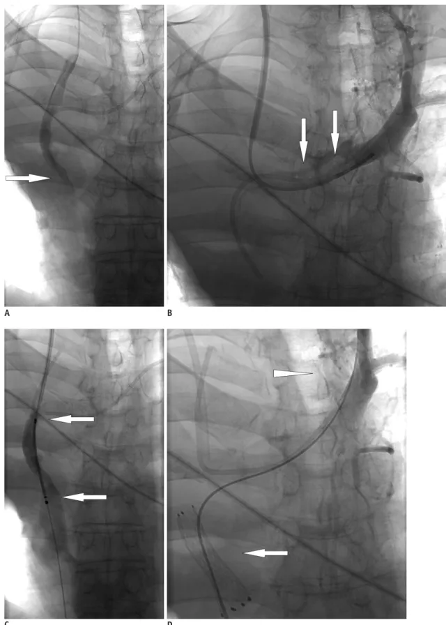
Fig. 1. Angiographic and schematic illustration of procedure.
A. Diagnostic angiography showing severe stenosis of superior vena cava (arrow). B. Diagnostic angiography of left brachiocephalic vein reveals subtotal stenosis (arrows). C. After repositioning tip of port-catheter into right brachiocephalic vein using 15 mm Andra-snare, self-expandable stent is placed in superior vena cava (arrows). D. Left jugular vein is punctured and 7 Fr introducer sheath is introduced (arrowhead). Released stent in superior vena cava (arrow).
one sheath for the snare and the stents, and this reduced the risk of pneumothorax, bleeding and air embolism.
Additionally, this procedure is cost-effective compared to the standard procedure since no new port system has to be implanted.
The first introducer sheath was inserted in the RJV because of the known SVC stenosis. Due to the lack of proper preinterventional imaging, the LBV stenosis was discovered on the first diagnostic angiograms. In retrospect, G
E F
H I J
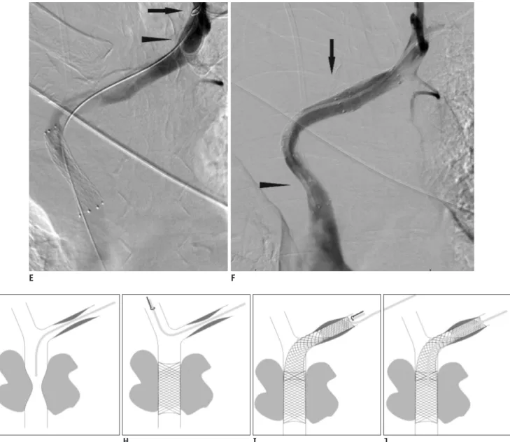
Fig. 1. Angiographic and schematic illustration of procedure.
E. Using Andra-snare (arrow), port-catheter (arrowhead) is repositioned in distal part of left brachiocephalic vein and is fixed there by keeping loop-wire of snare around port catheter, while snare’s catheter is removed. F. Left brachiocephalic vein stenosis is crossed with guide-wire via same sheath while snare’s loop wire remains in place and two 12 mm stents (arrow) are placed via same introducer sheath in left brachiocephalic vein, extending from superior vena cava to left brachiocephalic vein. Finally snare’s catheter is inserted again and port catheter is repositioned into superior vena cava (arrowhead). G-J. Schematic drawing of procedure. Initial situation with stenosis in superior vena cava and left brachiocephalic vein and in situ port catheter (G). Port catheter is repositioned in right brachiocephalic vein using snare before stent is placed in superior vena cava (H). Then port catheter is positioned in distal part of left brachiocephalic vein and two stents are placed in left brachiocephalic vein and superior vena cava via same sheath (I). Final situation with preserved port catheter (J).
the puncture of the RJV was not necessary since all the stent and snare insertions could have been performed via a single introducer sheath placed in the LBV, which highlights the value of adequate pre-interventional imaging, such as contrast-enhanced CT, for treatment planning. In contrast to the previous reports by Bovenschulte et al. (3) and Palmié et al. (4) we used an internal jugular venous access route instead of a femoral venous access route. This approach is known to reduce the rate of venous thrombosis
and post-punctional infections (10). It also facilitated proximal catheter displacement.
In conclusion, SVC and LBV stenting in a patient with CVS while preserving existing ventral venous catheters is feasible via a single venous access. The procedure is safe and it improves the patient’s quality of life, while omitting the need for a second intervention for central venous catheter re-insertion.
REFERENCES
1. Sheth S, Ebert MD, Fishman EK. Superior vena cava obstruction evaluation with MDCT. AJR Am J Roentgenol 2010;194:W336-346
2. Smayra T, Otal P, Chabbert V, Chemla P, Romero M, Joffre F, et al. Long-term results of endovascular stent placement in the superior caval venous system. Cardiovasc Intervent Radiol 2001;24:388-394
3. Bovenschulte H, Bangard C, Lackner KJ. [Superior vena cava stent implantation: preserving an indwelling port catheter by temporary shift of the brachiocephalic vein]. Rofo 2010;182:274-275
4. Palmié S, Heller M, Wetzel H. [PTA and stent placement in the
superior vena cava syndrome under the temporary salvage of a venous port system]. Rofo 1995;162:81-83
5. Qanadli SD, Mesurolle B, Sissakian JF, Chagnon S, Lacombe P. Implanted central venous catheter-related acute superior vena cava syndrome: management by metallic stent and endovascular repositioning of the catheter tip. Eur Radiol 2000;10:1329-1331
6. Stockx L, Raat H, Donck J, Wilms G, Marchal G. Repositioning and leaving in situ the central venous catheter during percutaneous treatment of associated superior vena cava syndrome: a report of eight cases. Cardiovasc Intervent Radiol 1999;22:224-226
7. Parish JM, Marschke RF Jr, Dines DE, Lee RE. Etiologic considerations in superior vena cava syndrome. Mayo Clin Proc 1981;56:407-413
8. Haage P, Vorwerk D, Piroth W, Schuermann K, Guenther RW.
Treatment of hemodialysis-related central venous stenosis or occlusion: results of primary Wallstent placement and follow- up in 50 patients. Radiology 1999;212:175-180
9. Kundu S. Review of central venous disease in hemodialysis patients. J Vasc Interv Radiol 2010;21:963-968
10. Szibor-Kriesen U, Rucker G, Vagts DA. [Central venous cannulation--the important things, you should know!].
Anasthesiol Intensivmed Notfallmed Schmerzther 2008;43:654- 663