Original Article
Predicting the optimal depth of left-sided central venous catheters
in children
H. Kim,
1C-H. Jeong,
2H-J. Byon,
3H. K. Shin,
4T. J. Yun,
5J-H. Lee,
6Y-H. Park
6and J-T. Kim
71 Clinical Assistant Professor, Department of Anesthesiology and Pain Medicine, Yonsei University College of Medicine, Seoul, Korea
2 Resident, 3 Clinical Assistant Professor, 4 Associate Professor, Department of Anesthesiology and Pain Medicine, Inha University Hospital, Incheon, Korea
5 Clinical Assistant Professor, Department of Radiology, 6 Clinical Assistant Professor, 7 Assistant Professor, Depart-ment of Anesthesiology and Pain Medicine, Seoul National University Hospital, Seoul, Korea
Summary
The aim of this study was to predict the optimal depth for insertion of a left-sided central venous catheter in chil-dren. Using 3D chest computed tomography angiography, we measured the distance from a point where the internal jugular vein is at the superior border of the clavicle, and from a point where the subclavian vein is inferior to the anterior border of the clavicle, to the junction of the superior vena cava and the right atrium in 257 children. Linear regression analysis revealed that the distances correlated with age, weight and height. Simple formulae for the depth of a central venous catheter via the left internal jugular vein (0.07 9 height (cm)) and the left subclavian vein (0.08 9 height (cm)) were developed to predict placement of the central venous catheter tip at the junction of the superior vena cava with the right atrium. Using these fomulae, the proportion of catheter tips predicted to be cor-rectly located was 98.5% (95% CI 96.8–100%) and 94.0% (95% CI 90.8–97.3%), respectively.
...
Correspondence to: H-J. Byon Email: [email protected] Accepted: 21 June 2013
Central venous catheter insertion into the superior vena cava is often performed in children who are criti-cally ill or who require major surgical procedures. Cor-rect positioning of the catheter tip is essential to obtain accurate haemodynamic information, adminis-ter vasoactive drugs and fluids and reduce complica-tions that may arise from misplacement of the tip within the right atrium such as arrhythmias, thrombo-sis, cardiac perforation and tamponade [1–5]. Although it is possible to identify the catheter tip posi-tion using radiographic evaluaposi-tion and echocardiogra-phy [6, 7], it is still important to decide the initial catheter depth clinically. Unlike adults, acceptable
catheter lengths in children vary according to their age, weight and height. Guidelines for optimal catheter depth have been published for children; however, these focus on right-sided placements [8–10]. The purpose of this study was to establish simple, clinically useful, guidelines for left-sided catheter depths in children based on the measured lengths of the internal jugular and subclavian veins using 3D computed tomography (CT) angiography.
Methods
This retrospective study was approved by the local Insti-tutional Review Board. Children under 13 years old
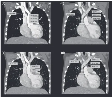
who had undergone chest CT angiography in the previ-ous 28 months were considered eligible for inclusion in the study. We selected patients whose CT scans con-tained contrast media within the internal jugular or sub-clavian veins. The scans of patients with cardiovascular or rotational abnormalities were excluded from the study. Three-dimensional angiography images were reconstructed from patients’ chest CT images using the Rapidiaâ program (Infinitt Healthcare Co. Ltd., Seoul, Korea). The 3D CT angiographies were reviewed to identify planes showing the entire course of each nal jugular and subclavian vein. The lengths of the inter-nal jugular and subclavian veins on both the left and the right were measured using the‘3D curve distance’ func-tion of the Rapidia program. Internal jugular vein length was defined as the distance from the superior border of the clavicle to the junction of the superior vena cava and right atrium. Subclavian vein length was defined as the distance from the anterior border of the clavicle to the junction of the superior vena cava and right atrium. Each measurement consisted of a curved line connected by points located within the centre of the vessel, the location of the connecting points being confirmed on
additive axial CT views provided by the Rapidia pro-gram. The junction of the superior vena cava with the right atrium was defined by the crista terminalis (Fig. 1). Information on patients’ age, weight and height was recorded at the time the CT scans were performed. Plots of the distance against these variables were generated, and linear regression analysis was performed to calculate regression lines and equations, coefficient of determina-tion (R2) and 95% CI. Statistical analysis was performed using SAS version 9.2 software (SAS Institute Inc., Cary, NC, USA) and a p value< 0.05 was considered signifi-cant.
Results
A total of 257 patients were included in the study. The left internal jugular vein length was measured in 198 scans, the left subclavian vein length in 202 scans, the right internal jugular vein length in 199 scans and the right subclavian vein length in 202 scans. Baseline characteristics are shown in Table 1. All the lengths for both sides showed a high correlation with the child’s age, weight and height, and regression equa-tions are shown in Table 2. There was greater
correla-(b) (a)
(d) (c)
Figure 1 3D chest CT angiography showing how the vein lengths were measured: (a) right subclavian vein; (b) left subclavian vein; (c) right internal jugular vein; (d) left internal jugular vein.
tion with height compared with age or weight. Based on these results, simple formulae using the child’s height were developed to place left internal jugular and subclavian vein catheter tips at the junction of the superior vena cava and right atrium: left internal jugu-lar vein catheter length = 0.07 9 height (cm); and left subclavian vein catheter length = 0.08 9 height (cm). When these were applied to the left internal jugular or subclavian vein lengths obtained from 3D CT angio-graphy, the proportion of catheter tips predicted to be correctly located was 98.5% (95% CI 96.8–100) for
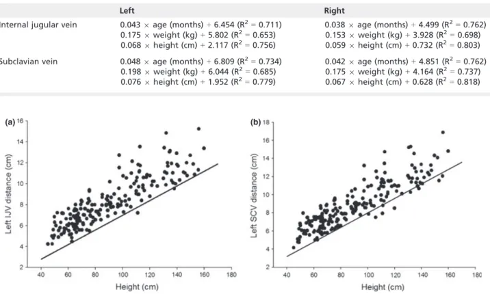
catheters inserted via the left internal jugular vein and 94.0% (95% CI 90.8–97.3) for catheters inserted via the left subclavian vein. Scatter plots of the left internal jugular and left subclavian vein lengths against height are shown in Fig. 2.
Discussion
Whilst previous studies have been conducted to predict right-sided catheter depth [8–10], less is known regard-ing left-sided catheters. This is because central venous catheters are more often inserted on the right side per-formed more often than left-sided insertions in order to avoid major vessel perforation or thoracic duct injury, which occurs with more frequency on the left [11–14]. Left-sided catheters may need to be inserted in certain situations, for example following failed inser-tion on the right, and therefore guidelines for the depth of left-sided catheters in children are warranted.
In the present study, instead of actual catheter inser-tion, the lengths of the left internal jugular vein and left
(b) (a)
Figure 2 Plots of measured vein length vs patient height for: (a) left internal jugular vein (IJV); and (b) left subcla-vian vein (SCV). Solid lines represent‘best fit’.
Table 2 Regression equations describing the distance of each vessel to the junction of the superior vena cava and the right atrium calculated with age, weight and height.
Left Right
Internal jugular vein 0.043 9 age (months) + 6.454 (R2= 0.711)
0.175 9 weight (kg) + 5.802 (R2 = 0.653) 0.068 9 height (cm) + 2.117 (R2 = 0.756) 0.038 9 age (months) + 4.499 (R2= 0.762) 0.153 9 weight (kg) + 3.928 (R2 = 0.698) 0.059 9 height (cm) + 0.732 (R2 = 0.803) Subclavian vein 0.048 9 age (months) + 6.809 (R2
= 0.734) 0.198 9 weight (kg) + 6.044 (R2 = 0.685) 0.076 9 height (cm) + 1.952 (R2 = 0.779) 0.042 9 age (months) + 4.851 (R2 = 0.762) 0.175 9 weight (kg) + 4.164 (R2 = 0.737) 0.067 9 height (cm) + 0.628 (R2 = 0.818)
Table 1 Baseline characteristics of the 257 children included in the study. Values are median (IQR [range]).
Age; months 19.5 (5.0–69.0 [0.1–155.0]) Weight; kg 9.8 (6.6–18.3 [1.8–57.4]) Height; cm 81.7 (64.9–110.5 [45.0–160.3])
subclavian vein measured from the clavicle to the junc-tion of the superior vena cava and right atrium were measured on 3D chest CT angiographies, and regression equations calculated by comparing these distances with the child’s age, weight and height. The purpose was to develop simple formulae that can easily be applied in clinical settings. Based on the regression equation, it was the height of the child that correlated most accurately, the simple formulae being 0.07 9 height (cm) for the left internal jugular vein (95% CI 96.8–100) and 0.089 height (cm) for the left subclavian vein (95% CI 90.8–97.3).
We also developed a formula for catheter depth (0.06 9 height (cm)) via the right internal jugular vein using 3D CT angiographies. This formula was compared with those from a previous study [8]. If the height of a child is 90 cm, the recommended catheter depth is 4.4 cm in our study and 8 cm in the study by Andropoulos et al. If the height of a child is 110 cm, the recommended catheter depth is 5.6 cm in our study and 9.4 cm in the study by Andropoulos et al. These discrepancies are explained by different mea-surement techniques; our distances were measured from the clavicle, whereas Andropoulos used the inser-tion point (the apex of the triangle formed by the two heads of the sternocleidomastoid muscle). The distance from the catheter insertion point on the skin to the clavicle should be added to the recommended depth based on the formulae developed in our study. Cathe-ter insertion points on the skin may be variable and the formulae developed in our study used a fixed ana-tomical landmark, the clavicle.
There are relatively few data available on catheter depth via the subclavian vein in children. In previous studies, catheters were placed via both the internal jugu-lar and subclavian veins without distinguishing between them, or via the subclavian vein only in children under 5 kg in weight [8, 9]. In adults, correct catheter length via the subclavian vein was reported to be approxi-mately 2 cm less than that via the internal jugular vein [15]. Catheter length via the subclavian vein is predicted to be greater than that via the internal jugular vein in our study because the clavicle was used as our landmark rather than the insertion point on the skin surface.
Our study has some limitations. First, the formulae used to predict left-sided catheter depth were
devel-oped using 3D chest CT angiography and not clinical measurements. Therefore, when using the formulae developed, physicians should be mindful that they have not been clinically validated and catheter tip posi-tion should be confirmed, if necessary, by chest radiog-raphy or echocardiogradiog-raphy. Second, in very small children whose height is less than 60 cm, the actual distance may be less than that based on the equation developed. The aim of the present study was to gener-ate simple formulae for left-sided catheter depth and additional formulae for very small children were not specifically developed. In these children, left-sided depths of 4 cm for the internal jugular vein and 4.5 cm for the subclavian vein may be appropriate.
In conclusion, the formulae proposed for left-sided central venous catheter depth in children, 0.079 height (cm) for the internal jugular vein and 0.089 height (cm) for the subclavian vein, can be used to prevent unintended catheter tip placement in the right atrium. The distance from the catheter inser-tion point to the clavicle should be added to the length generated by these formulae.
Acknowledgements
The authors thank Min Woong Kang, Biostatistician, Biostatistics Collaboration Unit, Yonsei University Col-lege of Medicine, for help with statistical analysis.
Competing interests
No external funding and no competing interests declared.
References
1. Hayashi Y, Maruyama K, Takaki O, Yamauchi J, Ohnishi Y, Kuro M. Optimal placement of CVP catheter in paediatric cardiac patients. Canadian Journal of Anesthesia 1995; 42: 479–82.
2. Currarino G. Migration of jugular or subclavian venous cathe-ters into inferior tributaries of the brachiocephalic veins or into the azygos vein, with possible complications. Pediatric Radiology 1996;26: 439–49.
3. Collier PE, Ryan JJ, Diamond DL. Cardiac tamponade from cen-tral venous catheters. Report of a case and review of the Eng-lish literature. Angiology 1984;35: 595–600.
4. McDonough JJ, Altemeier WA. Subclavian venous thrombosis secondary to indwelling cathers. Surgery, Gynecology and Obstetrics 1971;133: 397–400.
5. Nowlen TT, Rosenthal GL, Johnson GL, Tom DJ, Vargo TA. Peri-cardial effusion and tamponade in infants with central cathe-ters. Pediatrics 2002;110: 137–42.
6. Andropoulos DB, Stayer SA, Bent ST, et al. A controlled study of transesophageal echocardiography to guide central venous catheter placement in congenital heart surgery patients. Anes-thesia and Analgesia 1999;89: 65–70.
7. Yoon SZ, Shin JH, Hahn S, et al. Usefulness of the carina as a radiographic landmark for central venous catheter placement in paediatric patients. British Journal of Anaesthesia 2005;95: 514–7.
8. Andropoulos DB, Bent ST, Skjonsby B, Stayer SA. The optimal length of insertion of central venous catheters for pediatric patients. Anesthesia and Analgesia 2001;93: 883–6. 9. Kim JH, Kim CS, Bahk JH, et al. The optimal depth of central
venous catheter for infants less than 5 kg. Anesthesia and Analgesia 2005;101: 1301–3.
10. Yoon SZ, Shin TJ, Kim HS, et al. Depth of a central venous cathe-ter tip: length of insertion guideline for pediatric patients. Acta Anaesthesiologica Scandinavica 2006;50: 355–7.
11. Kwon SS, Falk A, Mitty HA. Thoracic duct injury associated with left internal jugular vein catheterization: anatomic con-siderations. Journal of Vascular and Interventional Radiology 2002;13: 337–9.
12. Mukau L, Talamini MA, Sitzmann JV. Risk factors for central venous catheter-related vascular erosions. Journal of Paren-teral and EnParen-teral Nutrition 1991;15: 513–6.
13. Teichgraber UK, Nibbe L, Gebauer B, Wagner HJ. Inadvertent puncture of the thoracic duct during attempted central venous catheter placement. Cardiovascular and Interventional Radiol-ogy 2003;26: 569–71.
14. Duntley P, Siever J, Korwes ML, Harpel K, Heffner JE. Vascular erosion by central venous catheters. Clinical features and out-come. Chest 1992;101: 1633–8.
15. Czepizak CA, O’Callaghan JM, Venus B. Evaluation of formulas for optimal positioning of central venous catheters. Chest 1995;107: 1662–4.