Original Articles 순환기:순환기:제순환기:순환기:제제제 2 7 권권권 제권 제제 6 호제 호호 1997호
경피적 관동맥 풍선확장 성형술후 재협착에 관한 Sulodexide의 효과에 대한 전향적, 무작위 비교연구
울산대학교 의과대학 서울중앙병원 내과학교실
김진우·이철환·정상식·강덕현·홍명기 송재관·김재중·박성욱·박승정
=
=
=
= Abstract = = = =
A Prospective, Randomized, Comparative Clinical Investigation of the Effects of Sulodexide on Restenosis after Percutaneous Transluminal
Coronary Balloon Angioplasty
Jin-Woo Kim, M.D., Cheol-Whan Lee, M.D., Sang-Sig Cheong, M.D., Duk-Hyun Kang, M.D., Myeong-Ki Hong, M.D.,
Jae-Kwan Song, M.D., Jae-Joong Kim, M.D., Ph.D, Seong-Wook Park, M.D., Ph.D, Seung-Jung Park, M.D., Ph.D.
Department of Internal Medicine, College of Medicine, University of Ulsan, Asan Medical Center, Seoul, Korea
Background:Restenosis remains as the major limitation of percutaneous transluminal coronary balloon angioplasty(PTCA). Although its mechanism remains incompletely understood, proliferative action of arterial smooth muscle cells has been found to play an important role on restenosis by neointimal formation after PTCA. Glycosaminoglycan-containing compounds, including Sulodexide (Vessel Due , ALFA, Wasserman, S.p.A., Italy), inhibit the proliferation and migration of vascular smooth muscle cells in vitro.
Objectives:This study was performed to assess the efficacy of Sulodexide, a glycosaminoglycan compound with antithrombotic and antiproliferative properties, in preventing restenosis after PTCA.
Method:Two hundred eighty-four patients with ischemic heart disease were randomized to receive either the standard PTCA without Sulodexide in 144 patients(control group, M:F=99:45, Age=58±9), 160 lesions or the standard PTCA with Sulodexide in 140 patients(treated group, M:
F=89:51, Age=58±10), 158 lesions. Successful angioplasties were performed in 258 atheromatous coronary lesions in 224 patients for whom follow-up angiographic data were obtained 6 months later.
Quantitative coronary angiographic analysis(QCA) was performed before, immediate after PTCA and 6-months later. Angiographic restenosis (>50% diameter stenosis at follow-up) was the primary end point;minimal luminal diameter at follow-up angiogram was the secondary end point.
Results:Successful PTCA was 97.6% and 97.5% in the standard PTCA with Sulodexide and the
standard PTCA without Sulodexide, respectively. Although reference vessel size and minimal luminal
diameter after PTCA were larger in the control group than in the Sulodexide group(2.94±0.11 vs 2.83±0.13mm and 2.26±0.12 vs 2.18±0.08mm, respectively, p=NS), there was a increased tendency of minimal lumen diameter at 6 months angiogram in the Sulodexide group than in the control group (1.12±0.50 vs 1.07±0.53mm, respectively, p=NS). Angiographic restenosis occurred in 42% of lesions in the Sulodexide group and 52% of the control group (p=NS).
Conclusions:Sulodexide treatment had a tendency to reduce restenosis rate in 6 months after coronary angioplasty. However, further study is necessary to verify the antiproliferative effect of Sul- odexide with much larger number of patients.
KEY WORDS:Sulodexide·Restenosis·Percutaneous transluminal coronary balloon angioplasty.
서 론
경피적 관동맥풍선확장성형술(percutaneous tran- sluminal coronary balloon angioplasty, 이하 PTCA 라 칭함)후 재협착은 현재 병리학적인 측면에서 2가 지 다른 기전이 관련있을 것이라고 생각하고있다. 첫 번째는 혈관내경을 침범하는 새로운 내막조직의 형성 (neointima formation)이며1), 두번째는 혈관의 병적 재형성(pathologic remodeling) 현상 즉, PTCA후 혈 관의 외막층(adventitia)이 작아지는 것이 재협착과 관 련이 있을 것이라고 생각되고 있다2).
Sulodexide(Vessel Due®, ALFA, Wasserman, S.
p.A., Italy)는 표준화된 추출 및 정제과정을 통하여 돼 지의 십이지장에서 얻은 glycosaminoglycan으로 de- rmatan sulfate와 heparin fraction이 1:4의 비율로 섞인 혼합물로 분자량은 약 8,000 daltons정도이고 생체내에서 Sulodexide는 지단백분해효소(lipoprotein- lipase)를 분비시켜 콜레스테롤이 풍부한 지단백(very low density lipoprotein, low density lipoprotein)을 분해시키므로써 항동맥경화효과가 증명된 바 있고3), 치 료용량에서 activated partial thromboplastin time과 thrombin time에 대한 영향없이 응고인자 Xa의 억제 시키며 antithrombin III와 heparin cofactor II에 대 한 촉매작용으로 항혈전효과가 있다4). 조직–플라즈 미노겐 활성제(tissue-plasminogen activator)를 활 성화시키고 플라즈미노겐 활성제 억제제(plasminogen activator inhibitor-1)를 억제하므로써 전섬유소용해 (profibrinolysis)작용도 나타낸다5-6). Sulodexide는 말초혈관의 동맥 경화성 패쇄질환을 가진 환자에서 임
상적으로 치료효과가 있는 것으로 증명되었으며7-8) 현 재 임상적 적용범위도 확대되어가고있는 것이 사실이 다9). 또한 여러 연구자들은 Sulodexide의 heparin- like compounds는 항혈전효과이외에도 동맥의 내막 하 평활근 세포의 증식을 억제하고 내막세포의 손상을 치유하여 혈관의 개존성을 유지하는데 기여한다고 보 고하였다10).
이에 저자들은 PTCA후 손상된 혈관의 중막(media) 에 있는 평활근 세포의 증식과 내막의 과증식(intimal hyperplasia)으로 인한 재협착에 대한 Sulodexide의 치료효과를 평가하고자 하였다.
연구대상 및 방법
1. 연구대상
본 연구는 서울중앙병원 심장내과에서 1994년 7월 부터 1995년 12월까지 관동맥질환으로 PTCA를 시 술받은 284명의 환자로서 무작위로 Sulodexide를 투 여한 140명(M:F=89:51, Age=58±10세), 158병 소와 Sulodexide를 투여하지 않은 144명(M:F=99:
45, Age=58±9세), 160병소를 6개월뒤 관동맥조영 술을 시행하여 양군간의 재협착여부 및 최소내강직경 (minimal luminal diameter, 이하 MLD라 칭함)을 측 정하였다. 혈관조영술상 육안적으로 병변의 내경이 70%
이상의 협착소견이 있는 경우에 PTCA를 시술하였으 며 PTCA의 성공은 시술과 관련된 주요 합병증없이 잔 존협착이 50%이하로된 경우로 정하였으며, 추적 관동 맥조영술전에 관동맥우회로술을 시행하였거나 좌주간 동맥에 심한 협착이 있는 경우와 심근경색관련 혈관에 PTCA를 시행한 경우는 본 연구에서 제외하였다.
2. 방 법
임상소견에 의한 초기의 진단에 따라 불안정형 협 심증, 안정형 협심증, 급성 심근경색증등으로 나누었고 진구성 심근경색증의 과거력도 조사하였다. 또한 위험 인자로서 고혈압, 당뇨병, 흡연력등을 조사하였고 혈중 cholesterol를 측정하여 240mg/dL 이상이면 고콜레 스테롤혈증으로 분류하였다. Sulodexide는 관동맥 성 형술을 시행하기 적어도 하루전에 600 lipoprotein- lipase-releasing units(이하 LRU라 칭함)를 정맥주 사하였고 PTCA후에는 정맥주사 및 450LRU를 하루 2번 복용하였고 퇴원후에는 정맥주사는 중지하고 450 LRU를 하루 2번 복용하였다. Aspirin 200mg/day을 관동맥조영술 수일전 부터 양군에 각각 투여하였다.
관동맥 조영술 결과의 분석으로는 내경협착(percent diameter stenosis)정도, 최소내강직경과 reference vessel 직경의 측정은 병소의 풍선확장전, 확장후 및 추적 관동맥 조영술시 협착이 가장 심하게 보이는 각 도에서 on-line quantitative coronary angiographic system(ANCOR V2.0, Simens)을 이용하여 측정하 였다. 혈관조영술상의 측정은 관동맥내 니트로글리세 린을 투여후 이완기에 이루어졌으며, 유도도자(guiding catheter)의 직경을 기준으로 삼았다. 조영소견은 2명 의 관찰자가 함께 분석하였으며 PTCA당시와 추적 관 동맥조영소견의 같은 투사면을 선택하여 직경의 협착 정도를 측정하였다. 조영술상의 재협착이란 추적조영 상 직경이 50% 이상 협착된 경우로 정의하였다. PTCA 를 시술한 병변의 형태는 American College of Car- diology/American Heart Association에 의한 분류에 따라 type A, B, C lesion으로 나누었다.
혈관확장을 위해서 풍선과 혈관의 직경비가 1:1이 되도록 풍선을 선택하였고 시술동안 환자는 heparin 10,000U를 정맥내 투여하였으며 activated clotting time을 250초 이상을 유지하기 위해서 필요하면 hep- arin 5,000U를 재차 투여하였다.
284명환자중 224명의환자(79%)가 6개월 후 관동 맥 조영술을 다시 시행하였고 양군간의 모든 측정된 자 료는 평균 +/- 표준오차로 표시하였으며 양군간의 검 사치를 비교하기 위해 two-tailed T test를 하였고 양 군사이의 임상적 혹은 혈관조영적 변수들과 재협착 및 장기 임상 추적결과와의 비교 분석을 위해 chi-square
test를 이용하였다.
3. 결 과
284명의 환자, 318병소에서 PTCA가 시술되었고, 평균 입원기간은 5±2일이었으며, 시술중 29명(10.2%) 의 환자에서 풍선확장후 관동맥박리로 스텐트를 삽입 했으며, 급성폐쇄나 심근경색증이 각각 3명(1.2%),5명 (1.8%)의 환자에서 발생하였고 입원기간중 반복 PTCA 나 관동맥우회로술이 필요한 경우는 각각 3명(1.2%), 2명(0.7%)명의 환자에서 발생하였다.
284명의 환자, 318병소중 시술중 스텐트를 삽입하 였거나 시술후 급성 폐쇄나 심근경색증이 생긴 경우 또 는 반복 PTCA나 관상동맥우회술이 필요하였던 환자 를 제외한 242명, 276병소중 224명(92.6%), 258(93.
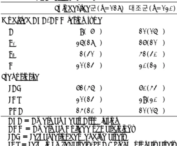
4%) 병소에서 6개월뒤 추적 관동맥조영술을 시행하 였다. 6개월뒤 추적 관동맥조영술을 시행한 224명 환 자들의 임상적 특성은 Table 1과 같다. 258개 병소부 위의 관동맥 조영소견의 특성은 Table 2와 같다. 양 군사이의 대상환자들간의 임상 및 조영술 소견의 특 성등에 유의한 차이는 없었다.
대상 병소에 대한 전체 관동맥조영술상의 재협착률 은 47%(122/258)였고, 이중 Sulodexide 투여군에서 는 42%(54/128), 대조군에서는 52%(68/130)로서 Sulodexide투여군에서 다소 낮았으나 통계학적인 차
Table 1. Baseline clinical characteristics of PTCA patients (n=224)
Sulodexide군 (n===110) =
대조군 (n====114) Age(years) 58±10 58±9 Male gender 74(67%) 84(74%) Risk factors
Hypertension 43(39%) 27(24%) Diabetes m ellitus 25(23%) 18(16%) Hyperlipidemia 8( 7%) 5( 4%) Current smoker 48(44%) 55(48%) Myocardial infarction
Previous 8( 7%) 18(16%)
Acute 20(18%) 32(28%)
Unstable angina 51(46%) 36(32%) Number of disease vessel
1 96(87%) 100(88%)
2 11(10%) 11(10%)
3 3( 3%) 3( 2%)
이는 없었다. 양군간의 임상(Table 3) 및 조영술 소견 의 특성(Table 4)에 따른 재협착률은 양군사이에 통 계학적인 차이는 없었다.
PTCA시 병변의 협착정도는 PTCA전 병변의 직경 협착(diameter stenosis)정도가 Sulodexide 투여군 이 74.6±9.6%에서 23.0±9.0%, 대조군은 73.2±
8.9%에서 21.0±10%로 줄었다. PTCA 시술직후의 최 소내강직경은 Sulodexide 투여군이 2.18±0.08mm, 대조군이 2.26±0.12mm였으며, 6개월뒤 추적검사의 최소내강직경은 Sulodexide 투여군이 1.12±0.50mm 로 1.07±0.53mm인 대조군에 비해 큰 경향을 가졌으 나 통계학적인 의의는 없었다(Table 5).
즉, 경피적 관동맥확장성형술후 6개월 추적조영상 Sulodexide 투여군이 대조군에 비해 재협착률을 감소 시키는 경향을 보였다.
고 안
정상적인 혈관의 평활근 세포와 내막 세포는 heparin 혹은 heparin sulfate를 생산하는데11) 이는 중막에 있 는 평활근 세포의 증식을 억제하고 수축성 표현형(con- tractile phenotype)으로 유지하는 기능을 가지고 있 다. 그러나 기계적, 염증성 또는 다른 원인으로 인해 혈 관벽이 손상을 입게 되면 혈소판에서 분비되는 platelet factor 4, heparitinase와 많은 성장자극물질들에 의해 평활근 세포는 증식하게 되며 합성형 표현형(synthetic phenotype)으로 변한다12).
Sulodexide는 돼지의 십이지장의 점막으로부터 추출 Table 2. Angiographic and procedural characteristics of
PTCA lesion(n=258)
Sulodexide군(n=128) 대조군(n=130) Modified AHA/ACC lesion type
A 9( 7%) 21(16%)
B1 36(28%) 27(21%)
B2 2(41%) 52(40%)
C 31(24%) 30(23%) Stenosis site
LAD 72(56%) 70(54%) LCX 31(24%) 39(30%) RCA 25(20%) 21(16%) AHA ==== American Heart Association
ACC ==== American College of Cardiology LAD === Left anterior descending artery =
LCX === Left circumflex artery, RCA = Right coronary artery = Table 3. The restenosis rate for clinical characteristics of
patients
Sulodexide군 (n=110)
대조군
(n=114) P value Risk factors
Diabetes mellitus 14(56%) 10(55%) NS Hypertension 18(42%) 12(44%) NS Hyperlipidemia 3(38%) 2(40%) NS Current smoking 21(44%) 28(51%) NS Clinical diagnosis
Unstable angina 20(39%) 16(44%) NS Myocadial infarction
Old 3(38%) 8(44%) NS
Acute 9(45%) 16(50%) NS
Number of diseased vessel
2 vessel disease 3(27%) 4(36%) NS 3 vessel disease 2(67%) 2(67%) NS
Table 4. The restenosis rate of PTCA lesions
*Total restenosis rate=47%
Sulodexide군 (n=128)
대조군
(n=130) P value Restenosis rate 54(42%) 68(52%) NS Modified AHA/ACC lesion type
A 15(60%) 11(56%) NS
B1 12(30%) 12(42%) NS
B2 23(45%) 32(61%) NS
C 14(47%) 13(46%) NS Stenosis site
LAD 34(41%) 41(32%) NS
LCX 8(29%) 13(11%) NS
RCA 12(43%) 14(11%) NS AHA === American Heart Association =
ACC === American College of Cardiology = LAD ==== Left anterior descending artery LCX === Left circumflex artery =
RCA ==== Right coronary Artery
Table 5. Quantitative angiographic measurement Sulodexide군
(n=128)
대조군
(n=130) P value Diameter
stenosis(%) 74.6±9.6 73.2±8.9 NS Reference vessel
diameter(mm) 2.83±0.13 2.94±0.11 NS Post MLD(mm) 2.18±0.08 2.26±0.12 NS Follow up MLD(mm) 1.12±0.50 1.07±0.53 NS Late loss(mm) 1.07±0.10 1.18±0.15 NS MLD === Minimal luminal diameter =
한 glycosaminoglycan으로 dermatan sulfate와 hep- arin 분획이 1:4의 비율로 섞인 혼합물로 분자량이 8,000 daltons의 medium molecular weight heparin 이다. Heparin은 위장관을 통한 흡수가 어려워 정맥 또는 피하주사로 사용되나 최근에 Mauro등은 derma- tan sulfate와 heparin이 혼합된 glycosaminoglycan (heparinoid)이 경구투여로 흡수될 수 있음을 보고하 였다13). Sulodexide는 antithrombin III와 응고인자 Xa에 작용하는 heparin-like substances(80%)와 he- parin cofactor II에 작용하는 dermatan fraction(20%) 모두 항혈전 작용을 가지나 특히 heparin-like subs- tance는 동맥의 평활근 세포에 대한 항증식 효과를 가
진다10,14,15). 1989년 태아의 생체외 실험에서 작용기전
은 밝혀지지 않았지만 배양 4일째 동맥 평활근 세포 의 증식과 내막으로의 이동을 억제하는 것이 현저하였 고 매우 적은 농도(5ug/ml)에서도 관찰되었는데 이는 약물농도 의존적이었으며 더 높은 농도, 즉 100ug/ml, 300ug/ml에서는 각각 32%, 48% 억제되었다10). Sulo- dexide는 약물투여 방법에 관계없이 효력(potency)은 비슷하나 정맥주사하면 Sulodexide의 반감기는 대략 16분에서 90분 사이이며 이는 약물용량에 따라 길어 지며 정맥주사5분내에 혈액응고검사에 변화가 오고 15 분에 최대 농도에 도달하게 된다16,17). 약물작용이 지 속되는 시간(elapsing time) 또한 약물의 용량에 의존 적인데 600LRU일때 120분, 1200LRU일때 300분간 지속되고 투여 48시간에 콩팥을 통해 완전히 배설된 다18). Sulodexide를 경구 투여하면(30~40% 흡수) 2 시간째 그리고 4시간에서 6시간 사이에 높은 농도를 보 이며 12시간뒤 혈중에서 사라지게 되고 약물 투여 4일 째에 일정한 치료농도(steady state)에 도달하게 된다18).
PTCA이후 6개월내 평활근 세포의 증식, 내막으로의 이동 및 분비기가 종결되므로 저자들은 6개월뒤 재협 착의 여부를 알기 위해서 관동맥조영술을 시행하였으 며 재협착은 추적조영상 내경협착정도(diameter sten- osis)가 50% 이상으로 된 경우로 하였다. 양군사이에 임상적, 동맥경화인자 및 관동맥조영인자등에 유의한 차 이가 없었고, 전체 재협착률은 47%였고 Sulodexide 투여군과 대조군의 재협착률이 각각 42%, 52%였지만 통계학적인 차이는 없었다. 양군간의 임상 및 조영술
소견의 특성에 따른 재협착률 또한 통계학적인 차이가 없었다. PTCA후 탄성반도(elastic recoil)에 의한 재 협착률은 양군에서 동일한 조건이므로 고려하지 않았 다. Heparin은 동물실험에서 혈관의 평활근 세포의 증 식과 이동을 억제하였다고 보고하였는데19), 비록 후향 적 아집단 분석이었지만 Hirshfeld등에 의해 혈관조영 상 재협착률이 감소되었다고 보고하였으나20) 보다 많은 환자를 대상으로 한 연구에서는 확인되지 않았다19,21). Low molecular weight heparin 또한 동물실험에서 평 활근 세포의 증식과 이동을 감소시켰으며 고지혈증 토 끼에서 혈관성형술후 재협착을 감소시켰다22). 그러나 인간을 대상으로 한 여러 연구(ERA Trial, EMPAR Trial and FACT)에서는 효과가 증명되지 않았다23-25). 동물실험에서의 효과에도 불구하고 인간에 있어 실망 스러운 결과는 정상적인 혈관에 풍선으로 인위적으로 손상을 입힌 실험동물보다 인간의 심하게 협착된 관동 맥에 풍선확장성형술을 시술시 더 크고 더 복합적인 손 상이 야기되어 더 넓은 표면적이 혈소판에 노출되므로 실험동물에서보다 평활근 세포를 더 강력하게 자극한 결과일 것으로 추정되며 아직까지 heparin의 항증식 효 과를 알기 위해 PTCA후 인간에게 오랜 기간동안 투 여된 연구가 없었다26)는 이유를 들 수 있겠다.
동물실험에 의하면, 평활근 세포가 내막에 도착해서 수주동안 계속해서 증식하여 신내막층의 두께는 8주째 (인간은 2~3개월)에 최대치에 도달했으며27) 혈소판 유 도성 성장인자(platelet-derived growth factor, PDGF) 는 중막에 있는 평활근 세포에 대한 화학적 인자 및 유 사분열물질(mitogen)로 알려져 있는데 PDGF에 대한 항체를 투여하여 내막으로의 이동을 억제하여 신내막 의 증식을 현저히 감소시켰다고 한다28). 풍선도자를 이 용한 혈관손상(balloon injury)후 2~3일 사이에 중 막의 평활근 세포는 성장하게 되고 7일내에 증식의 대 부분이 끝나게 된다29). 평활근 세포가 증식후 내막층 으로 이동(migration)하여 14일째 내막층에서 가장 많 은 수의 평활근 세포를 보게 되며 balloon injury후 1 년동안 동일한 수의 평활근 세포가 계속 존재하게 된 다30). Balloon injury후 14일째 초기의 신내막층의 두 께는 평활근 세포의 수에 의하지만 그 이후 신내막층 의 두께의 증가는 평활근 세포의 부피(volume) 그리고
세포외 기질의 축척에 의한다. 그러므로 내막에 존재하 는 평활근 세포의 대부분은 중막의 평활근 세포에서 유 래하므로 중막의 평활근 세포의 증식의 정도가 내막의 과증식(hyperplasia)의 정도를 결정할 것이라고 가정 할 때 많은 수의 평활근 세포의 증식과 이동이 PTCA 후 재협착의 발생에 관여하리라 생각된다. 본 연구의 혈 관조영상 정량적 측정의 결과에서는 PTCA전 Sulode- xide 투여군에서 diameter stenosis가 대조군에 비해 다소 심하게 좁았고, reference vessel diameter와 PTCA후 최소내강직경도 대조군에 비해 작았지만 6개 월 추적조영상 최소내강직경은 오히려 대조군보다 Sul- odexide 투여군에서 큰 경향을 나타내었다. 이러한 결 과는 Sulodexide가 중막의 평활근 세포의 증식과 이 동을 억제시켜 내막의 과증식을 억제시킨 데 기인하리 라 생각된다.
결론적으로 PTCA후 Sulodexide의 효과는 재협착 을 줄이는 경향을 보였으나, 내막의 과증식을 억제하 여 재협착을 감소시키는 기전에 대해서는 향후보다 광 범위한 연구가 진행되어야 하겠다.
요 약
연구배경:
Sulodexide는 heparin-like compound인 medium molecular weight glycosaminoglycans(80%)와 der- matan sulfate(20%)로 구성되어 있다. 응고인자 Xa 의 억제작용, 그리고 antithrombin III와 heparin cofa- ctor II에 대한 촉매작용에 의한 항혈전효과가 있고 동맥경화성 폐쇄질환에 임상적으로 치료효과가 증명되 었으며, 현재 임상적 적용범위도 확대되어가고 있는 것 이 사실이다. 여러 연구자들은 Sulodexide의 heparin- like compounds는 항혈전작용외에도 동맥의 내막하 평 활근 세포의 증식을 억제하여 내막의 과형성을 감소시 키므로써 혈관의 개존성을 유지하는데 기여한다고 하 였다.
경피적 관동맥풍선확장성형술후 재협착은 혈관의 중 막(media)에 있는 평활근 세포의 증식과 내막으로의 이 동 그리고 내막의 과증식에 의해서 올 수 있다고 알려 져 있다. 이에 저자들은 경피적 관동맥성형술후 재협착
에 관한 Sulodexide의 치료효과를 평가하고자 하였다.
대상 및 방법:
1994년 7월부터 1995년 12월까지 PTCA를 시술 받은 284명의 환자중 Sulodexide를 투여한 140명 (M:F=89:51, age=58±10), 158병소와 Sulode- xide를 투여하지않은 144명(M:F=99:45, age=58
±9), 160병소를 각각 Sulodexide 투여군과 대조군으 로 나누고 6개월뒤 관동맥조영술을 시행하여 양군간의 재협착 여부 및 최소내강직경(minimal luminal diam- eter)을 측정하였다.
Sulodexide는 관동맥성형술을 시행하기 적어도 하루 전에 600 lipoprotein-lipase-releasing units(LRU) 를 1회/일 정맥주사하였고 PTCA후에는 정맥주사 및 450LRU를 2번/일 복용하였고 퇴원후에는 정맥주사는 중지하고 450LRU를 2번/일 경구복용하였다. 또한 as- pirin 200mg/일과 diltiazem 180mg/일을 양군에 각 각 투여하였다. 6개월후 관동맥조영술을시행하였고 양 군사이의 비교는 chi-square test와 t-test를 이용하 였다.
결 과:
284명의 환자, 318병소에서 PTCA가 시술되었고, 시술중 29명(10.2%)의 환자에서 시술 관상동맥에서 풍 선확장후 동맥박리로 스텐트를 삽입했으며 급성폐쇄 나 심근경색증이 각각 3명(1.2%), 5명(1.8%)의 환자 에서 발생하였고 반복 PTCA나 관동맥우회로술이 필 요한 경우는 각각 3명(1.2%), 2명(0.7%)의 환자에서 발생하였다.
양군사이의 대상환자들의 임상 및 관동맥조영술 소 견상의 특성등에 유의한 차이는 없었고 대상 병소에 대한 전체 관동맥조영술상의 재협착률은 47%(122/
258)였고, 이중 Sulodexide 투여군에서는 42%(54/
124), 대조군에서는 52%(68/130)로써 Sulodexide 투여군에서 다소 낮았으나 통계학적인 차이는 없었다.
양군간의 임상 및 조영술 소견의 특성에 따른 재협착률 은 양군사이에 통계학적인 차이는 없었다.
PTCA시 병변의 협착정도는 PTCA전 내경협착정도 (diameter stenosis)가 Sulodexide 투여군이 74.6±
9.6%에서 PTCA후 23.0±9.0%, B군은 73.2±8.9%
에서 21.0±10%로 줄었다.
PTCA시술전과 시술후의 혈관조영상 정량적 측정의 결과는 시술직후의 최소내강직경이 2.18±0.08mm인 Sulodexide 투여군이 2.26±0.12mm인 대조군에 비해 작았으나, 6개월뒤 추적관찰시의 최소내강직경은 Sulo- dexide 투여군이 1.12±0.50mm였고 1.07±0.53mm 였다. 추적기간중 내막증식율은 Sulodexide를 투여하 지 않은 대조군에서 높은 경향을 보였으나 통계학적인 차이는 없었다.
결 론:
PTCA후 Sulodexide의 효과는 재협착을 줄이는 경 향을 보였으나, 내막의 과증식을 억제하여 재협착을 김 소시키는 기전에 대해서는 향후 보다 광범위한 연구가 진행되어야 하겠다.
References
1) Clowes AW, Schwartz SM:Significance of quiescent smooth muscle migration in the injured rat carotid artery.
Cir Res 56:139-145, 1985
2) Gibbons GH, Dzau VJ:The emerging conception of va- scular remodeling. N Engl J Med 330:1431-1438, 1994 3) Radakrishnamurthy B, Sharama C, Bandaru RR, Berenson GS, Stanzani L, Mastacchi R:Studies of chemical and biological properties of a fraction of Sulodexide, a hep- arin-like glycosaminoglycan. Atherosclerosis 60:141-9, 1986
4) Palazzini E, Procida C:Effect of some mucopolysacch- arides on activated factor X. Biochem Pharmacol 27: 608-10, 1975
5) Fiore G, Baraldi A, Gambarotta GC, Franco A, Liberati C:Inhibition of plasminogen activator inhibitor(PAI-1) by Sulodexide in post-thrombophlebitic patients. Drug Devel 3:173-8, 1991
6) Mannarino E, Pasqualini L, Ciuffetti G, Lombardini R:
Effects of oral administration of Sulodexide on fibrinoly- sis and plasma viscosity:A pilot study. Drug Invest 4: 346-50, 1992
7) Crepaldi G, Fellin R, Calabro A, et al:Double-blind mul- ticinter trial on a new medium-molecular weight glycos- aminoglycan:Current therapeutic effects and perspecti- ves for clinical use. Atherosclerosis 81:233-43, 1990 8) Bartolo M, Antignani PL, Eleuteri P:Experiences with
Sulodexide in the arterial peripheral diseases. Curr Ther Res 36:979-88, 1984
9) Santus G, Sottini G, Lombardi G, Rozzini R, Inzoli R:
Luso del Sulodexide nei vasculopatici cerebrali con dis- lipidemia e/o diabetes:Contributo clinico statistico. Ath- erosclerosis and Cardiovascular Diseases 415:20, 1984
10) Tiozzo R, Cingi MR, Pietrangelo A, Albertazzi L, Calan- dra S, Milani MR:Effect of heparin-like compounds on the in vitro proliferation and protein synthesis of various cell types. Arzneim-Forsch/Drug Res 39:15-20, 1989 11) Schwartz SM, Campbell GR, Campbell JH:Replication
of smooth muscle cells in vascular disease. Cir Res 58: 427-444, 1986
12) Chamley-Campbell JH, Campbell GR, Ross R:Phenot- ype-dependent response of cultured aortic smooth muscle to serum mitogen. J Cell Biol 89:379-383, 1981 13) Mauro M, Ferraro G, Palmieri G:Profibrinolytic and
antithrombotic effects of Sulodexide oral administration: A double-blind, crossover, placebo-controlled study. Cu- rrent Therapeutic Research 51:342, 1992
14) Tollefsen DM, Peska CA, Monafo WJ:Activation of he- parin cofactor II by dermatan sulfate. J Biol Chem 258: 6713, 1983
15) Clowes AW, Karnowsky MJ:Suppression by heparin of smooth muscle cell proliferation in injured arteries. Nat- ure 265:625, 1977
16) Silbert D:Structure and metabolism of proteoglycans and glycosaminoglycans. J. Invest Dermatol 79(supp. 1):31, 1982
17) Dol F, Houin G, Dupouy D, Cadroy Y, Caranobe C, Gab- aig AM, Mardiguian J, Sie P, Boneu B:Pharmacokine- tics of dermatan sulfate in the rabbit after intravenous injection. Thromb Haemost 59:255-258, 1988 18) Crepadi G, Fellin R, Calabro A, Rossi A:Double-blind
multicenter trial on a new medium weight glycosaminog- lycan-Current therapeutic effects and perspectives for clinical use. Atherosclerosis 81:223-243, 1990 19) Ellis SG, Roubin JW, Willentz J:Effect of 1824 hour
heparin administration for prevention of restenosis after uncomplicated coronary angioplasty. Am Heart J 117: 777-782, 1989
20) Hirshfeld JW, Schwartz JS, Jugo R, MacDonald RG, Goldbergs A, Savage MP:Restenosis after coronary ang- ioplasty:A multivariate statistical model to relate lesion and procedure variable to restenosis. J Am coll Cardiol 18(3):647-656, 1991
21) Hillegass WB, Ohman EM, Califf RM:Restenosis:The clinical issue. In Topol EJ, ed. Textbook of Interventioinal Cardiology. 2nd ed. Philadelphia:WB Saunders, 415- 435, 1994
22) Currier JW, Pow TK, Haudenschild CC, Minihan AC, Faxon DP:Low molecular weight heparin(Enoxaparin) reduces restenosis after iliac angioplasty in the hyperch- olesterolemic rabbit. J Am Coll Cardiol 17:118B-125B, 1991
23) Faxon D, Spiro T, Minors A, Cote G, Douglas J, Gottlieb R:Low molecular weight heparin in the prevention of restenosis after angioplasty. Results of enoxaparin rest- enosis(ERA)trial. Circulation 90:908-914, 1994 24) Cairns D, Gill J, Morton B, Roberts R, Gent M, Hirsh J:
Enoxaparin and Max EPA for the prevention of angiop- lasty restenosis(EMPAR)(abstr). Circulation 90:I-651, 1994
25) Gery RL, Koyama N, Wang TW, Vergels CD, Clowes AW:Failure of heparin to inhibit intimal hyperplasia in injured balloon arteries. The role of heparin-sensitive and -insensitive pathways in the stimulation of smooth mus- cle cell migration and proliferation. Circulation 91:27 72-2781, 1995
26) Palmieri G, Ambrosi G, Nazzari M, Palazzini, E:The influence of Sulodexide on some coagulation parameters: A pharmacokinetic study. Clot Hematol Malign 2:7-13, 1985
27) Serruys PW, Luijten HE, Beutt KJ, Geuskens R, Van de Brand M, Reiber JHC, Ten Katen HJ, Hugenholtz PG:
Incidence of restenosis after successful coronary angiop- lasty:A time-related phenomenon:A quantitative angi- ographic follow-up study in 342 patients at 1, 2, 3, and 4 months. Circulation 77:361-371, 1988
28) Ferns GAA, Raines EW, Sprugel KH, Motani AS, Reidy MA, Ross R:Inhibition of neointimal smooth muscle accumulation after angioplasty by an antibody to PDGF.
Science 253:1129-1132, 1991
29) Clows AW, Schwartz SM:Significance of quiescent smooth muscle migration in the injured rat carotid artery.
Cir Res 56:139-145, 1985
30) Clowes AW, Clowes HM, Reidy MA:Kinetics of cel- lular proliferation after arterial injury III. Endothelial and smooth muscle growth in chronically denuded ves- sels. Lab Invest 54:295-303, 1986