596
Open Access
Arterial Stiffness in Female Patients With Fibromyalgia and Its Relationship to Chronic Emotional and Physical Stress
Ji Hyun Lee, MD1, Kyoung Im Cho, MD2, Seong Man Kim, MD2, Hyeon Gook Lee, MD2, and Tae Ik Kim, MD2
1Divisions of Rheumatology and 2Cardiology, Maryknoll Medical Center, Busan, Korea
ABSTRACT
Background and Objectives: In patients with fibromyalgia (FM) syndrome, stress and pain may chronically enhance sym- pathetic activity, altering cardiovascular responses and inducing the arterial wall-stiffening process. We investigated arterial stiffness in FM patients using pulse wave velocity (PWV) and analyzed whether arterial stiffness was affected by the clinical parameters of FM. Subjects and Methods: This study included 108 female FM patients (51.5±8.9 years) without any known cardiovascular diseases and 76 healthy female controls (50.1±8.9 years). FM patients underwent a manual tender point survey for tender point counts, and completed the visual analogue scale (VAS) of pain and fibromyalgia impact ques- tionnaire (FIQ), which were composed of a physical and feel score. Brachial-ankle pulse-wave velocity (baPWV) was mea- sured with an automated device. The study participants were subdivided into 2 groups based on the sum of the FIQ score (group A: FIQ ≥50, group B: <50). Results: Patients with FM had significantly higher baPWV than the controls, and signif- icant increase were noted in baPWV values of group A compared with those of group B. BaPWV showed a significant posi- tive correlation (correlation coefficient=6.83, p=0.022) with severity of disease assessed by FIQ. Conclusion: The patients with FM showed significantly increased arterial stiffness, suggesting a pathophysiologic link between FM and endothelial dysfunction. This study provides a basis for clarifying the mechanism by which chronic pain syndrome is associated with an increased risk of vascular stiffness. (Korean Circ J 2011;41:596-602)
KEY WORDS: Fibromyalgia; Compliance; Stress; Pulse wave velocity.
Received: January 14, 2011 Revision Received: February 1, 2011 Accepted: February 7, 2011
Correspondence: Kyoung Im Cho, MD, Division of Cardiology, Maryk- noll Medical Center, 4-12 Daecheong-dong, Jung-gu, Busan 600-730, Korea
Tel: 82-51-461-2384, Fax: 82-51-465-7470 E-mail: [email protected]
• The authors have no financial conflicts of interest.
cc This is an Open Access article distributed under the terms of the Cre- ative Commons Attribution Non-Commercial License (http://creativecom- mons.org/licenses/by-nc/3.0) which permits unrestricted non-commer- cial use, distribution, and reproduction in any medium, provided the origi- nal work is properly cited.
Introduction
Fibromyalgia (FM) syndrome, characterized by chronic wi- despread pain and discomfort, has diverse characteristics in- cluding widespread musculoskeletal pain with discrete points of tenderness along with fatigue, sleep disturbance, cognitive dysfunction, and depressed mood. FM is considered to result from deregulation of pain modulator systems involving alter-
ed interactions between central and peripheral nervous sys- tems and immune systems.1) In patients with FM, stress and pain may chronically enhance sympathetic activity. Several lines of evidence indicate that sympathetic nervous system (SNS) function is altered in patients with FM.2)3) SNS activity is an important determinant of the arterial wall-stiffening pro- cess. Chronically enhanced sympathetic activity may alter cardiovascular responses and induce endothelial dysfunc- tion. Using a nonhuman primate model of atherogenesis, it has been observed that psychosocial stress can alter the au- tonomic balance towards a state of sympathetic arousal lead- ing to the development of coronary artery disease (CAD), per- haps through impairing endothelial function4) and intensific- ation of endothelium-mediated atherogenic processes.5) In recent years, several studies have shown a decrease in endo- thelial function in association with clinical depression or sub-clinical mood states or personality traits such as depres- sion, anxiety, type A personality (hostility) or anger.6)7)
Since stiffened arteries transmit pulse waves faster than the more elastic blood vessels, pulse wave velocity (PWV) is an
ideal indicator of arterial stiffness, and brachial artery pulse wave velocity (baPWV) is used to assess endothelial func- tion by measuring the status of large and small arteries in the lower extremity.8) We proposed that even in FM patients, we can observe a decrease in endothelial function, consider- ing that psychological symptoms including anxiety, stress, and depression are often observed in FM.9) This study inves- tigated arterial stiffness in FM patients using baPWV, and analyzed whether arterial stiffness was affected by the clini- cal parameters of FM, which were assessed by the fibromyal- gia impact questionnaire (FIQ).
Subjects and Methods
Study population
This study was conducted from January 2010 to December 2010, and enrolled 108 consecutive female FM patients and 76 healthy female controls that were matched to the FM pati- ents by their age, blood pressure (BP), heart rate, height, cho- lesterol and glucose levels. Diagnosis of FM was confirmed by a board-certified rheumatologist according to the guidelines outlined by the American College of Rheumatology.10) Pati- ents with history of major atherosclerotic risk factors includ- ing hypertension and diabetes, history of CAD, congestive he- art failure, history of neurological illness, smoking, obesity with body mass index above 26 and abnormal C-reactive pro- tein (CRP) were excluded from the study. Controls were re- cruited from the cardiology clinic and health screening cen- ter and included subjects without chronic widespread pain.
The study was approved by the Maryknoll medical center ins- titutional review board and all subjects gave their written in- formed consent.
Disease-specific evaluation
At each visit, patients with FM were asked to rate their cur- rent level of pain on the 10 cm Visual Analog Scales (VASs, 0=
no pain, 10=worst pain imaginable), a scale for rating pain intensity. Subjects then filled out the Korean Fibromyalgia Impact Questionnaire (kFIQ),11) which is a validated, self-ad- ministered test, scored out of 100, that evaluates physical func- tion, work status, depression, anxiety, sleep, pain, stiffness, fa- tigue, and well being. The study participants were also sub- divided into 2 groups based on the sum of the FIQ score (gr- oup A: FIQ ≥50, group B: <50). Subjects were then assessed for the number of positive tender points by digital palpation over the 18 characteristic tender point sites in the ACR cri- teria for the diagnosis of FM. The subjects were requested to identify if a given point was painful as slow steady digital pressure was applied. The self-administered questionnaire, the Beck Depression Inventory (BDI) scale, and Brief Fati- gue Inventory (BFI) scale, assessed symptoms of depression and fatigue, respectively.
Measurement of arterial stiffness
Arterial stiffness was assessed by measuring brachial-an- kle (ba) PWVs using an automatic waveform analyzer (VP- 1000; Colin Co., Komaki, Japan).12) The VP-1000 simultane- ously records pulse waves, BP (both arms and ankles), ankle- brachial pressure index (ABI), electrocardiography, and heart sounds, as described elsewhere.12)13) ABI was calculated by the ratio of the ankle systolic BP divided by the arm systolic BP, and the lower value of the ankle systolic BP was used for the calculation. For measuring baPWV, pulse waves obtain- ed from the brachial and tibial arteries were recorded simul- taneously, and the transmission time, which was defined as the time interval between the initial increase in brachial and tibial waveforms, was determined. The transmission distance from the arm to each ankle was calculated according to body height. The baPWV was automatically computed as the tr- ansmission distance divided by the transmission time. All participants included in the present study had a normal ABI (>0.9). A high baPWV was defined as the gender-specific highest quartile of the values among the study subjects {ba- PWV (the mean of the right and left values) ≥1,490 cm/s in females}.
Statistical analysis
All data were expressed as mean±standard deviation. Data were analyzed using standard statistical software (Statistical Package for the Social Sciences package version 11.0, Chica- go, IL, USA) and comparisons of all measurements were made with paired Student’s t-test for continuous variables and Pear- son correlation test for correlation. Multivariate analysis was performed with logistic regression for the associations be- tween demographic, atherosclerotic risk factors and baPWV.
P<0.05 was considered statistically significant.
Results
Clinical characteristics of subjects
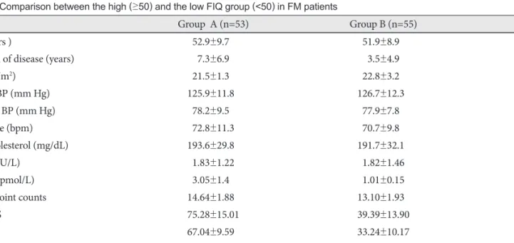
There were no significant differences between the FM and control groups in terms of age and the other parameters that are known to affect the PWV, including height, BP, heart rate and the total cholesterol levels (Table 1). There were also no significant differences in these variables except disease dura- tion of the FM syndrome (p=0.007) (Table 2) between the groups A and B (Table 2). The value of tender point counts, pain VAS, FIQ scores, scales of anxiety, fatigue and depression were significantly higher in group A, as compared to group B (Table 2).
Ankle-brachial pressure index and brachial artery pulse wave velocity values of patients and control group
The parameters of arterial stiffness, represented baPWV, of
the FM patients and controls are shown in Table 1. The baP- WV values of the FM and control groups (1,378.9±242.2 cm/s vs. 1,284.0±186.27 cm/s, p=0.005) were significantly differ- ent (Table 1). Significant increases were also noted in baPWV values of group A compared with those of group B (1,438.9±
281.7 vs. 1,330.4±184.3, p=0.013) (Table 3), and the same re-
sult was observed with the control group included (Table 3).
The baPWV value of the subgroup (less than 130 mm Hg of systolic BP, and less than 200 mg/dL of total cholesterol, and less than 60 years of age) were also significantly different in patients with FM, as compared to controls (n=55, 1,338.4±
228.6 vs. 1,241.9±181.9, p=0.005).
Table 1. Baseline demographic characteristics of FM patients and controls
FM patients (n=108) Control (n=76) p
Age (years) 51.5±8.9 50.1±8.9 0.32
BMI (kg/m2) 23.4±2.8 23.3±3.2 0.72
Systolic BP (mm Hg) 123.9±13.6 126.2±12.8 0.56
Diastolic BP (mm Hg) 77.9±8.2 76.8±9.6 0.64
Heart rate (bpm) 73.4±7.9 74.2±7.3 0.45
Total cholesterol (mg/dL) 192.0±31.7 192.3±28.2 0.95
TSH (mIU/L) 01.82±1.35 01.95±1.27 0.51
Free T4 (pmol/L) 01.92±0.20 01.04±0.13 0.43
ABI 01.08±0.07 01.10±0.07 0.044
baPWV (cm/s) 1378.9±242.2 1284.0±186.2 0.005
High baPWV (number, %) 34 (31) 8 (11) 0.029
All values are described as mean±SD. FM: fibromyalgia syndrome, BMI: body mass index, BP: blood pressure, TSH: thyroid stimulating hor- mone, ABI: ankle-brachial index, baPWV: brachial-ankle pulse wave velocity
Table 2. Comparison between the high (≥50) and the low FIQ group (<50) in FM patients
Group A (n=53) Group B (n=55) p
Age (years ) 52.9±9.70 51.9±8.90 0.300
Duration of disease (years) 7.3±6.9 3.5±4.9 0.001
BMI (kg/m2) 21.5±1.30 22.8±3.20 0.320
Systolic BP (mm Hg) 125.9±11.80 126.7±12.30 0.730
Diastolic BP (mm Hg) 78.2±9.50 77.9±7.80 0.520
Heart rate (bpm) 72.8±11.3 70.7±9.80 0.580
Total cholesterol (mg/dL) 193.6±29.80 191.7±32.10 0.740
TSH (mIU/L) 1.83±1.22 1.82±1.46 0.950
Free T4 (pmol/L) 3.05±1.40 1.01±0.15 0.310
Tender point counts 14.64±1.880 13.10±1.930 <0.001
Pain VAS 75.28±15.01 39.39±13.90 <0.001
FIQ 67.04±9.590 33.24±10.17 <0.001
Anxiety subscale 64.91±16.60 32.58±19.63 <0.001
Fatigue subscale 80.38±15.31 45.27±19.80 <0.001
Depression subscale 61.89±18.71 31.82±15.48 <0.001
All values are described as mean±SD. FIQ: Fibromyalgia Impact Questionnaire, FM: fibromyalgia syndrome, BMI: body mass index, BP: blood pressure, HR: heart rate, TSH: thyroid stimulating hormone, VAS : Visual Analog Scale
Table 3. Arterial stiffness parameters of subgroup A (FIQ ≥50) and B (FIQ <50) of FM subjects
Group A (n=53) Group B (n=55) p (A vs. B) Control (n=76) p (A vs. control) p (B vs. control)
ABI 01.07±0.08 01.08±0.07 0.680 01.10±0.07 0.047 0.16
baPWV (cm/s) 1438.9±281.7 1321.1±181.4 0.012 1284.0±186.2 0.001 0.17
High PWV (%) 20 (38) 10 (18) 0.018 8 (11) 0.006 0.34
All values are described as mean±SD. FIQ: Fibromyalgia Impact Questionnaire, FM: fibromyalgia syndrome, ABI: ankle-brachial index, baPWV: brachial-ankle pulse wave velocity
Fig. 1. Regression analysis between parameters of fibromyalgia functional status and brachial-ankle pulse wave velocity (baPWV). FIQ (A), pain VAS (B) and subscales of depression (C), anxiety (D), and fatigue (E) showed significant effect on baPWV. FIQ: fibromyalgia impact questionnaire, VAS: visual analog scales.
FIQ
Depression
Fatigue
Pain VAS
Anxiety
baPWVbaPWVbaPWV baPWVbaPWV
0 20 40 60 80 100
0 20 40 60 80 100
0 20 40 60 80 100
0 20 40 60 80 100
0 20 40 60 80 100 BaPWV=1,200.6+3.56×FIQ
R2=0.08, p=0.003
BaPWV=1,200.6+3.56×FIQ R2=0.08, p=0.003
BaPWV=1,200.6+3.56×FIQ R2=0.08, p=0.003
BaPWV=1,200.6+3.56×FIQ R2=0.08, p=0.003 2,400
2,200 2,000 1,800 1,600 1,400 1,200 1,000
2,400 2,200 2,000 1,800 1,600 1,400 1,200 1,000
2,400 2,200 2,000 1,800 1,600 1,400 1,200 1,000
2,400 2,200 2,000 1,800 1,600 1,400 1,200 1,000
2,400 2,200 2,000 1,800 1,600 1,400 1,200 1,000
A
C
E
B
D
baPWV=1,200.6+3.56×FIQ R2=0.08, p=0.003
baPWV=1,253.4+2.68×Depression R2=0.06, p=0.008
baPWV=1,255.1+1.98×Fatigue R2=0.04, p=0.034
baPWV=1,253.5+2.19×Pain VAS R2=0.04, p=0.030
baPWV=1,249.9+2.66×Anxiety R2=0.07, p=0.005
Correlation of the brachial artery pulse wave velocity values with clinical features in fibromyalgia patients
BaPWV showed a significant positive correlation with FIQ (r=0.29, p=0.003) (Fig. 1A) using Pearson’s correlation coefficient. Significant positive correlations were also found- ed between baPWV and parameters of physical and emotion- al stress such as pain VAS, depression, anxiety, and fatigue sub- scales (Fig. 1). Multiple regression analysis was used to ad- just for any potential confounding influences of age, chole- sterol, the duration of the disease and the clinical variables of FM. This relationship remained highly significant on multiva- riate regression analysis to determine the independent con- tribution FIQ score to baPWV (adjusted r=0.194, p=0.022).
However, grade of depression, fatigue scale, pain VAS, and tender point counts exhibited no significant effect on baPWV (Table 4). The proportion of patients with high baPWV was significantly greater in the patients group compared to the control, and group A compared to group B (all p<0.01).
Discussion
For over a century, it has been thought that abnormal acti- vity of the SNS may be involved in the pathogenesis of ch- ronic pain syndromes. This assumption was based on the observation that pain is spatially correlated with signs of au- tonomic dysfunction and that blocking the efferent sympa- thetic supply to the affected region relieves the pain.14) Several mechanisms of endothelial damage concomitant with, or downstream to, SNS activation have been proposed. Recent data suggest that elevated systemic levels of catecholamines are central to the pathophysiology of these kinds of disor- ders.15) The mechanisms of catecholamine-induced endo- thelial damage, however, are thought to be multifactorial, including persistent activation of calcium channels, membr- ane damage, and microvascular spasms.16) Microvascular en- dothelial dysfunction can sensitize the coronary circulation
to the vasoconstrictor effects of catecholamines.17) Microvas- cular spasm and cardiac syndrome X are also associated with female predominance, particularly in the postmenopausal years, congruent to the gender differences seen in transient left ventricular (LV) dysfunction.18) Previously, we examined myocardial function in patients with FM with 2-dimension- al strain echocardiography and assessed the relationship be- tween clinical parameters of FM and myocardial function.19) In that study, we suggested that the severity of distress in pati- ents with FM might be correlated with LV function and ch- ronic distress might reduce myocardial longitudinal defor- mation by possible microcirculatory impairment or en- dothelial dysfunction due to excessive activation of the SNS.
At that time, we expected that decrease in endothelial func- tion could be observed in patients with FM, and in the recent study,20) we evaluated endothelial dysfunction reflected by an impaired brachial artery flow-mediated dilatation (FMD) response in patients with FM. We showed that FM patients exhibited decreased endothelial-dependent vasodilatation and endothelial-independent vasodilatation, and these pa- rameters were in parallel with FIQ and pain parameters as well. Because FMD is endothelium-dependent and is largely controlled by the release of endothelial nitric oxide (NO),21) an impairment of endothelium-dependent FMD suggests that there is decreased endothelial NO activity, which has been found to directly regulate large artery stiffness in vivo.22)
In the present study, we compared ABI and baPWV in fe- male FM patients and evaluated the factors affecting arterial stiffness in patients with FM. ABI was reported to be a good marker for atherosclerosis and useful in the diagnosis of pe- ripheral artery occlusive disease.23)24) baPWV has been re- ported as a good marker for arterial stiffness.25) The results of smaller studies have shown that baPWV was an independent predictor of cardiovascular death and cardiac events in elderly persons in the community as well as in patients with CAD.26)27) Although data on the value of the baPWV for the prediction Table 4. Multiple regression analysis between FM functional parameters and vascular parameters
ABI baPWV
Coefficient r p Coefficient r p
Age 0.0020 0.260 0.012 12.50 0.51 <0.0001
Cholesterol 0.0001 0.110 0.560 -0.17 0.05 <0.790
Duration -0.0004 -0.030 0.730 3.51 0.28 <0.310
Pain VAS -0.0004 -0.150 0.390 -2.63 0.21 <0.220
FIQ -0.0280 -0.120 0.730 6.83 0.29 <0.022
FM TP 0.0010 -0.001 0.770 -14.30 0.07 <0.180
Fatigue 0.0007 -0.050 0.250 -2.03 0.20 <0.200
Depression 0.0001 -0.090 0.850 1.52 0.25 <0.440
Anxiety -0.0003 -0.090 0.720 0.66 0.27 <0.730
FM: fibromyalgia syndrome, ABI: ankle-brachial index, baPWV: brachial-ankle pulse wave velocity, VAS: Visual Analog Scales, FIQ: fibro- myalgia impact questionnaire, TP: tender point counts
of cardiovascular events is limited, the baPWV is easy to mea- sure and has the potential for screening applications.28) The ABI and baPWV measurement can be conducted at low cost using simple techniques and is non-invasive and not operator- dependent, like carotid ultrasound. In our study, FM patients showed decreased ABI values compared to the controls, but ABI showed no correlation with clinical parameters of FM, which suggests that the severity of disease is not associated with arterial stenosis. We found that the proportion of high baPWV was significantly higher in FM patients and baPWV was related to age and parameters of physical and emotional stress such as pain VAS, FIQ score, subscales of depression, anxiety, and fatigue. Increased arterial stiffness in FM patients suggested a pathophysiologic link between FM and endothe- lial dysfunction.
There were several limitations in our study. This study was performed in women, and the selected study population can be a limitation. However, male FM patients are more prone to smoking, old age, and dyslipidemia than female FM pa- tients, which were well known confounding factors on arte- rial stiffness. Besides considering the gender effect itself on arterial stiffness, we wanted to evaluate the pure contribu- tion of disease severity on arterial stiffness, so we excluded the male population. Second, we did not account for the men- strual cycle of patients during this study. Previous data has suggested that there are no effects of the different hormonal phases of the menstrual cycle on cardiovascular variables such as heart rate variability (HRV) and baroreflex sensitivity (BRS). Leicht et al.29) reported no alteration of HRV during different time points in a regular menstrual cycle. Similarly, Cooke et al.30) reported no alteration of BRS during different time points in a regular menstrual cycle. However, it is pos- sible that this difference masked some additional discrepan- cies between the women with FM and the healthy controls.
Finally, our participants were on a variety of medications. It is likely that antidepressants may have some effect on auto- nomic function, but we did not account for medication since the sample size was too small. Therefore, the findings of this study are only preliminary data, and large surveys with sam- pling of subjects by strict medical examination will be neces- sary in the future.
In conclusion, based on our findings, female FM patients had significantly increased arterial stiffness when compared with the healthy controls. This study provides a basis for clari- fying the mechanism by which FM is associated with in- creased risk of vascular stiffness, but longitudinal studies em- ploying a larger sample population are required to determine the pathophysiologic and prognostic implications of increas- ed arterial stiffness in FM.
REFERENCES
1) Wallace J, Clauw J. Epidemiology of chronic widespread pain and fi-
bromyalgia. In: Wallace DJ, Clauw DJ. Fibromyalgia and other Central Pain Syndromes. Philadelphia: Lippincott Williams & Wilkins;
2005. p.17-29.
2) Giske L, Vøllestad NK, Mengshoel AM, Jensen J, Knardahl S, Røe C. Attenuated adrenergic responses to exercise in women with fibro- myalgia: a controlled study. Eur J Pain 2008;12:351-60.
3) Martinez-Lavin M. Biology and therapy of fibromyalgia: stress, the stress response system and fibromyalgia. Arthritis Res Ther 2007;9:
216-22.
4) Strawn WB, Bondjers G, Kaplan JR, et al. Endothelial dysfunction in response to psychosocial stress in monkeys. Circ Res 1991;68:1270-9.
5) Manuck SB, Kaplan JR, Adams MR, Clarkson TB. Effects of stress and the sympathetic nervous system on coronary artery atherosclero- sis in the cynomolgus macaque. Am Heart J 1988;116:328-33.
6) Rajagopalan S, Brook R, Rubenfire M, Pitt E, Young E, Pitt B. Abnor- mal brachial artery flow-mediated vasodilation in young adults with ma- jor depression. Am J Cardiol 2001;88:196-8.
7) Harris KF, Matthews KA, Sutton-Tyrrell K, Kuller LH. Associations between psychological traits and endothelial function in postmeno- pausal women. Psychosom Med 2003;65:402-9.
8) Hirsch AT, Criqui MH, Treat-Jacobson D, et al. Peripheral arterial dis- ease detection, awareness, and treatment in primary care. JAMA 2001;
286:1317-24.
9) Wallace J, Clauw J. Symptoms and signs of Fibromyalgia syndromes:
an overview. In: Wallace DJ, Clauw DJ, editors. Fibromyalgia and Other Central Pain Syndromes. Philadelphia: Lippincott Williams &
Wilkins;2005. p.127.
10) Wolfe F, Smythe HA, Yunus MB, et al. The American College of Rh- eumatology 1990 criteria for the classification of fibromyalgia: re- port of the Multicenter Criteria Committee. Arthritis Rheum 1990;33:
160-72.
11) Bae SC, Lee JH. Cross-cultural adaptation and validation of the Ko- rean myalgia impact questionnaire in women patients with fibromy- algia for clinical research. Qual Life Res 2004;13:857-61.
12) Yamashina A, Tomiyama H, Takeda K, et al. Validity, reproducibility, and clinical significance of noninvasive brachial-ankle pulse wave velocity measurement. Hypertens Res 2002;25:359-64.
13) Tomiyama H, Yamashina A, Arai T, et al. Influences of age and gender on results of noninvasive brachial-ankle pulse wave velocity measure- ment: a survey of 12517 subjects. Atherosclerosis 2003;166:303-9.
14) Baron R, Levine JD, Fields HL. Causalgia and reflex sympathetic dy- strophy: does the sympathetic nervous system contribute to the gener- ation of pain? Muscle Nerve 1999;22:678-95.
15) Wittstein IS, Thiemann DR, Lima JA, et al. Neurohumoral features of myocardial stunning due to sudden emotional stress. N Engl J Med 2005;352:539-48.
16) Zaroff JG, Rordorf GA, Titus JS, et al. Regional myocardial perfusion after experimental subarachnoid hemorrhage. Stroke 2000;31:1136- 17) Vita JA, Treasure CB, Yeung AC, et al. Patients with evidence of coro-43.
nary endothelial dysfunction as assessed by acetylcholine infusion de- monstrate marked increase in sensitivity to constrictor effects of cat- echolamines. Circulation 1992;85:1390-7.
18) Mohri M, Koyanagi M, Egashira K, et al. Angina pectoris caused by coronary microvascular spasm. Lancet 1998;351:1165-9.
19) Cho KI, Lee JH, Kim SM, et al. Assessment of myocardial function in patients with fibromyalgia and the relationship to chronic emotional and physical stress. Korean Circ J 2010;40:74-80.
20) Cho KI, Lee JH, Kim SM, et al. Assessment of endothelial function in patients with fibromyalgia-cardiac ultrasound study. Clin Rheumatol 2011;30:647-54, doi: 10.1007/s10067-010-1599-8.
21) Joannides R, Haefeli WE, Linder L, et al. Nitric oxide is responsible for flow-dependent dilatation of human peripheral conduit arteries in vivo. Circulation 1995;91:1314-9.
22) Wilkinson IB, Qasem A, McEniery CM, Webb DJ, Avolio AP, Cock- croft JR. Nitric oxide regulates local arterial distensibility in vivo.
Circulation 2002;105:213-7.
23) Fishbane S, Youn S, Kowalski EJ, Frei GL. Ankle arm blood pressure index as a marker for atherosclerotic vascular diseases in hemodialy- sis patients. Am J Kidney Dis 1995;25:34-9.
24) Newman AB, Tyrrell KS, Kuller LH. Mortality over four years in SHEP participants with a low ankle arm index. J Am Geriatr Soc 1997;
45:1472-8.
25) Boutouyrie P, Tropeano AI, Asmar R, et al. Aortic stiffness is an inde- pendent predictor of primary coronary events in hypertensive patients:
a longitudinal study. Hypertension 2002;39:10-5.
26) Lee YS, Kim KS, Nam CW, Kim YN. Increased arterial stiffness in patients with cardiac syndrome X-pulse wave velocity in cardiac syn- drome X. Korean Circ J 2005;35:424-8.
27) Tomiyama H, Koji Y, Yambe M, et al. Brachial: ankle pulse wave ve- locity is a simple and independent predictor of prognosis in patients with acute coronary syndrome. Circ J 2005;69:815-22.
28) Rhee MY, Lee HY, Park JB. Measurements of arterial stiffness: meth- odological aspects. Korean Circ J 2008;38:343-50.
29) Leicht AS, Hirning DA, Allen GD. Heart rate variability and endoge- nous sex hormones during menstrual cycle in young women. Exp Phy- siol 2003;88:441-6.
30) Cooke WH, Ludwig DA, Hogg PS, Eckberg DL, Convertino VA.
Does the menstrual cycle influence the sensitivity of vagally mediated baroreflexes? Clin Sci (Lond) 2002;102:639-44.