INTRODUCTION
Large elastic arteries in the central region and medium-sized muscular arteries have two functions, i.e., they act as low resis- tance conduits and as flow pulsation buffers (1). Moreover, a reduction in buffering capacity may increase systolic blood pressure (BP), left ventricular afterload, and pulsatile flow in capillary beds and reduce the diastolic contribution to blood flow in the coronary artery (2). Arterial stiffness is determined by the properties of the arterial wall matrix and by vascular smooth muscle tone, and may be changed immediately by an alteration in vascular smooth muscle tone caused by exer- cise (3). Exercise training-induced alterations in arterial stiff- ness would be of great benefit to those with coronary artery disease (CAD), and would potentially reduce myocardial oxy- gen demand and ischemic symptoms (4).
In the present study, we investigated the effect of short- duration exercise on arterial stiffness in patients with coro- nary artery disease, by repeatedly measuring brachial-ankle (ba) pulse wave velocity (PWV); an established non-invasive means of assessing arterial stiffness.
MATERIALS AND METHODS Subjects
Fifty patients that underwent percutaneous coronary inter- vention (CAD group) and 50 patients without a history of cardiovascular disease (control group) who were referred for treadmill testing by physicians mostly due to atypical chest pain, were prospectively enrolled. Patients who were positive for myocardial ischemia on treadmill tests or those with comor- bid conditions that limited exercise were excluded to ensure adequate exercise duration. Thus, patients with residual ische- mia after PCI was excluded from CAD group and patients with overt clinical coronary artery disease was excluded from control group.
Measurements
Brachial-ankle PWV was measured using an automatic PWV measurement system (Form-PWV/ABI, Colin, Koma- ki, Japan) in both brachia and ankles before treadmill exer- cise testing. This instrument simultaneously records baPWV, and brachial and ankle blood pressures on left and right sides,
795
Jidong Sung, Jeong Hoon Yang, Soo Jin Cho, Sun Hee Hong, Eun Hee Huh, and Seung Woo Park
Division of Cardiology, Cardiac and Vascular Center, Department of Internal Medicine, Samsung Medical Center, Sungkyunkwan University School of Medicine, Seoul, Korea
Address for correspondence Seung Woo Park, M.D.
Department of Internal Medicine, Samsung Medical Center, Sungkyunkwan University School of Medicine, 50 Irwon-dong, Gangnam-gu, Seoul 135-710, Korea Tel : +82.2-3410-3419, Fax : +82.2-3410-3849 E-mail : [email protected]
The Korean Institute of Medicine supported this study.
DOI: 10.3346/jkms.2009.24.5.795
The Effects of Short-duration Exercise on Arterial Stiffness in Patients with Stable Coronary Artery Disease
Arterial stiffness is an important contributor to the development of cardiovascular disease. We investigated the effect of short duration exercise using the treadmill test on arterial stiffness in the presence of coronary artery disease. We enrolled patients with and without coronary artery diseases (CAD and control group, 50 patients each) referred for treadmill testing. Brachial-ankle pulse wave velocity (baPWV) were measured before and after treadmill testing. Values of baPWV were significantly reduced at 10 min after exercise in both groups, more in the CAD group than in the control group (baseline baPWV and post-exercise change [cm/sec]: 1,527±245 and -132±155 in the CAD group, 1,439±202 and -77±93 in the control group, respec- tively, P for change in each group <0.001, P for difference in changes between the two groups <0.001). These findings persisted after adjusting for age, body mass index, systolic blood pressure, mean arterial pressure (MAP), MAP decreases, and baseline baPWV. Significant post-exercise baPWV reductions were observed in both groups, and more prominently in the CAD group. This finding suggests that short-duration exercise may effectively improve arterial stiffness even in patients with stable coronary artery disease.
Key Words : Exercise; Coronary Artery Disease; Brachial Artery
Received : 8 April 2008 Accepted : 19 November 2008
Windows 9.00, Cary, NC, U.S.A.). The chi-square test and the unpaired t-test were used to assess differences between the two groups at baseline. Linear correlations between param- eters were established by Pearson correlation analysis. The paired t-test was used to compare pre- and post-exercise results, and the independent t-test was used to compare the effect of exercise in both groups. Multiple linear regression analysis was used to evaluate associations between baPWV changes and independent variables, and stepwise regression was used to select independent variables. P values of less than 0.05 were considered significant.
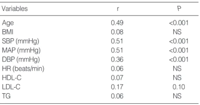
ues were found to correlate significantly with age, systolic BP (SBP), mean arterial pressure (MAP), and diastolic BP (DBP) (Table 2).
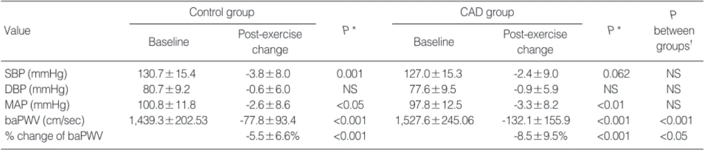
Brachial-ankle PWV values were significantly lower at 10 min after exercise than at baseline in both groups. However, this decrease was significantly larger in the CAD group, thus baPWV in the CAD group was initially higher than in the control group but became similar after exercise. In the con- trol group, SBP and MAP were significantly lower at 10 min after exercise than at baseline, but DBP was not. In the CAD group, MAP was significantly lower at 10 min after exercise than at baseline (Table 3), whereas SBP was marginally lower, and DBP was not significantly different. Heart rates were higher at 10 min after exercise than at baseline in both groups (Table 3). By multivariate analysis, the CAD group showed a larger decrease in baPWV after exercise than the control group after adjusting for age, BMI, SBP, MAP, MAP reduc- tion, and baseline baPWV (Table 4).
DISCUSSION
Arterial stiffness increases left ventricular afterload and alters coronary perfusion (5), and has been independently associat-
Variables (Mean±SD) Control group (n=50)
CAD group
(n=50) P value
Age (yr) 53.7±8.8 61.2±9.0 <0.001
Sex, male 33 (66%) 44 (88%) <0.01
BMI (kg/m2) 24.8±3.1 24.5±2.4 0.581
Diabetes mellitus 10 (20%) 12 (24%) 0.629
Current smoker 11 (22%) 13 (26%) 0.640
Hypertension 24 (48%) 34 (68%) 0.043
HDL-C (mg/dL) 51.2±13.8 48.8±14.2 0.410 LDL-C (mg/dL) 121.2±28.5 102.4±28.1 <0.01 Triglyceride (mg/dL) 159.6±82.7 153.8±106.3 0.777 Ejection fraction (%) 64.8±5.1 58.3±13.8 <0.05 Treadmill exercise time 10.3±1.3 9.3±1.9 <0.01
(min)
baPWV, averaged 1,359.3±188.8 1,448.9±230 <0.05 (cm/sec)
Medication
Aspirin 15 (30%) 49 (98%) <0.001
β-blocker 7 (14%) 33 (66%) <0.001
RAS inhibitors 12 (24%) 26 (52%) <0.01 HMG-CoA reductase 6 (12%) 41 (82%) <0.001
inhibitor
Calcium channel blocker 7 (14%) 17 (34%) <0.05
Diuretics 6 (12%) 7 (14%) 0.766
Table 1. Clinical characteristics and laboratory findings of study participants
BMI, body mass index; HDL-C, high density lipoprotein cholesterol; LDL- C, low density lipoprotein cholesterol; baPWV, brachial-ankle pulse wave velocity; RAS inhibitors, renin angiotensin system inhibitors.
Variables r P
Age 0.49 <0.001
BMI 0.08 NS
SBP (mmHg) 0.51 <0.001
MAP (mmHg) 0.51 <0.001
DBP (mmHg) 0.36 <0.001
HR (beats/min) 0.06 NS
HDL-C 0.07 NS
LDL-C 0.17 0.10
TG 0.06 NS
Table 2. Correlations between baPWV and clinical parameters at baseline
The values shown are Pearson correlation coefficients.
BMI, body mass index; SBP, systolic blood pressure; MAP, mean arte- rial pressure; DBP, diastolic blood pressure; HR, heart rate; HDL-C, high density lipoprotein cholesterol; LDL-C, low density lipoprotein cholesterol, TG, triglyceride; baPWV, brachial-ankle pulse wave velocity.
ed with target organ damage and increased cardiovascular morbidity and mortality (6). Brachial-ankle PWV is a sim- ple marker of arterial stiffness (7, 8) and mainly reflects large artery stiffness, although it has also been reported to reflect endothelium-dependent peripheral vasodilation.
Changes in vascular wall distensibility may be induced by changes in the quality and quantity of vascular fibrous matrix (e.g., elastic fibers and collagen fibers in media: an organic factor) and by changes in smooth muscle tone (a functional factor). Elastic fiber is the primary determinant of vascular distensibility under physiologic conditions (9, 10). Moreover, the elastin-collagen compositions of arterial walls represent a more chronic component of arterial stiffness and changes only over years, thus it is unlikely that short-duration aero- bic exercise changes these structural components (11). Instead, arterial compliance is probably altered in the short terms, or even acutely, via the modulation of the sympathetic-adren- ergic tone of smooth muscle cells in arterial walls (12), which is affected by autonomic nervous activity and vasoactive agents derived from vascular endothelial cells, e.g., nitric oxide (NO), prostacyclin, and endothelium-derived hyperpolarizing factor (13). In particular, the production of NO is important, because it is a potent endothelium-dependent vasodilator and reduces vasoconstrictor response to α-adrenergic receptor stimulation (14). Moreover, pulsatile flow in the aorta associated with exercise training might evoke the acute release of NO, upreg-
ulate NO production, and increase the productions of other vasodilatory factors (15-17). In CAD patients, endothelial dysfunction develops secondary to reduced NO production and early reactivation by reactive oxygen species (18). In the present study, baPWV, which was higher in the CAD group at baseline, was found to be reduced significantly at 10 min after exercise in both groups, and because this decrease was larger in the CAD group, no difference in baPWV was ob- served between the two groups after exercise. These observa- tions suggest that short-duration exercise affects arterial stiff- ness even in patients with CAD. We speculate that the mech- anism involved may be related to the restoration of an equi- librium between NO production and inactivation by reactive oxygen species, which also appears to be the primary mech- anism underlying exercise training-mediated perfusion im- provements in CAD patients (18).
BP and age have been reported to be important determi- nants of baPWV in healthy individuals (7, 19), which is con- sistent with the findings of the present study. Exercise dura- tion is significantly different between the two groups which might be a confounding factor. However, exercise duration is longer and the proportional change of baPWV is smaller in control group, and moreover exercise duration is not cor- related to both absolute and relative change of baPWV (data not shown). Thus it is not probable that different exercise dura- tion is a significant confounding factor.
Because, as mentioned above, CAD group members were taking more medications, it is unclear whether our findings suggest that patients with stable CAD have the potential to reverse arterial stiffness with exercise despite the presence of disease or whether they reflect an effect of the medications taken, such as aspirin, β-blocker, renin-angiotensin system inhibitor, and HMG-CoA reductase inhibitor. The lower level of LDL-cholesterol in the CAD group was probably due to HMG-CoA reductase inhibitors.
However, considering the higher baseline baPWV value in the CAD group, it appears that medications do not com- pletely normalize arterial stiffness in CAD patients and that short-duration exercise seems to independently improve arte- rial stiffness immediately. Because majority of patients in CAD group and only a small number of persons in control group
*P value for significant post-exercise change in each group; �P value for the difference between group post-exercise changes.
SBP, systolic blood pressure; DBP, diastolic blood pressure; MAP, mean arterial pressure; HR, heart rate; baPWV, brachial artery pulse wave velocity.
Control group
Value P *
Baseline Post-exercise change
CAD group
P *
P between
groups� Baseline Post-exercise
change
SBP (mmHg) 130.7±15.4 -3.8±8.0 0.001 127.0±15.3 -2.4±9.0 0.062 NS
DBP (mmHg) 80.7±9.2 -0.6±6.0 NS 77.6±9.5 -0.9±5.9 NS NS
MAP (mmHg) 100.8±11.8 -2.6±8.6 <0.05 97.8±12.5 -3.3±8.2 <0.01 NS
baPWV (cm/sec) 1,439.3±202.53 -77.8±93.4 <0.001 1,527.6±245.06 -132.1±155.9 <0.001 <0.001
% change of baPWV -5.5±6.6% <0.001 -8.5±9.5% <0.001 <0.05
Table 3. Changes in hemodynamic parameters and PWV due to exercise
Independent variables β P
Intercept -45.7±174.9
Age 4.23±1.69 0.014
BMI -7.8±4.6 0.10
SBP -4.3±2.3 0.07
MAP 8.7±3.0 0.005
Change of MAP 6.7±1.8 <0.001
Group (Control vs. CAD) -55.6±26.7 0.04
Baseline baPWV -0.25±0.08 0.002
Table 4. Predictors of baPWV change by exercise
β‚ regression coefficients. Model R2=0.31.
BMI, body mass index; SBP, systolic blood pressure; DBP, diastolic blood pressure; MAP, mean arterial pressure; baPWV, brachial artery pulse wave velocity.
of CAD may have more reversible component related to en- dothelial dysfunction and less irreversible component such as structural change of vascular wall. This group of patients has higher baseline baPWV due to the endothelial dysfunc- tion but much of this functional abnormality might be rever- sible by some intervention, such as short-duration exercise in this study. Patients in the CAD group had undergone per- cutaneous coronary intervention and those with findings of residual ischemia were excluded by exercise test. This exclu- sion may have resulted in selection of patients with lower risk and less extensive coronary artery disease.
However, as a limitation of this study, we do not have enough information on the duration and extent of CAD in the patient group. Also, it is not possible to investigate this speculation further by measuring biomarkers of NO production and oxida- tive stress. Another potential limitation is that only brachial- ankle PWV was measured in our study. If markers of central blood pressure (20, 21) had been measured, different findings might have been found. In previous studies, aerobic exercise improved peripheral arterial stiffness but not central arterial stiffness (22), and central arterial stiffness was a better prog- nostic factor than peripheral arterial stiffness (23). However, this does not mean that peripheral arterial stiffness is mean- ingless, which has been shown to have prognostic value in another study (24), and also shown to be correlated to central arterial stiffness (8). It is likely that central arterial stiffness is a better index but peripheral arterial stiffness measured by baPWV is a very convenient alternative. These points should be subjects of future studies.
Another weakness of the study is that we do not have data on the heart rate and blood pressure at the time of PWV mea- surement. Because these variables acutely influence baPWV, this can be a potential source of confounding. However, we assume that heart rate and blood pressure was probably not different from the baseline at 10 min post-exercise.
Though our study showed immediate short-term response to exercise, evidence is scarce on whether repeated short-dura- tion exercise may result in a persistent and long-term improve- ment of arterial stiffness. Further study is needed on this ques- tion, considering that frequent short-bout exercise is being discussed as a practical alternative to conventional long-dura- tion exercise (25, 26).
REFERENCES
1. Sugawara J, Otsuki T, Tanabe T, Maeda S, Kuno S, Ajisaka R, Mat- suda M. The effects of low-intensity single-leg exercise on regional arterial stiffness. Jpn J Physiol 2003; 53: 239-41.
2. Arnett DK, Evans GW, Riley WA. Arterial stiffness: a new cardio- vascular risk factor? Am J Epidemiol 1994; 140: 669-82.
3. Sugawara J, Maeda S, Otsuki T, Tanabe T, Ajisaka R, Matsuda M.
Effects of nitric oxide synthase inhibitor on decrease in peripheral arterial stiffness with acute low-intensity aerobic exercise. Am J Physi- ol Heart Circ Physiol 2004; 287: H2666-9.
4. Edwards DG, Schofield RS, Magyari PM, Nichols WW, Braith RW.
Effect of exercise training on central aortic pressure wave reflection in coronary artery disease. Am J Hypertens 2004; 17: 540-3.
5. Wada T, Kodaira K, Fujishiro K, Maie K, Tsukiyama E, Fukumoto T, Uchida T, Yamazaki S. Correlation of ultrasound-measured com- mon carotid artery stiffness with pathological findings. Arterioscler Thromb 1994; 14: 479-82.
6. Schiffrin EL. Vascular stiffening and arterial compliance. Implica- tions for systolic blood pressure. Am J Hypertens 2004; 17: 39S-48S.
7. Asmar R, Benetos A, London G, Hugue C, Weiss Y, Topouchian J, Laloux B, Safar M. Aortic distensibility in normotensive, untreated and treated hypertensive patients. Blood Press 1995; 4: 48-54.
8. Yamashina A, Tomiyama H, Takeda K, Tsuda H, Arai T, Hirose K, Koji Y, Hori S, Yamamoto Y. Validity, reproducibility, and clinical significance of noninvasive brachial-ankle pulse wave velocity mea- surement. Hypertens Res 2002; 25: 359-64.
9. Berry CL, Greenwald SE, Rivett JF. Static mechanical properties of the developing and mature rat aorta. Cardiovasc Res 1975; 9: 669-78.
10. Kakiyama T, Sugawara J, Murakami H, Maeda S, Kuno S, Matsuda M. Effects of short-term endurance training on aortic distensibility in young males. Med Sci Sports Exerc 2005; 37: 267-71.
11. Safar ME, Laurent S, Pannier BM, London GM. Structural and func- tional modifications of peripheral large arteries in hypertensive pa- tients. J Clin Hypertens 1987; 3: 360-7.
12. Boutouyrie P, Lacolley P, Girerd X, Beck L, Safar M, Laurent S.
Sympathetic activation decreases medium-sized arterial compliance in humans. Am J Physiol 1994; 267: H1368-76.
13. Ekblom B, Kilbom A, Soltysiak J. Physical training, bradycardia, and autonomic nervous system. Scand J Clin Lab Invest 1973; 32:
251-6.
14. Patil RD, DiCarlo SE, Collins HL. Acute exercise enhances nitric oxide modulation of vascular response to phenylephrine. Am J Phys- iol 1993; 265: H1184-8.
15. Delp MD, Laughlin MH. Time course of enhanced endothelium-medi- ated dilation in aorta of trained rats. Med Sci Sports Exerc 1997;
29: 1454-61.
16. Green D, Cheetham C, Mavaddat L, Watts K, Best M, Taylor R, O’Driscoll G. Effect of lower limb exercise on forearm vascular func- tion: contribution of nitric oxide. Am J Physiol Heart Circ Physiol 2002; 283: H899-907.
17. Joyner MJ. Effect of exercise on arterial compliance. Circulation 2000; 102: 1214-5.
18. Erbs S, Linke A, Hambrecht R. Effects of exercise training on mor- tality in patients with coronary heart disease. Coron Artery Dis 2006;
17: 219-25.
19. Nurnberger J, Dammer S, Opazo Saez A, Philipp T, Schafers RF.
Diastolic blood pressure is an important determinant of augmenta- tion index and pulse wave velocity in young, healthy males. J Hum Hypertens 2003; 17: 153-8.
20. Hirata K, Kawakami M, O’Rourke MF. Pulse wave analysis and pulse wave velocity: a review of blood pressure interpretation 100
years after Korotkov. Circ J 2006; 70: 1231-9.
21. Takaki A, Ogawa H, Wakeyama T, Iwami T, Kimura M, Hadano Y, Matsuda S, Miyazaki Y, Matsuda T, Hiratsuka A, Matsuzaki M.
Cardio-ankle vascular index is a new noninvasive parameter of arte- rial stiffness. Circ J 2007; 71: 1710-4.
22. Heffernan KS, Jae SY, Fernhall B. Racial differences in arterial stiff- ness after exercise in young men. Am J Hypertens 2007; 20: 840-5.
23. Pannier B, Guerin AP, Marchais SJ, Safar ME, London GM. Stiff- ness of capacitive and conduit arteries: prognostic significance for end-stage renal disease patients. Hypertension 2005; 45: 592-6.
24. Tomiyama H, Koji Y, Yambe M, Shiina K, Motobe K, Yamada J, Shido N, Tanaka N, Chikamori T, Yamashina A. Brachial-ankle pulse wave velocity is a simple and independent predictor of prognosis in patients with acute coronary syndrome. Circ J 2005; 69: 815-22.
25. Jakicic JM, Winters C, Lang W, Wing RR. Effects of intermittent exercise and use of home exercise equipment on adherence, weight loss, and fitness in overweight women: a randomized trial. JAMA 1999; 282: 1554-60.
26. Murphy MH, Hardman AE. Training effects of short and long bouts of brisk walking in sedentary women. Med Sci Sports Exerc 1998;
30: 152-7.