Metastasis of papillary thyroid carcinoma to the uvea is extremely rare, with only 6 patients reported in the literature
4
0
0
전체 글
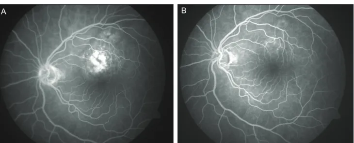
(2) Korean J Ophthalmol Vol.27, No.3, 2013. A. B. A. B. Fig. 1. (A) B-scan ultrasonogram of the right eye before enucleation shows the intraocular tumor with orbital shadowing which probably corresponds to the extraocular extension. (B) The enucleated right eye had a pigmented scleral nodule on the superior part.. A. B. Fig. 2. (A) Mid-phase fluorescein angiogram of the left fundus showing the relatively well-delineated lesion above the fovea with patchy areas of hyperfluorescence, before transpupillary thermotherapy. (B) Post-treatment early mid-phase fluorescein angiogram demonstrates the almost complete resolution of the lesion.. 11 mm and a thickness of 6 mm in the right eye (Fig. 1A). The left eye was normal. The right eye was enucleated, as there were no prospects of recovering useful vision. A dark, well-encapsulated scleral nodule was noted over the enucleated globe (Fig. 1B). A month later, the patient’s visual acuity in the left eye decreased to 20 / 200 with the new development of a solitary, reddish-pink choroidal mass that measured 6 × 4.5 mm in basal dimensions and 1.5 mm in thickness, superior to the fovea and within the temporal vascular arcades (Fig. 2A). There was minimal subretinal f luid over the lesion. This tumor was treated 216. with a single session of transpupillary thermotherapy using a 0.5 mm spot size, 500 mW power, and 29 overlapping spots each lasting 45 seconds. Six weeks later, the tumor in the left eye totally disappeared without any improvement in the patient’s visual acuity (Fig. 2B). Evidence for widespread metastatic involvement of the lungs was confirmed a month later and he expired from respiratory complications. Histopathological examination of the eye revealed that the choroidal tumor was composed of papillary excrescences and cystic spaces (Fig. 3A). The papillae were.
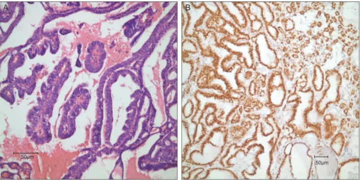
(3) H Kiratli, et al. Bilateral Uveal Papillary Thyroid Cancer Metastases. B. A. 50μm. 50μm. Fig. 3. Histopathological view of the specimen showing (A) papillary excrescences typical of papillary thyroid carcinoma (H&E, ×40) and (B) strong thyroid transcription factor 1 signals (×40).. covered by columnar and cuboidal cells with eosinophilic cytoplasms. These cells had elongated nuclei and irregular nuclear contour. In one area, the sclera was eroded in full thickness and tumor cells gained access into the subTenon’s space. Immunohistopathological studies showed strong signals of thyroglobulin and thyroid transcription factor 1 (Fig. 3B).. Discussion Papillary carcinoma constitutes 80% of all thyroid cancers in the human [1]. Forty-two percent of metastases develop in the regional lymph nodes. Hematogenous distant metastases occur in only 10% of patients, mainly to the lungs, bones, and brain [1,4]. Ocular metastasis is distinctly unusual and when present, the orbit is more commonly affected than the intraocular tissues [5]. Uveal metastatic thyroid carcinoma was found in patients who had their primary cancer diagnosed and treated for an average of 9.9 years (range, 4 to 30 years) [4-9]. In 2 cases, uveal metastases were the initial presentations of the papillary thyroid cancer [3,8]. Reported ocular signs and symptoms included a sudden loss of visual acuity, loss of color discrimination, visual field defects, and light flashes [3-9]. The majority of these patients had a solitary, amelanotic, orange-yellow choroidal mass sometimes associated with serous or hemorrhagic retinal detachment [3-7,9]. Three patients had bilateral choroidal involvement [3,7,8]. Extraocular extension, very similar to our case, was found. only once, in a blind and painful eye [8]. Because a very limited numbers of patients have been reported, no data exist describing the best and most effective treatment of this condition. External beam radiotherapy, I131 ablation therapy and enucleation have been employed based on the extent of the tumor, the potential for visual improvement and the systemic status of the patient [3-9]. In our patient, transpupillary thermotherapy resulted in satisfactory regression of the solitary choroidal metastatic tumor in the left eye.. Conflict of Interest No potential conflict of interest relevant to this article was reported.. References 1. Rosenbaum MA, McHenry CR. Contemporary management of papillary carcinoma of the thyroid gland. Expert Rev Anticancer Ther 2009;9:317-29. 2. Shields CL, Shields JA, Gross NE, et al. Survey of 520 eyes with uveal metastases. Ophthalmology 1997;104:1265-76. 3. Arat YO, Boniuk M. Red lesions of the iris, choroid, and skin secondary to metastatic carcinoma of the thyroid: a review. Surv Ophthalmol 2007;52:523-8. 4. Bucerius J, Meyka S, Michael B, et al. Papillary thyroid carcinoma with an uncommon spread of hematogenous metastases to the choroid and the skin. J Natl Med Assoc 2008;100:104-7. 5. Slamovits TL, Mondzelewski JP, Kennerdell JS. Thy-. 217.
(4) Korean J Ophthalmol Vol.27, No.3, 2013. roid carcinoma metastatic to the globe. Br J Ophthalmol 1979;63:169-72. 6. Ahmadi MA, Nicholes D, Esmaeli B. Late choroidal metastasis secondary to papillary thyroid carcinoma. Am J Ophthalmol 2001;132:796-8. 7. Avram AM, Gielczyk R, Su L, et al. Choroidal and skin metastases from papillary thyroid cancer: case and a review of the literature. J Clin Endocrinol Metab 2004;89:5303-7.. 218. 8. Singh U, Kaushik S, Pandav SS, et al. Papillary carcinoma thyroid presenting as a choroidal metastasis: report of a case and brief review of literature. Indian J Ophthalmol 2003;51:81-3. 9. Yunta Abarca PJ, Ponce JL, Prieto M, et al. Papillary thyroid carcinoma that metastasised to the choroid. Eur J Surg 1999;165:998-9..
(5)
수치
관련 문서
In differentiated thyroid carcinoma, follicular histology is a significant factor for survival, and the 3-year survival rate for papillary and follicular carcinomas with
Purpose: American Thyroid Association's guidelines (2015) recommend that papillary thyroid carcinomas (PTCs) ≤4 cm without extrathyroidal extension (ETE) and clinical lymph
Purpose: We aimed to determine the influencing factors for central neck lymph node metastasis (CLNM) in papillary thyroid microcarcinoma (PTMC) without clinical evidence of
We analyzed the risk of FNAB providing false negative results of lateral neck node metastasis, and evaluated diagnostic accuracy of FNAB, in patients with papillary thyroid
A histologi- cal analysis of the brain mass under a low-power view revealed a cystic and solid tumor mass that was infiltrating into the brain parenchyma with hem- orrhage
We aimed to develop a radiomics signature using US images of the primary tumor to preopera- tively predict lateral lymph node metastasis (LNM) in patients with conventional
Conclusion: Our results suggest that overexpression of p53 protein and Ki-67 in papillary thyroid carcinoma is associated with tumor progression and that IHC for these proteins
Tumor size(p=0.000), ETE(p=0.001), multifocality(p=0.014), and bilaterality(p=0.001) were significantly related factors for cervical lymph node metastasis clinically
