ABSTRACT
Purpose: We aimed to determine the influencing factors for central neck lymph node metastasis (CLNM) in papillary thyroid microcarcinoma (PTMC) without clinical evidence of metastasis on preoperative ultrasonography.
Methods: We retrospectively analyzed 625 patients with PTMC who underwent thyroid surgery at Chosun University Hospital from January 2002 to December 2012. A total of 575 patients who had no evidence of lymph node metastasis by preoperative ultrasonography were included in the study. Medical records, including clinical information and pathologic report, were reviewed.
Results: Central lymph node metastasis was found in 81 (14.1%) out of 575 patients.
Results of univariate analysis indicated that lymph node metastasis occurred frequently in patients with more than 0.5 cm largest tumor size by preoperative sonography and pathologic reports (P=0.048 and P=0.001, respectively) and lymphovascular invasion (LVI) (P<0.001). Multivariate analysis revealed that sex (female vs. male), pathologic tumor size (0.5–1.0 cm vs. <0.5 cm), and LVI (yes vs. no) were significantly associated with lymph node metastasis (odds ratio [OR], 0.498, 95% confidence interval [CI], 0.250–0.992, P=0.047;
OR, 2.450, 95% CI, 1.313–4.570, P=0.005; and OR, 24.954, 95% CI, 2.430–256.217, P=0.007, respectively).
Conclusion: Male sex, large tumor size (≥0.5 cm), and LVI were the risk factors for CLNM in patients with PTMC without clinical evidence of node metastasis. Prophylactic central neck lymph node dissection (CLND) might be required in these cases of PTMC.
Keywords: Thyroid neoplasms; Papillary carcinoma; Papillary thyroid microcarcinoma;
Lymph nodes
INTRODUCTION
Papillary thyroid carcinomas (PTCs) are the most common type of thyroid cancers and account for approximately 75%–85% of thyroid malignancies (1). According to the World Health Organization (WHO) classification system, papillary thyroid microcarcinomas (PTMCs) are PTCs that are 1 cm or less in diameter (2). The prevalence rate of PTCs and
Original Article
Received: May 17, 2017 Revised: Jun 27, 2017 Accepted: Jul 20, 2017 Correspondence to Kweon Cheon Kim
Department of Surgery, Chosun University College of Medicine, 365 Pilmun-daero, Dong-gu, Gwangju, Korea.
Tel: +82-62-220-3068 Fax: +82-62-228-3441 E-mail: [email protected] Copyright © 2017. Korean Association of Thyroid and Endocrine Surgeons; KATES This is an Open Access article distributed under the terms of the Creative Commons Attribution Non-Commercial License (https://
creativecommons.org/licenses/by-nc/4.0/).
ORCID iDs Su Yeon Jeong
https://orcid.org/0000-0003-4505-6887 Funding
This study was supported by grants from the Clinical Medicine Research Institute at Chosun University Hospital (2013).
Author Contributions
Conceptualization: Kweon Cheon Kim, Yoo Seok Kim; Data curation: Yoo Seok Kim, Su Yeon Jeong; Project administration: Kweon Cheon Kim, Yoo Seok Kim; Supervision: Kweon Cheon Kim; Writing - original draft: Su Yeon Jeong; Writing - review & editing: Yoo Seok Kim.
Su Yeon Jeong , Yoo Seok Kim, Kweon Cheon Kim
Department of Surgery, Chosun University College of Medicine, Gwangju, Korea
Predictive Factors for Central Neck Lymph Node Metastasis in Patients with Papillary Thyroid Microcarcinoma without Suspicious Metastasis by
Preoperative Ultrasonography
Conflict of Interest
No potential conflict of interest relevant to this article was reported.
PTMCs has recently increased. However, it does not reflect the actual increase of incidence rate itself. This phenomenon could be explained by the early detection of small-sized thyroid tumors through advanced diagnostic tools, such as thyroid ultrasonography and fine-needle aspiration (FNA) cytology (3,4).
Despite relatively good prognosis, with slow progression and high survival rate, several studies have reported recurrences and metastases in PTMCs (5), the most common of which is metastasis to the central lymph node of cervical neck (6,7). Given that central neck lymph node metastasis (CLNM) is the most significant risk factor in predicting locoregional recurrences (4,8,9), therapeutic central neck dissection is necessary if CLNM is detected in preoperative examination (10). However, whether prophylactic central neck lymph node dissection (CLND) improves survival rate and prognosis in patients without confirmed CLNM in preoperative examination is yet to be determined.
Given that accurate detection of CLNM via thyroid ultrasonography is challenging due to its anatomical structure (11), establishing factors that can be used in predicting CLNM is necessary. This study aims to determine the predictive factors of CLNM that can be used in preoperative patient evaluation and in determining the necessary extent of surgery.
METHODS
In this retrospective single-center study, 625 PTMC patients who underwent thyroid surgery at Chosun University Hospital from January 2002 to December 2012 were evaluated. Three types of surgery, namely, lobectomy, near-total thyroidectomy, and total thyroidectomy were performed.
Preoperative ultrasonography and FNA cytology results of the patients were reviewed, and of these, 575 patients were included in this study according to the following criteria: 1) tumor size of 1 cm or less in diameter, 2) no evidence of CLNM, and 3) diagnosis of PTC through FNA. All patients underwent ipsilateral or bilateral prophylactic CLND along with thyroid surgery according to the tumor location. Total or near-total thyroidectomy with bilateral CLND was performed in case of bilateral tumors and suspicious extra-thyroidal extension in preoperative ultrasonography, and lobectomy with unilateral CLND was performed in case of unilateral and intra-thyroidal tumor.
The medical records and postoperative pathologic reports of all the patients were reviewed, and the results were analyzed with respect to sex; location, number, and size of tumors; type of operation; capsular invasion; lymphovascular invasion (LVI); and recurrence.
According to the postoperative pathologic results, the patients were divided into 2 groups;
one with CLNM and the other without CLNM. Then, univariate and multivariate analyses of the correlation between CLNM and other clinicopathological features were conducted.
SPSS ver. 21.0 (SPSS Inc., Chicago, IL, USA) was used for statistical analyses. Pearson's χ2 test or Fisher's exact test was used for univariate analysis, and logistic regression analysis was used for multivariate analysis. Along with 95% confidence interval (CI), a P value of <0.05 was considered statistically significant.
RESULTS
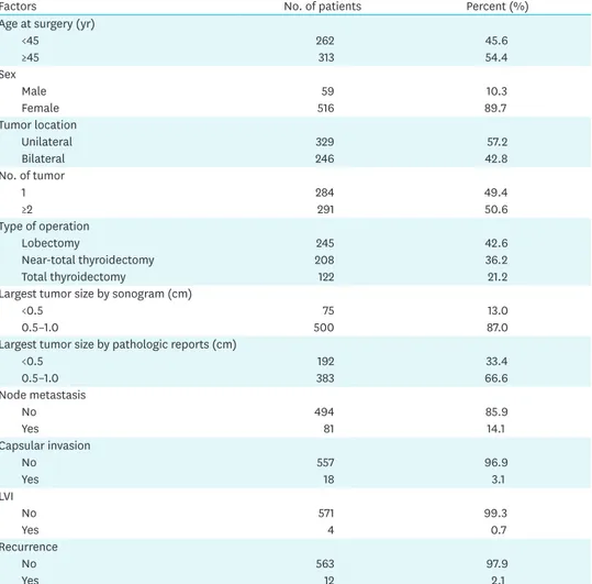
Mean follow-up periods were 109.4 months (range 43–168 months). A total of 262 patients (45.6%) out of the 575 were under 45 years of age, and 516 (89.7%) were female. Unilateral tumors were observed in 329 patients (57.2%), and multifocality (2 or more tumors) was observed in 291 patients (50.6%). In terms of tumor size, 383 cases (66.6%) of tumors larger than 0.5 cm and smaller than 1 cm in size were noted according to final biopsy results.
Capsular invasion occurred in 18 patients (3.1%), and LVI was observed in 4 patients (3.1%).
Recurrence was observed in 12 patients (2.1%), and all of the recurrences were lateral neck lymph node metastasis (Table 1).
Out of 575 patients, 81 (14.1%) who had no evidence of CLNM in preoperative
ultrasonography were confirmed to have CLNM in the postoperative pathologic examination.
Median number of metastasis was 2 (range 1–5). By analyzing the correlation between CLNM and each factors mentioned previously, tumor size larger than 0.5 cm and LVI showed statistically significant correlation, with a P value of <0.05. Recurrence during follow-up periods was more frequently observed in patients with CLNM with statistically significant difference (Table 2).
Table 1. Clinicopathologic features in patients with PTMC without suspicious lymph node metastasis on preoperative ultrasonography (n=575)
Factors No. of patients Percent (%)
Age at surgery (yr)
<45 262 45.6
≥45 313 54.4
Sex
Male 59 10.3
Female 516 89.7
Tumor location
Unilateral 329 57.2
Bilateral 246 42.8
No. of tumor
1 284 49.4
≥2 291 50.6
Type of operation
Lobectomy 245 42.6
Near-total thyroidectomy 208 36.2
Total thyroidectomy 122 21.2
Largest tumor size by sonogram (cm)
<0.5 75 13.0
0.5–1.0 500 87.0
Largest tumor size by pathologic reports (cm)
<0.5 192 33.4
0.5–1.0 383 66.6
Node metastasis
No 494 85.9
Yes 81 14.1
Capsular invasion
No 557 96.9
Yes 18 3.1
LVI
No 571 99.3
Yes 4 0.7
Recurrence
No 563 97.9
Yes 12 2.1
PTMC = papillary thyroid microcarcinoma; LVI = lymphovascular invasion.
In multivariate analysis using logistic regression, CLNM risk was higher in men than in women (odds ratio [OR], 0.498; 95% CI, 0.250–0.992; P=0.047). Tumor size (≤0.5 cm) (OR, 2.450; 95% CI, 1.313–4.570; P=0.005) and LVI (OR, 24.954; 95% CI, 2.430–256.217; P=0.007) were statistically significant associated factors (Table 3).
DISCUSSION
PTMC has good disease prognosis, with a 10-year disease-specific survival rate of up to 99.5%. Therefore, the aim should be not only to improve survival rate through simple treatments but also to improve quality of life of the patient after treatment.
Given that CLNM is among the most significant risk factors in predicting locoregional recurrences (4,8,9), the American Thyroid Association (ATA) strongly recommends therapeutic central neck dissection in PTMCs with evidence of CLNM (10). However, diagnosing CLNM only through preoperative imaging is difficult.
According to Hwang and Orloff (11) and Choi et al. (12), the sensitivity in predicting CLNM through preoperative ultrasonography is low (approximately 23.0%–53.2%). This value indicates Table 2. Comparison of the factors according to lymph node metastasis in patients with PTMC without suspicious lymph node metastasis on preoperative ultrasonography (n=575)
Factors No CLNM (n=494) CLNM (n=81) P value*
No. of patients (%) No. of patients (%)
Age at surgery (yr) 0.220
<45 220 (44.5) 42 (51.9)
≥45 274 (55.5) 39 (48.1)
Sex 0.064
Male 46 (9.3) 13 (16.0)
Female 448 (90.7) 68 (84.0)
Tumor location 0.124
Unilateral 289 (58.5) 40 (49.4)
Bilateral 205 (41.5) 41 (50.6)
No. of tumor 0.230
1 249 (50.4) 35 (43.2)
≥2 245 (49.6) 46 (56.8)
Type of operation 0.190
Lobectomy 218 (44.1) 27 (33.3)
Near-total thyroidectomy 174 (35.2) 34 (42.0)
Total thyroidectomy 102 (20.7) 20 (24.7)
Largest tumor size by sonogram (cm) 0.048
<0.5 70 (14.2) 5 (6.2)
0.5–1.0 424 (85.8) 76 (93.8)
Largest tumor size by pathologic reports (cm) 0.001
<0.5 178 (36.0) 14 (17.3)
0.5–1.0 316 (64.0) 67 (82.7)
Capsular invasion 0.157
No 481 (97.4) 76 (93.8)
Yes 13 (2.6) 5 (6.2)
LVI 0.010
No 493 (99.8) 78 (96.3)
Yes 1 (0.2) 3 (3.7)
Recurrence 0.017
No 487 (98.6) 76 (93.8)
Yes 7 (1.4) 5 (6.2)
PTMC = papillary thyroid microcarcinoma; CLNM = central neck lymph node metastasis; LVI = lymphovascular invasion.
*P<0.050, statistically significant.
a high possibility of false-negative results in imaging, resulting in insufficient treatment of patients who require central neck dissections. In this study, 14.1% of CLNM-negative patients in preoperative ultrasonography were diagnosed to have CLNM according to the final biopsy result.
Therefore, performing prophylactic central neck dissection is debated (13-15). Former studies reported transient or permanent hypoparathyroidism and recurrent laryngeal nerve injury as complications of central neck dissection. These complications could degrade the patient quality of life of after surgery (16). If the risk factors of CLNM in PTMCs can be determined, then surgeons can selectively require prophylactic central neck dissection in high-risk patients. As such, unnecessary treatments that might lead to possible complications can be avoided.
Given that age, sex, tumor size, tumor multifocality, and capsular invasion affect PTMC prognosis, these factors were analyzed in our study.
The correlation between age and CLNM is still controversial. Qu et al. (17) reported that the risk of CLNM increases in patients under 45 years of age; however, Lim et al. (18) and Roh et al. (19) reported no statistical significance between age and CLNM. Meanwhile, several researchers claim that performing prophylactic central neck dissection in patients older than 45 years of age results in good prognosis, citing the American Joint Committee on Cancer (AJCC) cancer staging manual that states that PTMC in patients with CLNM older than 45 years of age progresses to stage III and that mortality risk increases in this age group (20). The decreased effectiveness of additional radioactive iodine (RAI) treatment due to decreased RAI uptake ability is also speculated to result in poor prognosis in aging patients. However, the recently revised ATA guideline states that RAI treatment does not provide a significant therapeutic gain in PTMCs (10,21). Therefore, this treatment is not definitive. In our study, no statistical significance was noted between age and CLNM.
In general, PTMC is more prevalent in women than in men; however, studies reported that CLNM occurs more frequently in men (22,23). In this study, univariate analysis showed no statistical significance between sex and CLNM. However, multivariate analysis showed Table 3. Multivariate analysis of factors for central lymph node metastasis in patients with PTMC without suspicious lymph node metastasis on preoperative ultrasonography
Factors OR 95% CI P value*
Age at surgery (yr) 0.066
≥45 vs. <45 0.625 0.378–1.031
Sex 0.047
Female vs. male 0.498 0.250–0.992
Tumor location 0.425
Bilateral vs. unilateral 1.480 0.565–3.879
No. of tumor 0.924
≥2 vs. 1 0.954 0.363–2.508
Type of central neck lymph node dissection 0.070
Bilateral vs. unilateral 0.633 0.386–1.038
Largest tumor size by sonogram (cm) 0.173
0.5–1.0 vs. <0.5 2.034 0.733–5.643
Largest tumor size by pathologic reports (cm) 0.005
0.5–1.0 vs. <0.5 2.450 1.313–4.570
Capsular invasion 0.128
Yes vs. no 2.322 0.784–6.878
LVI 0.007
Yes vs. no 24.954 2.430–256.217
PTMC = papillary thyroid microcarcinoma; OR = odds ratio; CI = confidence interval; LVI = lymphovascular invasion.
*P<0.050, statistically significant.
higher CLNM risk in male population (OR, 0.498; 95% CI, 0.250–0.992; P=0.047). This result suggests that more thorough preoperative examinations and an aggressive recommendation of prophylactic central neck dissection are necessary in male PTMC patients.
Bilaterality and multifocality are risk factors that have consistently been studied. Several studies reported that the tumor recurrence rate increases in multifocal tumors (24,25). In one study, the risk of CLNM is higher in bilateral tumor because of the tendency of large tumor size and more frequent extrathyroidal invasions (26,27). However, according to Zhao et al.
(28), not only multifocality but both tumor size and multifocality are considered to be related to CLNM. In this study, neither location nor number of tumor showed statistically significant correlation to CLNM.
Meanwhile, tumor size was observed to be an important factor in predicting CLNM, and this result corresponds to that of several recent studies (29,30). Although a large tumor is associated with poor prognosis in PTC patients, some studies reported no significant difference in prognosis related to tumor size (≥0.5 vs. <0.5 cm) (31,32). However, we still consider tumor size to be a significant independent predictive factor of CLNM because our study is concerned with the overall mortality and tumor recurrences.
The correlation between capsular invasion and CLNM and between LVI and CLNM is also controversial. Although univariate and multivariate analyses indicated no correlation between capsular invasion and CLNM in this study, Kim et al. (33) reported otherwise. However, the proportion of patients with confirmed invasion was over 50% in Kim et al.'s study (33) and was only 3.1% in our study. This difference may have resulted from varying methods of histopathologic examination and the criteria of defining the invasion in each center.
LVI and CLNM showed a significant correlation in our study, similar to the result of Joo et al. (34,35) but different from that of Roh et al. (19). Considering the anatomical structure of thyroid gland and its well-developed lymphovascular structure that drains primarily to cervical lymph node (36), our result of a correlation between LVI and CLNM is logical. Arora et al. (35) reported that the incidence of LVI in PTMC was 6.1% (4 patients in total 66 patients). LVI was very low in our study, and further investigation is necessary with more patients.
CONCLUSION
Male sex, large tumor size, and LVI were found to be risk factors that can be used in detecting CLNM in PTMC patients who had no preoperative clinical evidence of CLNM. However, there is a limitation that we cannot confirm the LVI before surgery. So, further investigation should be performed to predict a CLNM in PTMC with available factors before surgery.
REFERENCES
1. Kumar V, Robbins SL. Robbins Basic Pathology. 8th ed. Philadelphia (PA): Saunders/Elsevier; 2007.
2. Hedinger C, Williams ED, Sobin LH. The WHO histological classification of thyroid tumors: a commentary on the second edition. Cancer 1989;63:908-11.
3. Chen AY, Jemal A, Ward EM. Increasing incidence of differentiated thyroid cancer in the United States, 1988–2005. Cancer 2009;115:3801-7.
PUBMED | CROSSREF
4. Hay ID, Hutchinson ME, Gonzalez-Losada T, McIver B, Reinalda ME, Grant CS, et al. Papillary thyroid microcarcinoma: a study of 900 cases observed in a 60-year period. Surgery 2008;144:980-7.
PUBMED | CROSSREF
5. Kuo EJ, Goffredo P, Sosa JA, Roman SA. Aggressive variants of papillary thyroid microcarcinoma are associated with extrathyroidal spread and lymph-node metastases: a population-level analysis. Thyroid 2013;23:1305-11.
PUBMED | CROSSREF
6. Yang Y, Chen C, Chen Z, Jiang J, Chen Y, Jin L, et al. Prediction of central compartment lymph node metastasis in papillary thyroid microcarcinoma. Clin Endocrinol (Oxf ) 2014;81:282-8.
PUBMED | CROSSREF
7. Kebebew E. Hereditary non-medullary thyroid cancer. World J Surg 2008;32:678-82.
PUBMED | CROSSREF
8. Pisanu A, Reccia I, Nardello O, Uccheddu A. Risk factors for nodal metastasis and recurrence among patients with papillary thyroid microcarcinoma: differences in clinical relevance between nonincidental and incidental tumors. World J Surg 2009;33:460-8.
PUBMED | CROSSREF
9. Usluogullari CA, Onal ED, Ozdemir E, Ucler R, Kiyak G, Ersoy PE, et al. A retrospective analysis of prognostic factors predictive of lymph-node metastasis and recurrence in thyroid papillary microcarcinoma. Minerva Endocrinol 2015;40:15-22.
PUBMED
10. Haugen BR, Alexander EK, Bible KC, Doherty GM, Mandel SJ, Nikiforov YE, et al. 2015 American Thyroid Association Management guidelines for adult patients with thyroid nodules and differentiated thyroid cancer: the American Thyroid Association guidelines task force on thyroid nodules and differentiated thyroid cancer. Thyroid 2016;26:1-133.
PUBMED | CROSSREF
11. Hwang HS, Orloff LA. Efficacy of preoperative neck ultrasound in the detection of cervical lymph node metastasis from thyroid cancer. Laryngoscope 2011;121:487-91.
PUBMED | CROSSREF
12. Choi JS, Kim J, Kwak JY, Kim MJ, Chang HS, Kim EK. Preoperative staging of papillary thyroid carcinoma:
comparison of ultrasound imaging and CT. AJR Am J Roentgenol 2009;193:871-8.
PUBMED | CROSSREF
13. Wada N, Duh QY, Sugino K, Iwasaki H, Kameyama K, Mimura T, et al. Lymph node metastasis from 259 papillary thyroid microcarcinomas: frequency, pattern of occurrence and recurrence, and optimal strategy for neck dissection. Ann Surg 2003;237:399-407.
PUBMED | CROSSREF
14. Giordano D, Gradoni P, Oretti G, Molina E, Ferri T. Treatment and prognostic factors of papillary thyroid microcarcinoma. Clin Otolaryngol 2010;35:118-24.
PUBMED | CROSSREF
15. Garrel R, Tripodi C, Cartier C, Makeieff M, Crampette L, Guerrier B. Cervical lymphadenopathies signaling thyroid microcarcinoma. Case study and review of the literature. Eur Ann Otorhinolaryngol Head Neck Dis 2011;128:115-9.
PUBMED | CROSSREF
16. Khairy GA, Al-Saif A. Incidental parathyroidectomy during thyroid resection: incidence, risk factors, and outcome. Ann Saudi Med 2011;31:274-8.
PUBMED | CROSSREF
17. Qu N, Zhang L, Ji QH, Chen JY, Zhu YX, Cao YM, et al. Risk factors for central compartment lymph node metastasis in papillary thyroid microcarcinoma: a meta-analysis. World J Surg 2015;39:2459-70.
PUBMED | CROSSREF
18. Lim YC, Choi EC, Yoon YH, Kim EH, Koo BS. Central lymph node metastases in unilateral papillary thyroid microcarcinoma. Br J Surg 2009;96:253-7.
PUBMED | CROSSREF
19. Roh JL, Kim JM, Park CI. Central cervical nodal metastasis from papillary thyroid microcarcinoma:
pattern and factors predictive of nodal metastasis. Ann Surg Oncol 2008;15:2482-6.
PUBMED | CROSSREF
20. Edge SB, Byrd DR, Compton CC, Fritz AG, Greene FL, Trotti A. AJCC Cancer Staging Handbook: from the AJCC Cancer Staging Manual. 7th ed. New York (NY): Springer; 2010.
21. Miccoli P, Minuto MN, Ugolini C, Panicucci E, Berti P, Massi M, et al. Intrathyroidal differentiated thyroid carcinoma: tumor size-based surgical concepts. World J Surg 2007;31:888-94.
PUBMED | CROSSREF
22. So YK, Son YI, Hong SD, Seo MY, Baek CH, Jeong HS, et al. Subclinical lymph node metastasis in papillary thyroid microcarcinoma: a study of 551 resections. Surgery 2010;148:526-31.
PUBMED | CROSSREF
23. Zhang L, Wei WJ, Ji QH, Zhu YX, Wang ZY, Wang Y, et al. Risk factors for neck nodal metastasis in papillary thyroid microcarcinoma: a study of 1066 patients. J Clin Endocrinol Metab 2012;97:1250-7.
PUBMED | CROSSREF
24. Mercante G, Frasoldati A, Pedroni C, Formisano D, Renna L, Piana S, et al. Prognostic factors affecting neck lymph node recurrence and distant metastasis in papillary microcarcinoma of the thyroid: results of a study in 445 patients. Thyroid 2009;19:707-16.
PUBMED | CROSSREF
25. Bansal M, Gandhi M, Ferris RL, Nikiforova MN, Yip L, Carty SE, et al. Molecular and histopathologic characteristics of multifocal papillary thyroid carcinoma. Am J Surg Pathol 2013;37:1586-91.
PUBMED | CROSSREF
26. Wang W, Zhao W, Wang H, Teng X, Wang H, Chen X, et al. Poorer prognosis and higher prevalence of BRAF (V600E) mutation in synchronous bilateral papillary thyroid carcinoma. Ann Surg Oncol 2012;19:31-6.
PUBMED | CROSSREF
27. Kim KE, Kim EK, Yoon JH, Han KH, Moon HJ, Kwak JY. Preoperative prediction of central lymph node metastasis in thyroid papillary microcarcinoma using clinicopathologic and sonographic features. World J Surg 2013;37:385-91.
PUBMED | CROSSREF
28. Zhao Q, Ming J, Liu C, Shi L, Xu X, Nie X, et al. Multifocality and total tumor diameter predict central neck lymph node metastases in papillary thyroid microcarcinoma. Ann Surg Oncol 2013;20:746-52.
PUBMED | CROSSREF
29. Lee HS, Park HS, Kim SW, Choi G, Park HS, Hong JC, et al. Clinical characteristics of papillary thyroid microcarcinoma less than or equal to 5 mm on ultrasonography. Eur Arch Otorhinolaryngol 2013;270:2969-74.
PUBMED | CROSSREF
30. Ghossein R, Ganly I, Biagini A, Robenshtok E, Rivera M, Tuttle RM. Prognostic factors in papillary microcarcinoma with emphasis on histologic subtyping: a clinicopathologic study of 148 cases. Thyroid 2014;24:245-53.
PUBMED | CROSSREF
31. Lee J, Park JH, Lee CR, Chung WY, Park CS. Long-term outcomes of total thyroidectomy versus thyroid lobectomy for papillary thyroid microcarcinoma: comparative analysis after propensity score matching.
Thyroid 2013;23:1408-15.
PUBMED | CROSSREF
32. Ito Y, Uruno T, Nakano K, Takamura Y, Miya A, Kobayashi K, et al. An observation trial without surgical treatment in patients with papillary microcarcinoma of the thyroid. Thyroid 2003;13:381-7.
PUBMED | CROSSREF
33. Kim BY, Jung CH, Kim JW, Lee SW, Kim CH, Kang SK, et al. Impact of clinicopathologic factors on subclinical central lymph node metastasis in papillary thyroid microcarcinoma. Yonsei Med J 2012;53:924-30.
PUBMED | CROSSREF
34. Joo JY, Park JY, Yoon YH, Choi B, Kim JM, Jo YS, et al. Prediction of occult central lymph node metastasis in papillary thyroid carcinoma by preoperative BRAF analysis using fine-needle aspiration biopsy: a prospective study. J Clin Endocrinol Metab 2012;97:3996-4003.
PUBMED | CROSSREF
35. Arora N, Turbendian HK, Kato MA, Moo TA, Zarnegar R, Fahey TJ 3rd. Papillary thyroid carcinoma and microcarcinoma: is there a need to distinguish the two? Thyroid 2009;19:473-7.
PUBMED | CROSSREF
36. Liu LS, Liang J, Li JH, Liu X, Jiang L, Long JX, et al. The incidence and risk factors for central lymph node metastasis in cN0 papillary thyroid microcarcinoma: a meta-analysis. Eur Arch Otorhinolaryngol 2017;274:1327-38.