Copyright © 2017 Korean Dementia Association 87
INTRODUCTION
The dot-like hippocampal signal intensity in diffusion- weighted images (DWI) is well-known as a characteristic im- aging feature in transient global amnesia (TGA).1 It is seen in single or multiple lesions that range in size from 1 mm to 5 mm and can be seen in both the unilateral and bilateral hip- pocampus.2 TGA is a neurological syndrome in which sud- den forward-and-backward memory loss occurs that is slow- ly recovered within 24 hours.3 We here report on two patients with these dot-like hippocampal hyperintensities who did not present with anterograde amnesia except for headaches.
CASE REPORT
Case 1
A 65-year-old woman without a specific medical history presented with a sudden onset of headache one day; the head- ache had developed suddenly while the patient had been watching TV, and the headache area was confined to the oc- ciput. She had experienced no trauma or infection history be- fore the symptom and no sudden emotional or postural chang- es as she watched TV. The pattern of headache was non-pulsatile, and it was the first headache the patient had experienced.
The pain intensity was not sufficient to interfere with daily life, and she had no accompanying symptoms such as nau- sea, vomiting, or dizziness. At the time of admission, the pa- tient’s physical signs were normal, and her neurological exami- nation revealed no specific findings. One week later, the patient revisited the clinic. Headache intensity had not improved, so we decided to conduct additional examination to differenti-
Hippocampal Lesions of Diffusion Weighted
Magnetic Resonance Image in Patients with Headache without Symptoms of Transient Global Amnesia
Jeong Hoon Park, Chung Geun Oh, Sung Hun Kim, Seung-Hwan Lee, Jae-Won Jang
Department of Neurology, Kangwon National University Hospital, Chuncheon, Korea
Background The dot-like hippocampal signal intensity in diffusion-weighted MR images is well-known as a characteristic imaging feature in transient global amnesia, a neurological syndrome in which sudden forward-and-backward memory loss occurs that is slowly recovered within 24 hours. We here report on patients with this dot-like hippocampal hyperintensity who did not present with anterograde amnesia ex- cept for headaches.
Case Report Two women without a specific medical history presented with sudden-onset headaches on the same day. Neither had any trauma or infection history before the symptom or any sudden emotional or postural changes. Brain MRI showed tiny hippocampal high sig- nal intensity on diffusion-weighted images (DWI).
Conclusions Dot-like hippocampal lesions seen on DWI may be present without memory impairment, and more studies are needed to determine whether there is any association with headache as in this case.
Key Words transient global amnesia, hippocampus, diffusion-weighted magnetic resonance image.
Received: August 4, 2017 Revised: August 14, 2017 Accepted: August 14, 2017
Correspondence: Jae-Won Jang, MD, Department of Neurology, Kangwon National University Hospital, 156 Baengnyeong-ro, Chuncheon 24289, Korea Tel: +82-33-258-9174, Fax: +82-33-258-2103, E-mail: [email protected]
cc This is an Open Access article distributed under the terms of the Cre- ative Commons Attribution Non-Commercial License (http://creative- commons.org/licenses/by-nc/4.0) which permits unrestricted non-com- mercial use, distribution, and reproduction in any medium, provided the ori- ginal work is properly cited.
DND
Print ISSN 1738-1495 / On-line ISSN 2384-0757
Dement Neurocogn Disord 2017;16(3):87-90 / https://doi.org/10.12779/dnd.2017.16.3.87
CASE REPORT
Jeong Hoon Park et al.
Hippocampal Dot-Like Lesion in Headache Patients
88 Dement Neurocogn Disord 2017;16(3):87-90
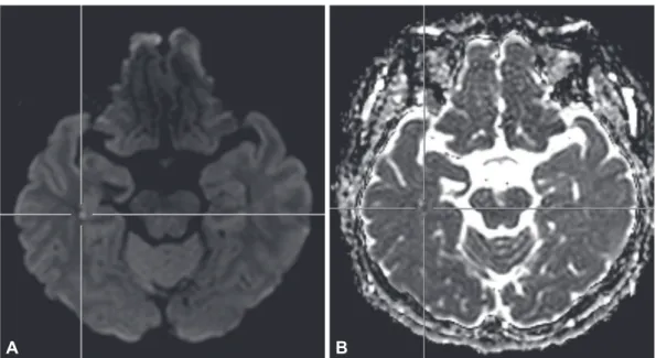
ate the secondary headache. There were no abnormal find- ings on the baseline blood test or electrocardiogram, but brain MRI showed dot-like hippocampal signal intensity on DWI with matching low apparent diffusion coefficient val- ues; there were no other brain lesions on T2-weighted images, and cerebral blood vessels were normal (Fig. 1). In a detailed discussion, the patient described all the events in chronologi- cal order with no deterioration in cognitive function includ- ing memory. The patient’s headache relieved gradually with administration of tricyclic antidepressants, and she is cur- rently being followed up with no headache recurrence.
Case 2
This patient visited the hospital with a headache that had started one week before. The headache started suddenly while she was washing, a pain that started at the back of the neck and spread around the eyes. The character of headache was non-pulsatile, and it was not a pain the patient had expe- rienced before. The headache was most painful within a few minutes, but it had gradually improved at the time of her vis- it; she had no associated symptoms. In the clinic, the patient’s physical signs were normal, and we found no specific findings on neurologic examination. The patient showed no cogni-
Fig. 1. Hippocampal hyperintensity in diffusion-weighted imaging in case 1. A: Diffusion-weighted images. B: Apparent diffusion coefficient.
Fig. 2. Hippocampal hyperintensity in diffusion-weighted imaging in case 2. A: Diffusion-weighted images. B: Apparent diffusion coefficient.
A B
A B
www.dnd.or.kr 89
DND
tive impairment, including memory, when she and her care- giver were interviewed. Because the patient’s headache had a thunderclap nature, we performed MRI to differentiate sec- ondary headache. Her brain DWI showed tiny high signal intensity in the right hippocampus (Fig. 2), but we observed no aneurysms or stenosis on cerebral angiography and no white degeneration or lacunar infarction on T2-weighted im- ages. The patient’s headache improved on its own with no medication, and we are following her with no recurrence.
DISCUSSION
TGA is a syndrome characterized by prospective memory impairment that disappears within 24 hours;4 its diagnosis is based on characteristic clinical features.5 In addition, DWI will show high signal intensity in the hippocampus in most patients from 24 hours to 72 hours after symptom onset, and these image findings are known to characterize TGA.6 Usually, the hippocampal lesions seen on DWI distinguish TGA from cerebral infarction, infection, and so on; however, dot-like lesions are rarely seen in other diseases (Table 1).1 In
general, TGA can be accompanied by characteristic antero- grade amnesia and other symptoms, the most common being headache.7 We searched the literature about the relationship between headache and hippocampal lesions, but we found no similar cases. Migraine is a well-known risk factor for TGA,8 but we did not diagnose this case as migraine because the headache’s features were different from migraine.
Two recent papers have been published for patients with dot-like hippocampal lesions but no prospective memory impairment. Foster et al.9 compared 12 TGA patients who had dot-like hippocampal lesions with 10 cerebral infarct patients who showed isolated punctuate hippocampal lesions.
The authors asserted that DWI alone cannot distinguish be- tween TGA and cerebral infarction and suggested that the ac- companying neurological symptoms and clinical features are important because neurologic abnormalities such as vertigo and dysarthria were more frequently observed in the cerebral infarction group. In contrast, Jeong et al.10 reported eight pa- tients with dot-like hippocampal lesions without anterograde amnesia, and these lesions can be seen in patients with a history of Valsalva manipulation. Kim et al. argued that this phenome-
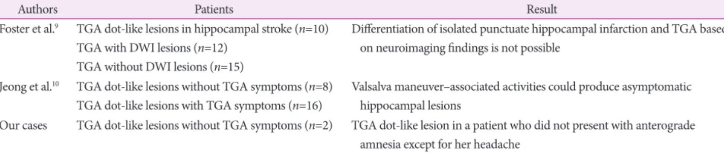
Table 2. TGA dot-like lesions without TGA symptoms
Authors Patients Result
Foster et al.9 TGA dot-like lesions in hippocampal stroke (n=10) TGA with DWI lesions (n=12)
TGA without DWI lesions (n=15)
Differentiation of isolated punctuate hippocampal infarction and TGA based on neuroimaging findings is not possible
Jeong et al.10 TGA dot-like lesions without TGA symptoms (n=8) TGA dot-like lesions with TGA symptoms (n=16)
Valsalva maneuver–associated activities could produce asymptomatic hippocampal lesions
Our cases TGA dot-like lesions without TGA symptoms (n=2) TGA dot-like lesion in a patient who did not present with anterograde amnesia except for her headache
DWI: diffusion-weighted images, TGA: transient global amnesia.
Table 1. Differential diagnosis for hippocampal lesions with a high diffusion signal
Etiology Clinical presentation DWI findings
Stroke 1) Visual field defects 2) Motor weakness 3) Sensory loss 4) Hemisensory deficit
1) Multiple lesions involving extrahippocampal tissue in the PCA territory
2) Patterns involving hippocampus, complete, dorsal, lateral, dot-like lesions (rare)
TGA 1) Sudden onset of anterograde amnesia 2) Complete resolution within 24 h 3) No disturbance of consciousness 4) No other neurological deficit
1) Small circumscribed lesions of 1–2 mm in diameter 2) Lateral part of the hippocampus, CA-1 region 3) Delayed occurrence
Seizure 1) Nonspecific symptoms
2) Single seizure (febrile, psychomotor, generalized tonic-clonic) 3) Repeated seizures
1) Involvement of the lateral hippocampus
2) Additional hyperintensities in the pulvinar or cortex Limbic encephalitis 1) Subacute confusion
2) Psychiatric symptoms 3) Seizures
1) Uncus, amygdale, hippocampus
2) Extending to the entorhinal cortex and subiculum DWI: diffusion-weighted images, PCA: posterior cerebral artery, TGA: transient global amnesia.
Jeong Hoon Park et al.
Hippocampal Dot-Like Lesion in Headache Patients
90 Dement Neurocogn Disord 2017;16(3):87-90
non was attributable to individual sensitivity to hippocam- pal lesion manifestations on Valsalva manipulation (Table 2).
In this case, the absence of accompanying neurologic abnor- malities is explained by a mechanism similar to that of TGA, but the absence of Valsalva manipulation requires a differential diagnosis for cerebral infarction and other diseases. One un- usual thing is that both published papers described accompa- nying symptoms, but no patients had accompanying head- aches. This patient in our study is thought to be the first to show dot-like hippocampal lesions on DWI with only headache and no symptoms of TGA.
The pathophysiology of TGA is still hypothesized to be hemodynamic abnormalities such as cerebral infarction, dif- fusion of cortical inhibition, or venous congestion; of these, venous congestion is the most likely.11-13 If veins are clogged, intravenous hypertension increases the risk of venous con- gestion and subsequent ischemic injury,14 although if these ischemic injuries are not sufficiently severe to cause memory impairment, clinicians may only observe imaging findings without memory disturbances, as in this case. However, wheth- er venous congestion can cause headache or whether the stress caused by headache causes hippocampal lesions is still a question that needs to be resolved.
In conclusion, dot-like hippocampal lesions seen on DWI may be present without memory impairment, and addition- al studies are needed to determine whether there is any as- sociation with headache as in case 2 in this study.
Conflicts of Interest
The authors have no financial conflicts of interest.
REFERENCES
1. Förster A, Griebe M, Gass A, Kern R, Hennerici MG, Szabo K. Dif-
fusion-weighted imaging for the differential diagnosis of disorders affecting the hippocampus. Cerebrovasc Dis 2012;33:104-115.
2. Weon YC, Kim JH, Lee JS, Kim SY. Optimal diffusion-weighted im- aging protocol for lesion detection in transient global amnesia. AJNR Am J Neuroradiol 2008;29:1324-1328.
3. Bartsch T, Deuschl G. Transient global amnesia: functional anatomy and clinical implications. Lancet Neurol 2010;9:205-214.
4. Kritchevsky M, Zouzounis J, Squire LR. Transient global amnesia and functional retrograde amnesia: contrasting examples of episodic memory loss. Philos Trans R Soc Lond B Biol Sci 1997;352:1747-1754.
5. Quinette P, Guillery-Girard B, Dayan J, de la Sayette V, Marquis S, Viader F, et al. What does transient global amnesia really mean? Review of the literature and thorough study of 142 cases. Brain 2006;129(Pt 7):
1640-1658.
6. Bartsch T, Alfke K, Stingele R, Rohr A, Freitag-Wolf S, Jansen O, et al. Selective affection of hippocampal CA-1 neurons in patients with transient global amnesia without long-term sequelae. Brain 2006;129 (Pt 11):2874-2884.
7. Williamson J, Larner AJ. Transient global amnesia. Br J Hosp Med (Lond) 2015;76:C186-C188.
8. Donnet A. Transient global amnesia triggered by migraine in a French Tertiary-Care Center: an 11-year retrospective analysis. Headache 2015;55:853-859.
9. Förster A, Al-Zghloul M, Wenz H, Böhme J, Groden C, Neumaier- Probst E. Isolated punctuate hippocampal infarction and transient global amnesia are indistinguishable by means of MRI. Int J Stroke 2017;12:292-296.
10. Jeong M, Jin J, Kim JH, Moon Y, Choi JW, Kim HY. Incidental hippo- campal hyperintensity on diffusion-weighted MRI: individual suscep- tibility to transient global amnesia. Neurologist 2017;22:103-106.
11. Frisoni GB. PET and 18F ligands in the diagnosis of Alzheimer’s dis- ease. Lancet Neurol 2011;10:397-399.
12. Pantoni L, Lamassa M, Inzitari D. Transient global amnesia: a review emphasizing pathogenic aspects. Acta Neurol Scand 2000;102:275- 13. Menéndez González M, Rivera MM. Transient global amnesia: in-283.
creasing evidence of a venous etiology. Arch Neurol 2006;63:1334- 1336.
14. Park YH, Jang JW, Yang Y, Kim JE, Kim S. Reflections of two paral- lel pathways between the hippocampus and neocortex in transient global amnesia: a cross-sectional study using DWI and SPECT. PLoS One 2013;8:e67447.