785
INTRODUCTION
Interference in modern immunoassays caused by heterophilic antibodies (HA) is well documented [1].
According to various published reports, in the general population these antibodies can be found in up to 40% of clinical samples [2]. In many cases, these antibodies have nonspecific affinities and do not interfere with different immunoassays. However, for reasons not well understood, they can cause false-positive results in the analysis of different markers, even in the absence of antigen.
Interference by HA is described in the analysis of different tumor markers (e.g., prostate-specific antigen [PSA], human
Repeated spurious elevation of serum
prostate-specific antigen values solved by chemiluminescence analysis: A possible interference by heterophilic antibodies
Arturo Domínguez, Miquel Bayó
1, Jesús Muñoz-Rodríguez, Jose Antonio Bellido, Jose María Abascal-Junquera, Naim Hannaoui, Josep Maria Banús
Urology Department, Institut Català d´Urologia i Nefrologia, Barcelona, 1Laboratori d´anàlisis M. Bayó, Terrassa, Spain
Heterophilic antibodies are human immunoglobulins directed against various animal antigens. They can produce false-positive results in the analysis of different tumor markers, including prostate-specific antigen. This interference can lead to misdiagnosis, unnecessary tests, and overtreatment in some cases. We present herein the case of a 52-year-old man with repeated spurious el- evation of prostate-specific antigen, reaching levels of 108.7 ng/mL, that were suspected to be caused by heterophilic antibodies.
The interference was solved by changing the analysis technique. Real values of prostate-specific antigen were less than 1 ng/mL.
Keywords: Analysis; Diagnosis; Heterophile antibodies; Immunoassay; Prostate-specific antigen
This is an Open Access article distributed under the terms of the Creative Commons Attribution Non-Commercial License (http://creativecommons.org/licenses/by-nc/4.0) which permits unrestricted non-commercial use, distribution, and reproduction in any medium, provided the original work is properly cited.
Korean J Urol 2015;56:785-787.
http://dx.doi.org/10.4111/kju.2015.56.11.785 pISSN 2005-6737 • eISSN 2005-6745
Case Report
Received: 27 August, 2015 • Accepted: 22 September, 2015 Corresponding Author: Arturo Domínguez
Urology Department, Institut Català d´Urologia i Nefrologia, c/ Brusi 45 Bajos, CP 08006, Barcelona, Spain TEL: +34 934145806, FAX: +34 934142231, E-mail: arturodom1980@hotmail.com
ⓒ The Korean Urological Association, 2015
chorionic gonadotropin, alpha-fetoprotein, cancer antigen 125, and calcitonin), infectious diseases, hormones, drugs, and cardiac markers (e.g., troponin-I) [3-10].
In this report, we describe an unusual case of a repeated spurious elevation of PSA possibly caused by HA.
CASE REPORT
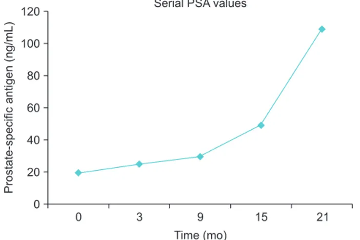
A 52-year-old man presented with repeated elevation of PSA serum values (initial value, 19.7 ng/mL). During 2 years, the patient had a progressive increase in PSA levels reaching 108.7 ng/mL (PSA doubling time, 9.8 months; PSA velocity, 3.6 ng/mL/mo; Fig. 1). Controls were performed
www.kjurology.org
786 www.kjurology.org
Domínguez et al
http://dx.doi.org/10.4111/kju.2015.56.11.785 every 6 months with PSA. All analyses were performed by
enzyme-linked immunosorbent assay (ELISA). The results of a digital rectal examination were not suspicious for prostate cancer and the results of three transrectal ultrasound-guided biopsies performed were all negative. Moreover, abdominal computed tomography and bone scans did not show any extraprostatic disease. Subsequently, a new serum test was performed in another laboratory using a chemiluminescent enzyme immunoassay (CHL). Contrary to the previous analysis, the new recorded PSA value was 1.02 ng/mL. This number was confirmed by a second determination.
As a result of the discrepancy in values, a false-positive result of the initial PSA tests was suspected. To evaluate the possibility of an interfering factor in the immunoassays, we studied the same sample divided in 3 aliquots that were analyzed by three different laboratories using different techniques (Table 1): ELISA (DRG PSA equimolar, DRG International Inc., Springfield, NJ, USA; PSA values were 110.3 ng/mL and 114.1 ng/mL), immunochromatography (VEDALAB PSA-CHECK, VEDALAB, Alencon, France;
PSA values were 115.7 ng/mL and 98.7 ng/mL), and chemilu- minescence (MAGLUMI Total PSA, Snibe Diagnostic, Shenzhen, China; PSA values were 0.8 ng/mL and 0.9 ng/
mL).
Because the medical literature describes the existence of spurious high PSA concentrations because of HA, a possible test interference was investigated by two different
laboratories. Rheumatoid factor (RF) and human antimouse antibodies (HAMA) were studied as possible causes of falsely raised PSA levels. The value of RF (24 UI/L) was obtained by immunoturbidimetry and the HAMA value (<40 mcg/L) was determined by ELISA. On the basis of the preceding results, RF and HAMA values could not explain the observed interference.
Despite our failure to validate our working hypothesis by measuring RF and HAMA levels, we cannot rule out that other serum HA may contribute to causing false-positive PSA determinations if the analysis is not performed by use of the chemiluminescence technique.
DISCUSSION
HA are human immunoglobulins directed against various animal antigens. Most of them are natural antibodies with polyspecific characters that are derived from B cells; less frequently they are auto-antibodies. It is rarely possible to determine the etiology of HA, but their appearance is often described after contact with animals or after therapeutic treatments with drugs containing animal immunoglobins (e.g., iatrogenic immunization, in vivo diagnostic tests, or immunoglobin therapy) [3]. Within specific antianimal antibodies, there exist different types: HAMA, human antirabbit antibodies, and human antigoat antibodies. RF is an auto-antibody that can also have HAMA-like activity, but its concentration in plasma is not high enough to cause significant interference [1]. Levinson and Miller [1] reported that HA interference in a healthy population is mainly due to natural polyspecific and idiotypic antibodies. By contrast, in allergic or diseased patients, the auto-antibody-type polyspecific or RF may be found more frequently.
The prevalence of spurious elevated PSA values is around 0.3%. There are eight cases in the medical literature of falsely elevated PSA due to HA. Six of these patients were diagnosed with prostate cancer and continued to have detectable false values of PSA after radical prostatectomy [6-10]. In some of these patients, an unnecessary salvage treatment was performed [6,8,9]. In the other two patients, the interference was detected during the screening [4,5], presenting with significantly high PSA values (up to 83 ng/
Table 1. Results of the values of PSA in three differents laboratories (A, B, and C) using enzyme-linked immunosorbent assay (ELISA), immuno- chromatography (IC), and chemiluminescence (CHL) techniques
Laboratory
A B A B B C
Technique ELISA ELISA IC IC CHL CHL
PSA (ng/mL) 110.3 114.1 115.7 98.7 0.9 0.8
Fig. 1. Evolution of prostate-specific antigen (PSA) values.
0 3 9 15
120 100 80 60 40 20
21
Prostate-specificantigen(ng/mL)
Time (mo) 0
Serial PSA values
787
Korean J Urol 2015;56:785-787. www.kjurology.org
PSA interference by heterophilic antibodies mL), but only one patient did not receive an unnecessary
therapeutic treatment [4].
In this case, we employed three "sandwich" immunoassays.
These techniques use two monoclonal anti-PSA antibodies:
a capture antibody (immobilized on a solid phase) and a detector antibody coupled to a signal transducer, such as an enzyme (ELISA) or a CHL. In ELISA, the enzyme substrate is added to produce a visible color. The intensity of the color produced is measured by spectrophotometry and indicates the amount of PSA in the sample. In CHL, there is an emission of light as the result of a chemical reaction and this intensity is also measured. The presence of HA links capture and detector antibodies in the absence of the antigen, creating a test interference and a false-positive result. The use of blocking agents could prevent this crosslink. The CHL technique is faster, is less expensive, and has higher sensitivity than ELISA. It may be more accurate at detecting false-positive results of elevated PSA due to HA, as was observed in the patient reported previously. Further studies would be required to support this assumption.
HA represents a challenge for the laboratory analytical staff and remains an unpredictable problem. Different options can be considered to solve the HA interference.
The simplest approach is to analyze the sample in another laboratory using a different formulation [1]. Another option for removing or identifying HA is the use of blocking agents.
With use of this technique, the incidence of HA interference has been reduced from the 2%–5% observed in unblocked assays [1,3]. Unfortunately, those investigators were unable to completely eliminate the problem because the antibodies have significant polyclonality and natural variability.
Therefore, it is complicated to identify the specific type of HA responsible for the interference, but in clinical practice this point is not essential [5]. In the case described herein, despite our efforts to find the HA responsible for the observed interference, we were unable to obtain a positive outcome. In our case, changing the analysis technique was sufficient to confirm that the initial PSA value was a false- positive result.
To the best of our knowledge, this is the second case in the medical literature reporting spurious elevation of PSA values diagnosed before unnecessary therapy. We recommend a close communication between the urologist and laboratory staff in cases in which the results do not correlate with the clinical scenario in order to avoid unnecessary overtreatment by misdiagnosis.
CONFLICTS OF INTEREST
The authors have nothing to disclose.
ACKNOWLEDGMENTS
The authors would like to thank Carlos García- Echeverría for review of the report and technical support.
REFERENCES
1. Levinson SS, Miller JJ. Towards a better understanding of het- erophile (and the like) antibody interference with modern im- munoassays. Clin Chim Acta 2002;325:1-15.
2. Weber TH, Kapyaho KI, Tanner P. Endogenous interference in immunoassays in clinical chemistry: a review. Scand J Clin Lab Invest Suppl 1990;201:77-82.
3. Kricka LJ, Schmerfeld-Pruss D, Senior M, Goodman DB, Kala- das P. Interference by human anti-mouse antibody in two-site immunoassays. Clin Chem 1990;36:892-4.
4. Camacho T, Mora J, Segura A, Guitian J, Lema F, Bandín J, et al. Falsely increased prostate-specific antigen concentration attributed to heterophilic antibodies. Ann Clin Biochem 2002;
39(Pt 2):160-1.
5. Henry N, Sebe P, Cussenot O. Inappropriate treatment of pros- tate cancer caused by heterophilic antibody interference. Nat Clin Pract Urol 2009;6:164-7.
6. Fritz BE, Hauke RJ, Stickle DF. New onset of heterophilic an- tibody interference in prostate-specific antigen measurement occurring during the period of post-prostatectomy prostate- specific antigen monitoring. Ann Clin Biochem 2009;46(Pt 3):
253-6.
7. Park S, Wians FH Jr, Cadeddu JA. Spurious prostate-specific antigen (PSA) recurrence after radical prostatectomy: interfer- ence by human antimouse heterophile antibodies. Int J Urol 2007;14:251-3.
8. Morgan BR, Tarter TH. Serum heterophile antibodies interfere with prostate specific antigen test and result in over treatment in a patient with prostate cancer. J Urol 2001;166:2311-2.
9. Kummar S, Shafi NQ. False elevations in prostate-specific an- tigen levels affecting patient management. Clin Adv Hematol Oncol 2004;2:599-601.
10. Poyet C, Hof D, Sulser T, Muntener M. Artificial prostate- specific antigen persistence after radical prostatectomy. J Clin Oncol 2012;30:e62-3.