Vol. 19, No. 10 pp. 506-514, 2018
심근 경색 환자에서의 손의 근력과 심폐기능의 연관성
김지희
원광대학교 의과대학 재활의학과교실
Relationship between Handgrip Strength and Cardiopulmonary Fitness in Patients with Myocardial Infarction
JiHee Kim
Department of Rehabilitation Medicine, Wonkwang University School of Medicine
요 약 본 이 연구의 목적은 심근 경색증 환자에서 손의 악력과 심폐 기능의 관련성을 알아보는 것이다. 본 후향적 연구에서 는 10개월간 심근 경색을 경험한 총 67 명의 자료를 분석하였다. 손의 악력은 휴대용 동력계로 측정되었다. 운동신경감소증 (dynapenia)는 우성 손의 악력을 기준으로 남성의 경우 30kg 미만, 여성의 경우 20kg 미만일 경우 진단하였다. 심폐 운동 검사는 트레드밀과 수정 된 Bruce 프로토콜에 따라 수행되었다. 신체활동척도로는 국제 신체활동설문을 사용하여 평가 하였 다. 67 명의 대상자 중 60 명은 남성, 7 명은 여성이었고 평균 연령은 56 ± 9 세였다. 운동신경감소증군과 정상인군을 t-test와 Mann-Whitney test를 이용하여 심폐기능검사 결과를 분석하였고, 최대 산소 소모량, 최대 운동시의 신진대사 해당치, 운동시 간 등이 운동신경감소증군에서 의미있게 낮았다(p<0.05). 상관관계 분석에서 손의 악력이 심폐능력과 유의한 관련이 있음을 보여 주었다. 최대산소 소모량, 최대 신지대사 해당치, 운동 시간은 고 신체활동 군에서 저 신체 활동군에 비해 유의하게 높았다(p<0.05). 심근 경색 환자에서 손의 악력은 잠재적 심폐 기능의 지표로 사용될 수 있다. 환자들의 손의 악력과 신체 활동정도를 측정하여, 적극적으로 심장 재활에 참여하도록 권장할 수 있다.
Abstract This study was conducted to investigate the relationship between hand grip strength and cardiopulmonary fitness in patients with myocardial infarction. In this retrospective study, 67 patients who experienced myocardial infarction for 10 months were analyzed. Hand grip strength was measured using a handheld dynamometer. Dynapenia was diagnosed based on a dominant hand grip strength of less than 30 kg for males and 20 kg for females. A cardiopulmonary exercise test was performed using a treadmill. Physical activity status was also evaluated.
Cardiorespiratory fitness parameters were analyzed using a t-test and a Mann-Whitney test. VO2max, METmax, and exercise time significantly decreased in the dynapenia group compared with the non-dynapenia group. Correlation analysis revealed that dominant handgrip strength was significantly related to cardiorespiratory fitness parameters.
Moreover, VO2max, METmax, and exercise time were significantly increased in patients with vigorous activity compared with the sedentary group. These findings indicate that handgrip strength could potentially be used as a marker of cardiorespiratory functions. Accordingly, patients with myocardial infarction should be evaluated for grip strength and physical activity, and we can encourage patients to participate actively in cardiac rehabilitation.
Keywords : cardiorespiratory fitness, hand grip strength, rehabilitation, myocardial infarction, physical activity
이 논문은 2018학년도 원광대학교의 교비지원에 의해 수행됨
*Corresponding Author : Ji Hee Kim(Wonkwang Univ.) Tel: +82-63-859-1610 email: [email protected]
Received July 19, 2018 Revised (1st August 20, 2018, 2nd September 13, 2018) Accepted October 5, 2018 Published October 31, 2018
1. 서론
심장 재활은 심혈관 질환의 유병률과 사망률을 현저
히 감소시키는 치료로, 심근 경색 후 심장 재활을 시행하면 사망률을 약 20-30 % 낮춘다고 보고되어있다[1]. 미국 심장 학회는 심근경색증 환자에게 다학제(multidisciplinary)
치료인 심장재활을 최고 수준인 I 급으로 권장하고 있다 [2]. 그럼에도 불구하고, 심장재활프로그램에 등록하는 비율은 캐나다에서 20-30 % [3] 및 아시아 12 %로 차선 책으로 남아 있으며[4], 국내에서 네 곳의 병원이 참여한 연구에서는 31%의 참여율이 보고되었다[5]. 많은 환자 가 거리의 문제, 시간의 부족, 참여 필요성 결여 등 여러 가지 이유로 심장 재활을 중단하는 것으로 보고되고 있 다[6].
심폐기능은 심혈관 질환과 밀접한 관련을 가지므로, 심폐기능을 향상시키고, 심혈관 질환 재발의 예방을 위 해 개별화된 맞춤운동 처방하는 것은 매우 중요하다[7].
14.5 년간 시행한 일반인 추적조사 연구에서, 심폐기능 이 높을수록 심혈관 질환의 모든 원인으로 인한 사망 위 험이 낮아지는 것으로 보고되었다[8]. 심장 재활에서 운 동 부하검사는 정확한 심폐 기능을 측정하고, 적합한 강 도의 운동을 처방하는 데 사용되는 중요한 도구이다[9].
그러나 노인이나 동반 질환이 있는 환자의 경우 운동부 하 검사의 참여가 어려우며, 다른 환자들도 경제적인 이 유, 거리, 시간 등의 문제로 검사에 잘 참여하지 않는다.
그런데 운동부하검사에서 운동능력을 잘 반영하며, 심장 재활을 시행하면 향상되는 것으로 알려진[10] 최대 산소 섭취량(maximal oxygen uptate)은 사망률을 예측할 수 있는 강력한 지표이므로[11], 이를 평가하고 개선시키는 것은 중요하다.
근감소증(sarcopenia)은 근육양의 감소와 함께 수반되 는 근력의 저하와 기능의 저하를 포함한 개념으로, 나이 와 관계된 키에 대한 상대적인 근육양의 소실로 정의된 다[12]. 근육량은 심폐기능과 중요한 연관성이 있으며, 심폐기능이 낮을수록 적은 근육 양을 가지는 것으로 보 고되었다[13]. 근육량과 근력은 연관성이 있으며, 이에 근력의 감소는 운동신경감소증(dynapenia)이라는 용어 로 명명하였다[14]. 신체기능은 근력과 연관성이 있으며, 근력의 저하는 사망률을 증가시키는 것으로 알려져 있다 [15]. 4년간 약 50만 명을 대상으로 시행한 연구에서는, 손의 악력이 사망률과 관련된 위험 인자이며, 신체활동 정도와 손의 악력이 연관성이 높음이 보고되었다[16].
그러나 심근경색증의 환자에서 운동신경감소증과 심폐 능력의 연관성에 대한 연구는 부족하다.
심장재활은 평가, 치료, 교육 등으로 이루어진 다학제 치료로서, 심장환자의 생활양식을 개선하는 것은 중요하 다[17]. 급성 관상동맥증후군의 예후와 관련한 연구에서,
신체활동이 높을수록 급성 관상동맥증후군의 중증도가 감소하고, 예후가 좋은 것으로 보고되고 있다[18]. 또 다 른 문헌에서는 신체활동이 높을수록 급성관상동맥 환자 의 입원 기간 동안의 합병증 등을 감소함을 보고하고 있 다[19].
이에 본 연구는 심근경색 환자들의 손의 악력 및 심폐 기능을 측정하고, 신체활동을 조사하며, 악력과 심폐기 능과의 연관성 및 신체활동과 심폐기능의 연관성을 알아 보고자 한다, 이를 통해 여러 이유로 운동 부하검사에 참 여하지 못하는 환자의 심폐기능을 예측하고, 환자들의 근력 및 신체활동을 증진시키기 위한 심장재활프로그램 의 참여를 더 높이고자 한다.
2. 본론
2.1 연구대상 및 방법 2.1.1 연구대상
2017년 2월부터 2017년 11월까지 급성심근경색으로 oo대학교 병원에서 경피적 관상동맥중재술을 받은 후 심장재활 프로그램에 의뢰된 환자 중 심장 재활 치료 프 로그램에 참여한 환자들을 대상으로 의무기록 검토를 통 한 후향적 연구를 시행하였다. 그 기간 동안 심장재활에 의뢰된 환자는 총 211명이었다. 의뢰된 환자 중 중환자 실에 있거나, 의사소통이 되지 않거나, 걸을 수 없는 27 명의 환자들은 연구대상에서 제외하였다. 악력 및 운동 부하 검사에 모두 참여한 인원은 총 67명이었다. 평균 연령은 56.2±9.2세, 남자가 60명, 여자가 7명이었다. (Fig. 1)
Fig. 1. Study flow chart.
2.1.2 연구방법
모든 대상자는 경피적 관상동맥 중재술을 받은 후, 재 활의학과로 심장재활이 의뢰되어 입원기간 중 소집단 교 육을 통해 재활의학과 의사에게 심근경색에 대한 설명 및 위험인자 관리에 대한 교육 및 약물요법과 운동요법 에 대하여 교육을 받았고, 심장재활 전문 간호사에 의해 금연교육, 영양상담 등을 시행 받았으며, 심장재활 치료 실에서 스트레칭 및 실내 보행 운동을 시행하였다. 실내 보행 운동은 8시간 이내에 가슴통증 혹은 호흡곤란이 없 었던 환자에 한해서 심전도 감시 하에 분당 120회 이하 의 심박수를 유지하거나 안정 시 심박수가 높은 경우 안 정 시 심박수에서 20회를 넘기지 않도록 유지하며 시행 하였고, 환자의 상태에 따라 13 미만의 운동자각도를 유 지하며 2분에서 5분간 하루 2회에서 4회를 실시하였다.
환자들의 악력을 평가하기 위하여 심장재활 치료실에서 악력기(Jamar Hydraulic Hand Dynamometer, Pennsylvania, U.S.A.)를 통해 악력을 평가하였다, 평가 방법은 팔은 몸 통과 손바닥은 어깨 라인에 수직으로 적어도 2초간 측정 하며, 각각의 손에 대해 3번의 시도가 이루어진 뒤 평균 을 내서 기록하였다. 운동신경감소증(dynapenia)은 우성 손의 악력에 따라 남자는 30kg, 여자는 20kg 보다 작은 경우로 진단하였다[20].
신체활동척도로는 일반적으로 널리 사용되는 국제 신 체활동설문(The International Physical Activity Questionnaire (IPAQ))을 사용하였다[21]. 이 국제적 신 체활동 기준은 1998년 제네바에서 개발되었으며, 12개 국 이상에서 신뢰도와 타당성이 검증되었으며, 2007년 가정의학회에서 한국어 버전의 적합성이 평가되었다 [22]. 본 연구에서 사용된 신체활동척도는 국제 신체활 동설문 중 단문형자가기입식 설문지를 이용하며, 공식 인정받은 한국어 버전이었다. 이 설문지는 크게 7가지 문항으로 이루어졌으며, 설문을 작성하기 전의 일주일 동안 시행한 활동에 대하여 물어보고 있다. 10분 이상 시행한 격렬한 활동, 중간 정도의 신체 활동, 걸은 시간 이 각각 며칠, 평균 몇 시간인지 기입하고, 앉아서 보낸 시간이 얼마나 되는지 대답하게 한 후, 이 자료들을 분석 하였다. 조사대상자의 신체활동도를 평가 점수화 체계에 따라 세 가지 그룹으로 분류하였다. 저신체활동군 (I:inactive)은 신체활동을 적게 하는 군으로 아무 활동도 적지 않았거나, 다른 군에 해당되지 않는 사람들이었다.
중신체활동군(II: minimally active )은 하루에 최소한 30
분의 중간정도 활동이나 걷기를 5일 이상 하는 경우 등을 포함한다. 고신체활동군(III:health enhancing physical activity)은 주3일 이상 격렬한 활동을 하는 사람들이다.
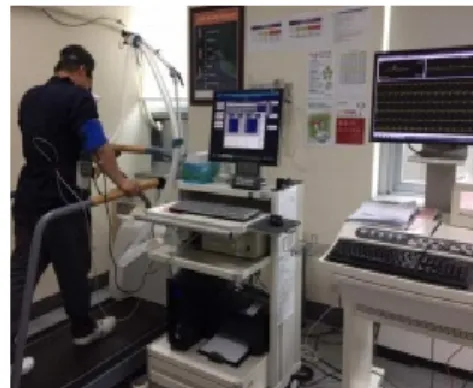
이후 재활의학과 외래에 내원하여 수정된 브루스 프 로토콜(modified Bruce protocol)을 이용한 증상제한 (symptom limited) 운동부하검사 및 호흡가스분석을 시 행하였다(Fig. 2). 검사는 12채 널 실시간 운동부하검사용 심전도 검사기 (Quinton Q-stress, Mortara Instrument, INC, USA) 및 운동부하검사용 트레드밀 (Q-stress TM55, Mortara Instrument, INC, USA), 자동 혈압 및 맥박 측정기 (247BP, SunTech Medical, USA), 호흡가 스 분석기(TrueOne 2400, ParvoMedics, INC, USA)를 사용하였다. 운동부하검사용 심전도 검사기와 자동 혈압 및 맥박 측정기를 이용하여 최대 운동검사 가능시간, 최 대하(3단계) 심근부담률, 안정 및 최대 심박수, 안정 및 최대 수축기혈압을 측정하였으며, 호흡가스 분석기를 이 용하여 최대 운동 시의 신진대사 해당치(METs), 최대 산소소모량을 측정하였다. 대상자들은 초기 운동부하검 사 결과를 바탕으로 심장재활 치료실에 방문하여 심전도 감시 하에 유산소 운동요법을 시행하였다. 운동요법 시 에는 Q-Tel RMS (Mortara Instrument, INC, USA)와 JT-4000M (SUNGDOMC, CO., Korea)를 이용하였고, 운동의 강도는 최초 시행한 운동부하 검사를 통해 얻은 각 대상자의 안정 심박수와 최대 심박수를 기준으로 여 유 심박수를 계산하여 40%에서 85%까지 점진적으로 운동 강도를 조절 하였다.
Fig. 2. Cardiopulmonary exercise test
2.1.3 통계 분석
통계학적 분석은 SPSS 통계프로그램을 이용하였으며 평균 및 표준편차를 이용하였다. 남녀의 성별에 따른 차
이를 보기위해 두 군간의 연령, 체질량지수, 손의 악력 등은 독립표본 t 검정을, 두 군 간의 약물 복용력 및 사 회적 과거력 등 은 Mann-Whitney test를 이용하였다.
악력검사를 통해 나눈 운동신경감소증환자와 정상군 두 군별 약물 복용력 및 사회적 과거력 등은 Mann-Whitney test를 이용하였다. 두 군별 심폐기능의 비교를 위해 각 지표들의 평균값을 비교하여 정규분포가 확인된 변수는 독립표본 t 검정을 확인되지 않은 것은 Mann-Whitney test 검정을 이용하여 분석하였다.
악력과 심폐기능 및 신체활동척도와 상관관계를 알아 보기 위하여 스피어만의 상관분석(Spearman’s correlation) 을 통해 분석하였다. 심폐기능과 신체활동정도와 관련성 이 있는지를 알아보기 위해 신체활동정도에 따라 두 군 으로 나누어 독립표본 t 검정을 이용하여 분석하였다. 통 계적 유의수준은 p값을 0.05 미만으로 하였다.
2.2 결과
2.2.1 환자군의 특성
남성과 여성으로 나누어 비교하였을 때, 다른연구와 마찬가지로 남성에서 여성에 비해 골격근량이 높았고, 손의 악력도 유의하게 높았다 (p<.05). 남성과 여성에서 고혈압, 고지혈증 및 당뇨의 진단여부, 알코올 및 흡연 여부, 있어서 두군 간의 차이가 없었다(p>.05)(Table 1).
운동신경감소증의 유무에 따른 고혈압, 고지혈증 및 당 뇨의 진단여부, 알코올 및 흡연 여부, 있어서 두군 간의 차이가 없었다(p>.05)(Table 2).
Table 1. Baseline characteristics of the subjects.
Parameters Total
(n=67) Male
(n=60) Female (n=7) p-value
Age (years) 56±9 55±10 61±4 0.103
Height (cm) 168.0±7.7 169.5±6.4 155.1±6.0 <0.01**
Weight (kg) 78.3±39.2 80.4±4.9 60.7±5.3 0.211 BMI (kg/㎡) 26.3±3.4 26.4±3.5 25.3±2.4 0.389 Skeletal muscle mass (kg) 27.3±7.2 28.0±7.2 21.4±3.8 <0.05*
HGS(D) (kg) 34.2±8.6 35.8±7.1 20.2±7.5 <0.01**
HGS(ND) (kg) 32.2±8.8 33.7±7.8 19.7±6.1 <0.01**
Past medical history
HTN, n (%) 32 29 (48.3) 3 (42.9) 0.784
DM, n (%) 14 12 (20) 2 (28.6) 0.598
Hyperlipidemia, n (%) 16 14 (23.3) 2 (28.6) 0.758 Social history
Alcohol, n (%) 27 26 (43.3) 1 (14.3) 0.228 Smoking, n (%) 31 30 (50) 1 (14.3) 0.113 Values are presented as the mean±standard deviation or number (%).
*p<0.05, **p<0.01, significant difference between male and female.
BMI, body mass index; HGS(D), dominant hand grip strength;
HGS(ND), non-dominant hand grip strength; HTN, hypertension;
DM, diabetes mellitus.
Table 2. Baseline characteristics of the subjects.
Parameters Total
(n=67) Dynapenia (n=18)
Non- dynapenia
(n=49) p-value Past medical history
HTN,
n (%) 32 8
(44.4) 24
(49.0) 0.742 DM,
n (%) 14 3
(16.7) 11
(22.4) 0.606 Hyperlipidemia,
n (%) 16 3
(16.7) 13
(26.5) 0.401 Social history
Alcohol,
n (%) 27 7
(38.9) 20
(40.8) 0.887 Smoking,
n (%) 31 8
(44.4) 23
(46.9) 0.856 Values are presented as the mean±standard deviation or number (%).
2.2.2 운동신경감소증의 유무에 따른 운동부하검 사의 결과
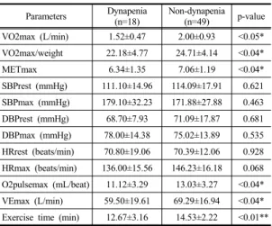
운동신경감소증의 유무에 따른 가스분석 운동부하검 사 결과 비교에서 운동신경감소증환자에서 최대 산소 소 모량, 단위체중당 산소섭취량, 환기 역치, 최대 신진대사 해당치, 최대 산소맥, 분당 최대 환기량, 운동시간이 유 의하게 낮게 관찰되었고(p<0.05) 그 외 혈압이나 심박수 에서는 유의한 차이는 관찰되지 않았다(Table 3).
Table 3. Comparison of cardiorespiratory fitness between the groups.
Parameters Dynapenia
(n=18) Non-dynapenia (n=49) p-value VO2max (L/min) 1.52±0.47 2.00±0.93 <0.05*
VO2max/weight 22.18±4.77 24.71±4.14 <0.04*
METmax 6.34±1.35 7.06±1.19 <0.04*
SBPrest (mmHg) 111.10±14.96 114.09±17.91 0.621 SBPmax (mmHg) 179.10±32.23 171.88±27.88 0.463 DBPrest (mmHg) 68.70±7.93 71.09±17.87 0.681 DBPmax (mmHg) 78.00±14.38 75.02±13.89 0.535 HRrest (beats/min) 70.80±19.06 70.39±12.06 0.928 HRmax (beats/min) 136.00±15.56 146.23±16.18 0.068 O2pulsemax (mL/beat) 11.12±3.29 13.03±3.27 <0.04*
VEmax (L/min) 59.50±19.61 69.29±16.94 <0.04*
Exercise time (min) 12.67±3.16 14.53±2.22 <0.01**
Values are presented as the mean±standard deviation.
*p<0.05, **p<0.01, significant difference between dynapenia group and non-dynapenia group.
VO2max, maximal oxygen consumption;VT, ventilatorythreshold;
METmax, maximal metabolic equivalents; SBPrest, resting systolic blood pressure; SBPmax, maximal systolic blood pressure; DBPrest, resting diastolic blood pressure; DBPmax, maximal diastolic blood pressure; HRrest, resting heart rate; HRmax, maximal heart rate;
O2pulsemax, maximal oxygen pulse; VEmax, maximal minute ventilation.
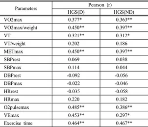
2.2.3 악력과 심폐기능과의 상관관계
측정한 악력과 가스분석 운동부하검사 결과를 통해 측정한 심폐기능사이의 관계를 알기 위해, 시행한 상관 분석 검사에서 최대 산소 소모량, 단위체중당 산소섭취 량, 환기역치, 단위체중단 환기역치. 최대 신진대사 해당 치, 최대 산소맥, 분당 최대 환기량, 운동시간이 손의 악 력과 유의한 상관관계를 보였으며, 그외 혈압이나 심박 수에서 유의한 상관성은 관찰되지 않았다(Table 4).
Table 4. Relationship between dominant handgrip strength and cardiorespiratory fitness parameters measured by a cardiopulmonary exercise test
Parameters Pearson (r)
HGS(D) HGS(ND)
VO2max 0.377* 0.363**
VO2max/weight 0.450** 0.397**
VT 0.321** 0.312*
VT/weight 0.202 0.186
METmax 0.450** 0.397**
SBPrest 0.069 0.038
SBPmax 0.114 0.044
DBPrest -0.092 -0.056
DBPmax -0.022 -0.046
HRrest -0.035 -0.058
HRmax 0.220 0.182
O2pulsemax 0.485** 0.386**
VEmax 0.453** 0.297*
Exercise time 0.464** 0.467**
*p<0.05, **p<0.01,
HGS(D), dominant handgrip strength; HGS(ND), non-dominant handgrip strength; VO2max, maximal oxygen consumption; VT, ventilatory threshold; METmax, maximal metabolic equivalents;
SBPrest, resting systolic blood pressure; SBPmax, maximal systolic blood pressure; DBPrest, resting diastolic blood pressure; DBPmax, maximal diastolic blood pressure; HRrest, resting heart rate;
HRmax, maximal heart rate; O2pulsemax, maximal oxygen pulse;
VEmax, maximal minute ventilation.
2.2.4 신체 활동 정도에 따른 환자의 임상검사 및 운동부하검사의 결과
측정한 신체활동 정도와 운동부하검사, 손의 악력 사 이의 관계를 알기 위해, 환자들을 IPAQ 점수에 따라 두 군으로 나누었다. 고 신체활동 환자들에게서 단위체중당 산소섭취량, 최대 신진대사 해당치, 운동시간이 유의하 게 높게 나타났으며, 최대 심박수, 손의 악력 검사결과에 서 유의한 차이는 관찰되지 않았다(Table 5).
Table 5. Comparison of cardiorespiratory fitness between the groups.
Parameters inactivity
(n=29) Active
(n=24) p-value
Grip strength 33.4±8.8 34.3±8.6 0.70
VO2max/weight 22.89±4.37 25.60±4.59 <0.03*
METmax 6.54±1.24 7.38±1.32 <0.03*
HRmax(beats/min) 140.31±18.61 145.50±12.31 0.25 Exercise time (min) 13.18±2.93 14.69±2.38 <0.05*
Values are presented as the mean±standard deviation.
*p<0.05, **p<0.01, significant difference between dynapenia group and non-dynapenia group.
VO2max, maximal oxygen consumption; METmax, maximal metabolic equivalents: HRmax, maximal heart rate.
2.3 고찰
심장재활은 모든 사망률 및 심혈관관련 사망률을 예 측할 수 있는 강력한 지표인 최대 산소섭취량을 증가시 키며, 심혈관 질환의 사망률을 유의하게 감소시키는 중 요한 치료이다[10,23,24]. 본 연구에서는 심근경색환자 에서 손의 악력과 심폐기능과의 연관성을 밝히기 위해, 심폐기능을 나타내는 다양한 척도와 손의 악력을 분석 하였다. 손의 악력이 낮은 운동신경감소증(dynapenia)군 에서 심폐기능이 낮았고, 심폐기능과 손의 악력은 연관 성이 있음을 나타냈다. 또한 평소 신체 활동이 높을수록 심근경색증 환자에서 심폐기능이 높음이 나타났다.
운동부하 검사는 심장환자에서 환자의 평가 및 예후 설정, 그리고 개인별 운동 처방에 사용할 수 있는 최대 산소섭취량을 직접 측정할 수 있는 중요한 방법이다 [25,26]. 낮은 심폐기능은 다른 심혈관 위험인자와 비교 하여 심혈관질환과 사망률을 예측할 수 있는 강력한 지 표로 보고되고 있으므로[27], 환자의 정확한 심폐기능을 아는 것은 매우 중요하다. 그러나 환자가 평가에 참여하 지 않아도, 손의 악력측정 및 평소 신체활동 설문을 통해 심폐기능을 예측할 수 있으므로, 이를 통해 운동부하를 시행하지 않은 환자들의 심장재활 참여를 유도할 수 있 을 것으로 생각된다.
현재까지 근육의 감소와 심장기능의 감소에 관해서는 명확히 밝혀지지 않았다. 근육의 양은 매우 중요한데, 이 는 총 지방질량이 최대산소소모량과 음의 관계를 가진다 는 연구와[28], 심부전 환자에서도 체지방을 보정한 수 치가 체중보다 최대 산소소모량과 더 연관성이 있다는 연구를 통해서 알 수 있다[29]. 안정된 심부전환자에서 운동능력의 저하는 근력과 강력한 연관 관계를 가지는
것으로 보고되었다[30]. 대퇴사두근의 근력은 심혈관 환 자의 운동능력을 예측하는 지표로 사용되었으며[31], 사 망률을 예측할 수 있는 지표로도 이용할 수 있음이 보고 되었다[32]. 심혈관 환자의 근력은 나이가 같은 지역거 주자에 비해 낮은 것으로 보고되었다[33]. 심장재활치료 시행 후 심폐기능의 개선과 함께 근력이 개선되었음이 보고되었다[34,35]. 본 연구에서도 운동신경감소증 환자 의 심폐기능이 정상군에 비해 의미 있게 낮은 것으로 나 타났다. 악력을 통해 근력을 측정하는 것은 섬유근육통, 만성 심부전과 같은 만성 환자에서 흔히 사용되는 방법 이다. 17개국의 14만명을 대상으로 4년 동안 추적 조사 한 연구에서 손의 악력은 수축기 혈압보다 사망률을 예 측하는데 더 강력한 인자이며, 손의 악력이 5KG 줄 때 마다 심혈관 질환으로 인한 사망률은 17% 증가하는 것 으로 보고하였다[36]. 손의 악력을 측정하는 것은 도구 가 간단하며, 소요되는 시간도 짧다. 손의 악력은 하지 근육의 힘과 강한 연관성이 있으며, 낮은 손의 악력은 근 육의 양이 낮은 것보다 이동성의 저하에 더 관계가 깊은 예측 인자로 보고되었다[37]. 손의 악력측정은 하지의 근력과 연관성을 가지면서 쉽고, 경제적인 근력 측정 방 법이다. 이에 하지의 근력 검사에 비해, 간단한 손의 악 력 측정을 통해 근력을 측정하며, 악력이 낮은 경우 예측 되는 낮은 심폐기능을 개선시키기 위해 심장재활 참여시 키는 것은 중요하다.
심근경색은 재발 위험성이 높은 질병으로[38], 재발을 막기 위해 평생 약물 치료를 받게 된다. 심장재활을 시행 할 경우 재발률을 20-30% 줄일 수 있는 것으로 알려져 있다[1]. 그러므로 심장재활을 통해 환자의 심폐기능 및 근력을 개선하는 것은 매우 중요하다. 그러나 심장재활 치료의 참여율은 낮으며, 환자들은 거리나 비용, 또는 참 여의 필요성을 인식하지 못하는 등의 문제로 참여하지 않는다[3,4]. 이에 여러 이유로 운동부하검사에 참여하 지 않은 환자라 할지라도 손의 악력 측정을 통해 환자의 심폐기능을 예측하여, 낮은 근력을 가진 환자에게 위험 요인을 설명할 수 있다.
심장재활치료는 운동치료로만 구성되지 않는다. 심근 경색 치료의 연장성상에서 다양한 프로그램 및 다학제적 접근을 통해, 환자에게 심혈관 질환에 대하여 환자가 가 지고 있는 위험요인을 인식시키고, 개선을 위해 환자의 생활습관 전체를 바꾸도록 교육 하는 것을 포함하고 있 다[16,39]. 발병 초기의 환자들은 삶의 방식을 바꾸는 것
에 대해 수용적인 태도를 가지므로[40], 발병 초기에 환 자에게 현재의 상태를 잘 설명하고, 평소 신체 활동과 현 재 심폐능력을 설명하여주고, 이를 개선하기 위해 삶의 방식을 바꿀 수 있도록 도와 줘야 한다. 정확한 심폐기능 이 평가되지 않은 환자라 할지라도, 낮은 근력으로 평가 된 환자에게 위험성을 설명하고, 심장재활에 참여하는 것의 중요성을 설명할 수 있다.
본 연구의 결과에서도 신체활동점수가 높은 군에서 심폐기능이 의미 있게 높게 나타났다. 다른 연구에서도 신체활동이 증가할수록 심근경색 환자의 예후가 개선되 는 것으로 보고하고 있다[18,19]. Chomistek 등은 긴 앉 아있는 시간이 높은 심혈관 질환의 위험과 연관성이 있 으며, 낮은 신체활동과 앉아있는 시간의 증가의 조합은 심혈관 질환의 위험을 증가시킨다고 보고하였다[41]. 그 러나 심근 경색 이후의 신체활동에 관한 연구를 살펴보 면 그들이 심장재활에서 처방 받은 신체활동을 발병 6개 월째에 수행하는 경우는 55%에 불과하며, 수치는 시간 이 갈수록 감소한다[42]. Izawa등은 심근경색 발병 18개 월 이상의 환자에서도 운동의 유지는 삶의 질과 신체 활 동 상태를 개선시키기 위한 요소 중 하나라고 보고하였 다[43]. 하버드 연구팀 보고에서 높은 신체활동을 가지 는 남성 사람에서 낮은 신체 활동을 갖는 사람과 비교하 였을 때 의미 있게 낮은 심혈관 질환의 위험성을 갖는 것으로 보고하였다[44]. 또한 중년이상의 여성에서도 평 소 높은 에너지 소비를 하는 여성에서 관상동맥질환의 상대적인 위험도가 낮은 것이 보고되었다[45]. 심뇌혈관 질환 예방을 위해 격렬한 운동을 일주일에 75분 이상 또 는 중간강도의 운동을 150분 이상 시행할 것을 권고되 고 있다[17]. 이에 심혈관 환자에게 평소 신체활동 점수 를 측정하는 것은 중요하며, 삶의 방식을 바꾸어 심혈관 질환의 재발을 줄이도록 교육 하여야 한다.
본 연구의 제한 점으로는 첫째, 연구에 포함된 환자의 수가 충분히 많지 않았다. 둘째, 모든 환자가 심근경색 환자에 국한되었으며, 모두 심장재활에 의뢰되기 전에 경피적 관상동맥 중재술을 받은 환자였기 때문에 관상동 맥 우회술을 받은 환자에게 본 연구 결과를 적용하기 어 렵다. 따라서 앞으로의 연구에서는 더 많은 수의 환자와 장기적인 연구가 필요할 것이다.
3. 결론
심근경색 환자에서 심폐기능의 다양한 척도와 손의 악력을 분석하여, 최대 산소 소모량과 손의 악력이 상관 관계가 있음을 보고하였다. 또한 운동신경감소증의 유무 에 따라 심폐기능의 차이가 있음을 보고하였으며, 신체 활동점수가 높은 군에서 심폐기능이 의미 있게 높게 나 타났음을 보고하였다. 이에 심근경색환자에게 환자의 악 력 및 신체활동척도를 측정하고, 결과를 통해 예후를 설 명하고, 환자에게 심장재활에 참여하도록 권장할 수 있다.
References
[1] A. M. Clark, L. Hartling, B. Vandermeer, F. A.
McAlister, “Meta-Analysis: Secondary Prevention Programs for Patients with Coronary Artery Disease”, Annals of Internal Medicine, Vol.143, No.9, pp.659-72, 2005.
DOI:https://dx.doi.org/10.7326/0003-4819-143-9-200511010- 00010
[2] G. J. Balady, P. A. Ades, V. A. Bittner, A. Franklin, N.
F. Gordon, R. J. Thomas, G. F. Tomaselli, C. W. Yancy,
“Referral, Enrollment, and Delivery of Cardiac Rehabilitation/Secondary Prevention Programs at Clinical Centers and Beyond”, Circulation, Vol.124, No.25, pp.2951-60, 2011.
DOI: https://dx.doi.org/10.1161/CIR.0b013e31823b21e2 [3] S. L. Grace, K. L. Angevaare, R. D. Reid, P. Oh, S.
Anand, M. Gupta, S. Brister, D. E. Stewart,
“Effectiveness of inpatient and outpatient strategies in increasing referral and utilization of cardiac rehabilitation: a prospective, multi-site study”, Implementation Science, Vol.7, No.1, Article ID 120, 2012.
DOI:https://dx.doi.org/10.1186/1748-5908-7-120 [4] R. Poh, H. N. Ng, G. Loo, L. S Ooi, T. J. Yeo, R.
Wong, C. H. Lee, “Cardiac Rehabilitation After Percutaneous Coronary Intervention in a Multiethnic Asian Country: Enrollment and Barriers”, Archives of Physical Medicine and Rehabilitation, Vol.96, No.9, pp.1733-1738, 2015.
DOI:https://dx.doi.org/10.1016/j.apmr.2015.05.020 [5] H. W. Im, S. Baek, S. Jee, J. M. Ahn, M. W. Park, W.
S. Kim, “Barriers to Outpatient Hospital-Based Cardiac Rehabilitation in Korean Patients With Acute Coronary Syndrome”, Annals of Rehabilitation Medicine, Vol.42, No.1, pp.154-165, 2018.
DOI:https://dx.doi.org/10.5535/arm.2018.42.1.154 [6] D. M. Resurrección, P. Moreno-Peral, M. Gómez-
Herranz, M. Rubio-Valera, L. Pastor, J. Miguel, C. de Almeida, E. Motrico, “Factors associated with non-participation in and dropout from cardiac rehabilitation programmes: a systematic review of prospective cohort studies”, European Journal of Cardiovascular Nursing, pp.1-10, 2018.
DOI:https://dx.doi.org/10.1177/1474515118783157 [7] C. E. Garber, B. Blissmer, M. R. Deschenes, B. A.
Franklin, M. J. Lamonte, I. M. Lee, D. C. Nieman, D.
P. Swain, “Quantity and Quality of Exercise for Developing and Maintaining Cardiorespiratory, Musculoskeletal, and Neuromotor Fitness in Apparently Healthy Adults: Guidance for Prescribing Exercise”, Medicine & Science in Sports & Exercise, Vol.43, No.7, pp.1334-1359, 2011.
DOI: https://dx.doi.org/10.1249/MSS.0b013e318213fefb [8] E. G. Artero, A. S. Jackson, X. Sui, D. C. Lee, D. P.
O’Connor, C. J. Lavie, T. S. Church, S. N. Blair,
“Longitudinal Algorithms to Estimate Cardiorespiratory Fitness: Associations With Nonfatal Cardiovascular Disease and Disease-Specific Mortality”, Journal of the American College of Cardiology, Vol.63, No.21, pp.2289-96, 2014.
DOI: https://dx.doi.org/10.1016/j.jacc.2014.03.008 [9] GF Fletcher, PA Ades, P Kligfield, R Arena, GJ Balady,
et al. “Exercise standards for testing and training: a scientific statement from the American Heart Association”, Circulation, Vol.128, No.8, pp.873-934, 2013.
DOI: https://dx.doi.org/10.1161/CIR.0b013e31829b5b44 [10] H. Valkeinen, S. Aaltonen, U. M. Kujala, “Effects of
exercise training on oxygen uptake in coronary heart disease: a systematic review and meta-analysis”, Scandinavian Journal of Medicine & Science in Sports, Vol.20, No.4, pp.545-55, 2010.
DOI:https://dx.doi.org/10.1111/j.1600-0838.2010.01133.x [11] S. J. Keteyian, C. A. Brawner, P. D. Savage, J. K.
Ehrman, J. S. Schairer, G. Divine, H. Aldred, K.
Ophaug, P. A. Ades, “Peak aerobic capacity predicts prognosis in patients with coronary heart disease”, American Heart Journal, Vol.156, No.2, pp.292-300, 2008.
DOI: https://dx.doi.org/10.1016/j.ahj.2008.03.017 [12] I. H. Rosenberg, “Summary comments”, The American
Journal of Clinical Nutrition, Vol.50, No.5, pp.1231-1233, 1989.
DOI: https://dx.doi.org/10.1093/ajcn/50.5.1231
[13] T. N. Kim, M. S. Park, Y. J. Kim, E. J. Lee, M. K. Kim, J. M. Kim, K. S. Ko, B. D. Rhee, J. C. Won,
“Association of Low Muscle Mass and Combined Low Muscle Mass and Visceral Obesity with Low Cardiorespiratory Fitness”, PLOS ONE, Vol.9, No.6, Article ID e100118, 2014.
DOI: https://dx.doi.org/10.1371/journal.pone.0100118 [14] B. C. Clark, T. M. Manini, “Sarcopenia=/=dynapenia”, J
Gerontol A Biol Sci Med Sci, Vol.63, No.8, pp.829-34, 2008.
[15] Q. L. Xue, B. A. Beamer, P. H. M. Chaves, J. M.
Guralnik, L. P. Fried, “Heterogeneity in Rate of Decline in Grip, Hip, and Knee Strength and the Risk of All‐ Cause Mortality: The Women's Health and Aging Study II”, Journal of American Geriatric Society, Vol.58, No.11, pp.2076-84, 2010.
DOI:https://dx.doi.org/10.1111/j.1532-5415.2010.03154.x [16] C. A. Celis-Morales, D. M. Lyall, J. Anderson, S.
Iliondromiti, Y. Fan, U. E. Ntuk, D. F. Mackay, J. P.
Pell, N. Sattar, J. M. R. Gil, “The association between physical activity and risk of mortality is modulated by grip strength and cardiorespiratory fitness: evidence from 498 135 UK-Biobank participants”, European Heart
Journal, Vol.38, No.2, pp.116-122, 2017.
DOI:https://dx.doi.org/10.1093/eurheartj/ehw249 [17] M. F. Piepoli, A. W. Hoes, S. Agewall, C. Albus, C.
Brotons, A. L. Catapano, M. T. Cooney, U. Corrà, B.
Cosyns, C. Deaton, I. Graham, M. S. Hall, F. D. R.
Hobbs, M. L. Løchen, H. Löllgen, P. Marques-Vidal, J.
Perk, E. Prescott, J. Redon, D. J. Richter, N. Sattar, Y.
Smulders, M. Tiberi, H. B. van der Worp, I. van Dis, W.
M. M. Verschuren, S. Binno, “2016 European Guidelines on cardiovascular disease prevention in clinical practice: The Sixth Joint Task Force of the European Society of Cardiology and Other Societies on Cardiovascular Disease Prevention in Clinical Practice (constituted by representatives of 10 societies and by invited experts)Developed with the special contribution of the European Association for Cardiovascular Prevention & Rehabilitation (EACPR)”, European Heart Journal, Vol.37, No.29, pp.2315-2381, 2016.
DOI:https://dx.doi.org/10.1093/eurheartj/ehw106 [18] C. Pitsavos, S. A. Kavouras, D. B. Panagiotakos, S.
Arapi, C. A. Anastasiou, S. Zombolos, P. Stravopodis, Y. Mantas, Y. Kogias, A. Antonoulas, C. Stefanadis,
“Physical Activity Status and Acute Coronary Syndromes Survival: The GREECS (Greek Study of Acute Coronary Syndromes) Study”, Journal of the American College of Cardiology, Vol.51, No.21, pp.2034-2039, 2008.
DOI:https://dx.doi.org/10.1016/j.jacc.2008.01.053 [19] R. S. Taylor, B. Unal, J. A. Critchley, S. Capewell,
“Mortality reductions in patients receiving exercise-based cardiac rehabilitation: how much can be attributed to cardiovascular risk factor improvements?”, Eur J Cardiovasc Prev Rehabil, Vol.13, No.3, pp.369-74, 2006.
[20] A. J. Cruz-Jentoft, J. P. Baeyens, J. M. Bauer, Y. Boirie, T. Cederholm, F. Landi, F. C. Martin, J. P. Michel, Y.
Rolland, S. M. Schneider, E. Topinková, M.
Vandewoude, M. Zamboni, “Sarcopenia: European consensus on definition and diagnosis: Report of the European Working Group on Sarcopenia in Older People”, Age and Ageing, Vol.39, No.4, pp.412-23, 2010.
DOI:https://dx.doi.org/10.1093/ageing/afq034 [21]www.ipaq.ki.se
[22] J. Y. Oh, B. S. Kim, J. H. Kang, Y. Y. Yang, “Validity and Reliability of Korean Version of International Physical Activity Questionnaire (IPAQ) Short Form”, The Korean Academy of Family Medicine, Vol.28, No.7, pp.532-541, 2007.
[23] B. A. Franklin, C. J. Lavie, R. W. Squires, R. V. Milani,
“Exercise-Based Cardiac Rehabilitation and Improvements in Cardiorespiratory Fitness: Implications Regarding Patient Benefit”, Mayo Clinic Proceedings, Vol.88, No.5, pp.431-437, 2013.
DOI:https://dx.doi.org/10.1016/j.mayocp.2013.03.009 [24] K. Goel, R. J. Lennon, R. T. Tilbury, R. W. Squires, R.
J. Thomas, “Impact of Cardiac Rehabilitation on Mortality and Cardiovascular Events After Percutaneous Coronary Intervention in the Community”, Circulation, Vol.123, No.21, pp.2344-952, 2011.
DOI: https://dx.doi.org/10.1161/CIRCULATIONAHA.110.983536 [25] L. Vanhees, R. Fagard, L. Thijs, J. Staessen, A. Amery,
“Prognostic significance of peak exercise capacity in
patients with coronary artery disease”, Journal of American College Cardiology, Vol.23 No.2, pp.358-363, 1994.
DOI: https://dx.doi.org/10.1016/0735-1097(94)90420-0 [26] A. Mezzani, P. Agostoni, A. Cohen-Solal, U. Corra, A.
Jegier, E. Kouidi, S. Mazic, P. Meurin, M. Piepoli, A.
Simon, C. Van Laethem, L. Vanhees, “Standards for the use of cardiopulmonary exercise testing for the functional evaluation of cardiac patients: a report from the Exercise Physiology Section of the European Association for Cardiovascular Prevention and Rehabilitation”, European Journal of Preventive Cardiology, Vol.16 No.3, pp.249-67, 2009.
DOI: https://dx.doi.org/10.1097/HJR.0b013e32832914c8 [27] J. Myer, M. Prakash, V. Froelicher, D. Do, S. Partington,
J. E. Atwood, “Exercise Capacity and Mortality among Men Referred for Exercise Testing”, The New England Journal of Medicine, Vol.346, No.11, pp.793-801, 2002.
DOI: https://dx.doi.org/10.1056/NEJMoa011858
[28] J. P. Clausen, “Effects of Physical Conditioning a Hypothesis Concerning Circulatory Adjustment to Exercise”, Scandinavian Journal of Clinical and Laboratory Investigation, Vol.24, No.4, pp.305-13, 1969.
DOI: https://dx.doi.org/10.3109/00365516909080167 [29] A. F. Osman, M. R. Mehra, C. J. Lavie, E. Nunez, R. V.
Milani, “The incremental prognostic importance of body fat adjusted peak oxygen consumption in chronic heart failure”, J Am Coll Cardiol, Vol.36, No.7, pp.2126-2131, 2000.
[30] M. Cicoira, L. Zanolla, L. Franceschini, A. Rossi, G.
Golia, M. Zamboni, P. Tosoni, P. Zardini, “Skeletal muscle mass independently predicts peak oxygen consumption and ventilatory response during exercise in noncachectic patients with chronic heart failure”, Journal of the American College of Cardiology, Vol.37 No.8, pp.2280-85, 2001.
DOI: https://dx.doi.org/10.1016/s0735-1097(01)01306-7 [31] K. Kamiya, A. Mezzani, K. Hotta, R. Shimizu, D.
Kamekawa, C. Noda, M. Yamaoka-Tojo, A. Matsunaga, T. Masuda, “Quadriceps isometric strength as a predictor of exercise capacity in coronary artery disease patients”, European Journal of Preventive Cardiology, Vol.21, No.10, pp.1285-1291, 2014.
DOI: https://dx.doi.org/10.1177/2047487313492252 [32] K. Kamiya, T. Masuda, S. Tanaka, N. Hamazaki, Y.
Matsue, A. Mezzani, R. Matsuzawa, K. Nozaki, E.
Maekawa, C. Noda, M. Yamaoka-Tojo, Y. Arai, A.
Matsunaga, T. Izumi, J. Ako, “Quadriceps Strength as a Predictor of Mortality in Coronary Artery Disease”, The American Journal of Medicine, Vol.128, No.11, pp.1212-1219, 2015.
DOI: https://dx.doi.org/10.1016/j.amjmed.2015.06.035 [33] S. Yamamoto, A. Matsunaga, K. Kamiya, K. Miida, Y.
Ebina, K. Hotta, R. Shimizu, R. Matsuzawa, Y. Abe, M.
Kimura, S. Shimizu, H. Watanabe, C. Noda, M.
Yamaoka-Tojo, T. Masuda, T. Izumi, “Walking Speed in Patients With First Acute Myocardial Infarction Who Participated in a Supervised Cardiac Rehabilitation Program After Coronary Intervention”, International Heart Journal, Vol.53, No.6, pp.347-52, 2012.
DOI: https://dx.doi.org/10.1536/ihj.53.347
[34] K. Izawa, Y. Hirano, S. Yamada, K. Oka, K. Omiya, S.
Iijima. “Improvement in Physiological Outcomes and Health-Related Quality of Life Following Cardiac Rehabilitation in Patients With Acute Myocardial Infarction”, Circulation Journal, Vol.68, No.4, pp.315-20, 2004.
DOI:https://dx.doi.org/10.1253/circj.68.315
[35] K. J. Adams, K. L. Barnard, A. M. Swank, E. Mann, M.
R. Kushnick, D. M. Denny, “Combined High-Intensity Strength and Aerobic Training in Diverse Phase II Cardiac Rehabilitation Patients”, Journal of Cardiopulmonary Rehabilitation, Vol.19, No.4, pp.209-15, 1999.
DOI:https://dx.doi.org/10.1097/00008483-199907000-00001 [36] D. P. Leong, K. K. Teo, S. Rangarajan, P.
Lopez-Jaramillo, A. Avezum Jr, A. Orlandini, P. Seron, S. H. Ahmed, A. Rosengren, R. Kelishadi, O. Rahman, S. Swaminathan, R. Iqbal, R. Gupta, S. A. Lear, A.
Oguz, K. Yusoff, K. Zatonska, J. Chifamba, E. Igumbor, V. Mohan, R. M. Anjana, H. Gu, W. Li, S. Yusuf,
“Prognostic value of grip strength: findings from the Prospective Urban Rural Epidemiology (PURE) study”, The Lancet, Vol.386, No.9990, pp.266-73, 2015.
DOI:https://dx.doi.org/10.1016/s0140-6736(14)62000-6 [37] F. Lauretani, C. R. Russo, S. Bandinelli, B. Bartali, C.
Cavazzini, A. M. Corsi, T. Rantanen, J. M. Guralnik, L.
Ferrucci, “Age-associated changes in skeletal muscles and their effect on mobility: an operational diagnosis of sarcopenia”, Journal of Applied Physiology, Vol.95, No.5, pp.1851-1860, 2003.
DOI:https://dx.doi.org/10.1152/japplphysiol.00246.2003 [38] A. A. Motivala, U. Tamhane, V. S. Ramanath, F. Saab,
D. G. Montgomery, J. Fang, E. Kline-Rogers, N. May, G. Ng, J. Froehlich, H. Gurm, K. A. Eagle, “A prior myocardial infarction: how does it affect management and outcomes in recurrent acute coronary syndromes?”, Clinical Cardiology, Vol.31, No.12, pp.590-596, 2008.
DOI:https://dx.doi.org/10.1002/clc.20356
[39] JCS Joint Working Group, “Guidelines for rehabilitation in patients with cardiovascular disease(JCS 2012)”, Circulation Journal, Vol.78, No.8, pp.2022-2093, 2014.
DOI:https://dx.doi.org/10.1253/circj.CJ-66-0094 [40] A. H. Herdy, F. Lopez-Jimenez, C. P. Terzic, M. Milani,
R. Stein, T. Carvalho, S. Serra, C. G. Araujo, P. C.
Zeballos, C. V. Anchique, G. Burdiat, K. González, G.
González, R. Fernández, C. Santibáñez, J. P.
Rodríguez-Escudero, H. Ilarraza-Lomelí, “South American guidelines for cardiovascular disease prevention and rehabilitation”, Arquivos Brasileiros de Cardiologia, Vol.103, No.2, Suppl 1, pp.1-31, 2014.
DOI:https://dx.doi.org/10.5935/abc.2014S003
[41] A. K. Chomistek, J. E. Manson, M. L. Stefanick, B. Lu, M. Sands-Lincoln, S. B. Going, L. Garcia, M. A.
Allison, S. T. Sims, M. J. LaMonte, K. C. Johnson, C.
B. Eaton, “Relationship of Sedentary Behavior and Physical Activity to Incident Cardiovascular Disease:
Results from the Women’s Health Initiative”, Journal of the American College of Cardiology, Vol.61, No.23, pp.2246-54, 2013.
DOI:https://dx.doi.org/10.1016/j.jacc.2013.03.031 [42] R. D. Reid, L. I. Morrin, A. L. Pipe, W. A. Dafoe, L.
A. Higginson, A. T. Wielgosz, P. W. McDonald, R. C.
Plotnikoff, K. S. Courneya, N. B. Oldridge, L. J. Beaton, S. Papadakis, M. E. S. D’Angelo, H. E. Tulloch, C. M.
Blanchard, “Determinants of physical activity after hospitalization for coronary artery disease: the Tracking Exercise After Cardiac Hospitalization (TEACH) Study”, European Journal of Preventive Cardiology, Vol.13, No.4, pp.529-537, 2006.
DOI:https://dx.doi.org/10.1097/01.hjr.0000201513.13343.97 [43] K. P. Izawa, S. Yamada, K. Oka, S. Watanabe, K.
Omiya, S. Iijima, Y. Hirano, T. Kobayashi, Y. Kasahara, H. Samejima, N. Osada, “Long-Term Exercise Maintenance, Physical Activity, and Health-Related Quality of Life After Cardiac Rehabilitation”, American Journal of Physical Medicine & Rehabilitation, Vol.83, No.12, pp.884-892, 2004.
DOI:https://dx.doi.org/10.1097/01.phm.0000143404.59050.11 [44] H. D. Sesso, R. S. Paffenbarger, I. M. Lee, “Physical
Activity and Coronary Heart Disease in Men: The Harvard Alumni Health Study”, Circulation, Vol.102, No.9, pp.975-980, 2000.
DOI: https://dx.doi.org/10.1161/01.CIR.102.9.975 [45] I. M. Lee, K. M. Rexrode, N. R. Cook, J. E. Manson, J.
E. Buring, “Physical activity and coronary heart disease in women: Is “No Pain, No Gain” Passé?”, JAMA, Vol.285, No.11, pp.1447-1454, 2001.
DOI: https://dx.doi.org/10.1001/jama.285.11.1447
김 지 희(Ji Hee Kim) [정회원]
•2008년 2월 : 원광대학교 의과대학 의학과 (의학사)
•2012년 2월 : 원광대학교 대학원 재활의학 (의학석사)
•2014년 3월 ~ 2017년 2월 : 원광 대학교 의과대학 재활의학교실 임 상조교수
•2017년 3월 ~ 현재 : 원광대학교 의과대학 재활의학교실 교수
<관심분야>
임상의학, 뇌신경재활, 심장재활