Journal of Korean Arthroscopy Society abbreviation (by Index Medicus): J Korean Arthrosc Soc Volume 16, Number 2, August, 2012
The radial tears of the meniscus are more biome- chanically detrimental than other types of tears.1) Moreover, radial meniscal tears in young patients are serious problems due to concerns about future degenerative disease. Preservation of meniscal tissue, whenever possible, results in better clinical outcomes.
Most radial tears are treated by partial meniscectomy, because radial tears can be difficult to repair.2) Meniscal repair may be an alternative treatment for radial tears, involving the red-red or red-white zone, to preserve important functions of the meniscus.
But, meniscal repairs of radial tears of the meniscus are rarely reported.2,3)Traditionally, repair is not con- sidered for chronic degenerative tears. There are sev-
eral studies describing the repair of chronic meniscal tears.4,5)However, according to our knowledge, there are few reports describing the repair of chronic degenerative radial tears of the discoid lateral meniscus. We present a case of a chronic degenerative radial tear of an incomplete discoid lateral meniscus in a patient who had undergone meniscal repair.
CASE
A 22-year-old male presented with left knee pain after a soccer injury, sustained 1 week prior to pre- sentation. He had previously sustained an basketball injury 2 years prior to presentation. At the time of the basketball injury, conservative treatment was prescribed without further evaluation secondary to patient preference. Since then, the patient experienced several episodes of pain on the lateral side of the knee before his most recent injury. Physical examination revealed tenderness and a McMurray test elicited pain on the lateral side of the knee in conjunction
젊
젊은 은 환 환자 자의 의 외 외측 측 불 불완 완전 전 원 원판 판형 형 연 연골 골판 판의 의 만 만성 성 방 방사 사상 상 파 파열 열의 의 관
관절 절경 경적 적 봉 봉합 합술 술
성가롤로병원 정형외과 송지훈∙임영진∙박진영∙허순호
Arthroscopic Meniscal Repair in a Young Patient with a Chronic Radial Tear of the Incomplete Discoid Lateral Meniscus
Ji Hun Song, M.D., Young Jin Lim, M.D., Jin Yeong Park, M.D., Soon Ho Huh, M.D.
Department of Orthopedic Surgery, Saintcarollo Hospital, Suncheon, Korea
The objective of this case report was to evaluate meniscal suturing for a young patient with a chronic radial tear of the incomplete discoid lateral meniscus. The patient underwent saucerization in conjunction with repair of the displaced radial tear of the discoid meniscus. Six-months after surgery, arthroscopic examination showed the repaired meniscus to be healed well with good continuity.
Repair of radial tears, even chronic tears, should be considered for young patients with torn discoid lateral menisci.
KEY WORDS: Discoid meniscus, Chronic radial tear, Repair
�Address reprint request to Jin Yeong Park, M.D.
Department of Orthopedic Surgery, Saintcarollo Hospital, 1742, Joray-dong, Suncheon, Jeonnam 540-718, Korea Tel: 82-61-720-2130, Fax: 82-61-720-6000
E-mail: [email protected]
접수일: 2012년 7월 2일 게재심사일: 2012년 7월 17일 게재승인일: 2012년 8월 10일
the anterior horn of the lateral meniscus. Arthroscopic nations were made using a spinal needle to facilitate
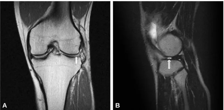
Fig. 1. (A) Coronal magnetic resonance images (MRI) view revealed the absent midbody of the lateral meniscus (arrow) due to a radial tear (ghost sign). (B) Sagittal MRI demonstrated a radial tear (arrow) of the midbody of the lateral meniscus.
A B
Fig. 2. A arthroscopic view showed a complete radial tear in the midbody of the lateral meniscus resembling a cyclops lesion extending to the peripheral meniscocap- sular junction.
Fig. 3. The radial tear of the midbody was approximated with 2-sutures by all-inside and inside-out techniques in con- junction with saucerization.
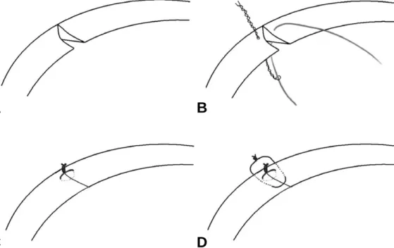
postoperative bleeding. We performed 2-suture repair of the radial tear of the midbody with an all- inside suture using No. 1 PDS (Ethicon, Somerville, NJ, USA) at the red-red zone and an inside-out technique using zone-specific curved cannulae (Linvatec, Largo, FL, USA) at the red-white zone (Fig. 3, 4). After repair of the radial tear, we performed saucerization of the meniscus in conjunction with 2- suture repair of the longitudinal tear using an outside- in technique.
The knee was immobilized in a brace immediately after the operation. Range of motion exercise was allowed from 0。to 90。of flexion for 6 weeks, toe- touch weight bearing was recommended 6 weeks and full weight bearing was permitted 8 weeks after surgery. Full squatting and return to sports activities were allowed after 6 months.
Six-months after surgery, a second-look arthroscopy with informed consent revealed that the free edge of the saucerized meniscus was smooth and the sutured tear was healed completely (Fig. 5). At final follow- up at 2 years, the patient reported no joint pain during participation in sports activities. He had a Tegner activity score of 8 and a Lysholm score of 88. He had full range of motion compared with his contralateral
side, from 0。to 140。. The final radiographic evaluations showed no degenerative changes.
DISCUSSION
With the advent of improved arthroscopic techniques, the generally recommended procedure for symptomatic stable, complete, or incomplete discoid meniscus is an Fig. 4. Schematic drawing of the meniscal repair. (A) A complete radial tear of the midbody of the lateral meniscus. (B, C) All-inside suture was performed with a suture hook loaded with suttle relay and No. 1 PDS (Ethicon, Somerville, NJ, USA) at red-red zone. (D) Inside-out suture was performed at red-white zone.
C A
D B
Fig. 5. An arthroscopic view 6 months after index surgery showed smoothing at the free edge of the saucerized meniscus and connecting tissue at the previous tear site.
the discoid meniscus are most commonly treated with partial meniscectomy.7)Partial meniscectomy has been shown to greatly increase contact pressure.
Meniscectomy for radial tears within the vascular zone of the meniscus transects circumferential fibers, leaving the meniscus with reduced function.
Therefore, meniscal repair should be considered for the treatment of radial tears involving the red-red or red-white zones. Meniscal repairs are rarely performed for radial tears. Choi et al.2)reported fourteen cases with good clinical results after all inside repairs or the radial tears of the lateral meniscus. Noyes and Barber-Westin3) reported that 4 patients had no complaints of pain after inside-out repairs of radial tears involving the avascular zone and confirmed partial healing of a repaired meniscus by second- look arthroscopy in 1 patient. There are few reports in the literature addressing the repair of chronic meniscal tears.4,5)According to Ozkoc et al.,6)one limb of a radial tear resembles a cyclops lesion resulting from repetitive high mechanical stresses of that por- tion of the meniscus, and such lesions are considered to be chronic degenerative tears. The techniques to promote a healing response are considered in the management of meniscal injuries. A fibrin clot has been used in order to provide a reparative scaffold that supplies growth factors to promote chemotaxis, cell proliferation, and matrix synthesis to the tear site. Another thehnique is trephination, in which channels to the peripheral vascular supply are created to encourage vascular and cell migration to the tear site. Rasping promotes an injury response to assist healing. In the present case, trehination and rasping were performed to promote healing of the repaired site.
In the present case, meniscal transplantation after total meniscectomy may be considered. However, results of mensical transplantation have not yet been
even if they are radial tears that extend to the outer third of the meniscus and periphery of the meniscal attachment, meniscal repair should be considered.
Evaluating menisci that have previously been repaired can be difficult because MRI method continue to show an abnormal meniscal signal, even when the meniscus is successfully healed. Therefore MRI examination was not performed in our case. Second-look arthroscopic examination was performed 6 months postoperatively and revealed complete healing of the repaired meniscus. At the final follow-up, the patient reported and demonstrated favorable functional outcomes.
According to Choi et al.,2)because radial tears heal with intervening scars, progressive spreading at the repaired site can alter normal meniscal geometry, adversely influencing mechanical function.
Therefore, our patient will require a longer follow-up period to clarify whether the repaired meniscus remains effective in preventing degenerative osteoarthritis.
REFERENCES
01. Wojtys EM, Chan DB. Meniscus structure and function.
Instr Course Lect. 2005;54:323-30.
02. Choi NH, Kim TH, Son KM, Victoroff BN. Meniscal repair for radial tears of the midbody of the lateral meniscus. Am J Sports Med. 2010;38:2472-6.
03. Noyes FR, Barber-Westin SD. Arthroscopic repair of meniscus tears extending into the avascular zone with or without anterior cruciate ligament reconstruction in patients 40 years of age and older. Arthroscopy.
2000;16:822-9.
04. Kotsovolos ES, Hantes ME, Mastrokalos DS, Lorbach O, Paessler HH. Results of all-inside meniscal repair with the FasT-Fix meniscal repair system. Arthroscopy. 2006;22:3-9.
05. Ponce de Leon JC, Sierra Suarez L, Almazan Diaz A, et al. [Meniscal repair in patients with chronic lesions]. Acta Ortop Mex. 2008;22:12-8.
06. Ozkoc G, Circi E, Gonc U, Irgit K, Pourbagher A, Tandogan RN. Radial tears in the root of the posterior horn of the medial meniscus. Knee Surg Sports Traumatol Arthrosc. 2008;16:849-54.
07. Fritschy D, Gonseth D. Discoid lateral meniscus. Int Orthop. 1991;15:145-7.
08. Yaniv M, Blumberg N. The discoid meniscus. J Child Orthop. 2007;1:89-96.
09. Kim JM, Bin SI. Meniscal allograft transplantation after total meniscectomy of torn discoid lateral meniscus.
Arthroscopy. 2006;22:1344-50 e1.
본 증례 보고의 목적은 외측 불완전 원판형 연골판의 만성 방사상 파열이 있는 젊은 환자에서 시행한 봉합술의 결과를 보고자 한다. 원판형 연골판의 전위된 방사상 파열을 배형 성형술과 봉합술로 치료하였다. 술 후 6개월에 시행한 2차 관 절경 검사에서 봉합된 연골판은 연속성을 유지한 상태로 치유 되었음을 확인하였다. 만성 방사상 파열이라 하더라도 젊은 환자의 경우 봉합술이 고려될 수 있을 것으로 생각된다.
색인 단어: 원판형 연골판, 만성 방사상 파열, 봉합술 초 록