https://doi.org/10.4174/astr.2018.95.5.278 Annals of Surgical Treatment and Research
Kidney transplantation using expanded criteria deceased donors with terminal acute kidney injury: a single center experience in Korea
Kyung Jai Ko1, Young Hwa Kim1, Mi Hyeong Kim1, Kang Woong Jun1, Kyung Hye Kwon1, Hyung Sook Kim2, Sang Dong Kim1, Sun Cheol Park1, Ji Il Kim1, Sang Seob Yun1, In Sung Moon1, Jeong Kye Hwang1,3
1Division of Vascular and Transplant Surgery, Department of Surgery, College of Medicine, The Catholic University of Korea, Seoul, Korea
2Organ Transplant Center, Seoul St. Mary’s Hospital, The Catholic University of Korea, Seoul, Korea
3Department of Surgery, Daejeon St. Mary’s Hospital, College of Medicine, The Catholic University of Korea, Daejeon, Korea
INTRODUCTION
The process of deceased donor recovery and procurement in Asia is not as well structured as are those systems in Europe and America [1]. Confucianism-based thinking in East Asia has led to some hesitance to donate one’s organs, given the belief that one’s body is a gift from one’s parents that should not be damaged [2]. The mean wait time for a deceased donor
kidney transplant (KT) in South Korea is 1934 days [3]. In order to expand the kidney donor pool, the use of organs has been extended to include expanded criteria donors (ECDs) and donors after circulatory death (DCD). Receiving a KT from an ECD or DCD is considered more beneficial than is remaining on the waiting list and continuing dialysis [4,5].
The perception that acute kidney injury (AKI) disqualifies a potential donor kidney has led to loss of many viable organs
Received January 16, 2018, Revised May 22, 2018, Accepted June 8, 2018
Corresponding Author: Jeong Kye Hwang
Department of Surgery, Daejeon St. Mary’s Hospital, College of Medicine, The Catholic University of Korea, 64 Daeheung-ro, Jung-gu, Daejeon 34943, Korea
Tel: +82-42-220-9235, Fax: +82-42-220-9565 E-mail: jjungyong@catholic.ac.kr
ORCID code: https://orcid.org/0000-0001-7146-6957
Copyright ⓒ 2018, the Korean Surgical Society
cc Annals of Surgical Treatment and Research is an Open Access Journal. All articles are distributed under the terms of the Creative Commons Attribution Non- Commercial License (http://creativecommons.org/licenses/by-nc/4.0/) which permits unrestricted non-commercial use, distribution, and reproduction in any medium, provided the original work is properly cited.
Purpose: We investigated the clinical outcomes of deceased donor kidney transplantation (KT) using kidneys with terminal acute kidney injury (AKI).
Methods: Between February 2000 and December 2013, we performed 202 deceased donor renal transplants from 159 brain dead donors. According to the expanded criteria donor (ECD) and AKI network criteria, we divided 202 recipients into 4 groups: Group I: Non-AKI & standard criteria donor (SCD) (n = 97); group II: Non-AKI & ECD (n = 15); group III: AKI & SCD (n
= 52); and group IV: AKI & ECD (n = 38).
Results: The incidence of delayed graft function (DFG) was significantly higher in patients with AKI than it was in the non- AKI group (P = 0.008). There were no significant differences among the 4 groups in graft survival (P = 0.074) or patient survival (P = 0.090). However, the long-term allograft survival rate was significantly lower in group IV than it was in other groups (P = 0.024).
Conclusion: Allografts from deceased donors with terminal AKI had a higher incidence of DGF than did those from donors without AKI. However, there is no significant difference in graft and patient survival rates among the groups. So, the utilization of renal grafts from ECDs with terminal AKI is a feasible approach to address the critical organ shortage.
[Ann Surg Treat Res 2018;95(5):278-285]
Key Words: Acute kidney injury, Donor selection [E04.936.537.500], Kidney transplantation
[6]. However, a donor kidney with AKI from a toxic or ischemic insult can fully recover its renal function [7]. Therefore, the use of deceased donor kidneys with AKI has been considered an alternate plan to using a healthy organ. Prior studies have shown that the outcomes in recipients of kidneys with AKI are comparable [8,9]. Several standardized classification systems for AKI status have been developed in clinical practice [10]. The Acute Kidney Injury Network (AKIN) criteria for the diagnosis of AKI in deceased donor transplantation are useful to predict the development of delayed graft function (DGF) in KT [11].
In this study, we investigated the clinical outcomes of KT from deceased donors with terminal AKI, which was defined using the AKIN criteria and ECD classification.
METHODS
In this retrospective study, we reviewed the medical records and electronic transplant registry of KT recipients at Seoul St.
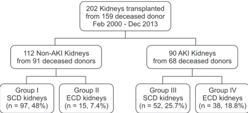
Mary's Hospital, The Catholic University of Korea, between February 2000 and December 2013. This study was conducted according to the Declaration of Helsinki and approved by the Institutional Review Board of The Catholic University of Korea (No. KC17RESI0397). We analyzed 202 deceased donor KTs from 159 brain dead donors. The data included information regarding 53 ECD grafts and 90 grafts with AKI. Based on the ECD classification and AKIN criteria, the recipients were divided into 4 groups. In group I, 97 recipients received non-AKI and SCD kidneys. Fifteen recipients received ECD kidneys without AKI in group II. In group III, 52 recipients received kidneys with AKI and SCD, and 38 recipients received ECD kidneys with AKI in group IV (Fig. 1).
Definitions and assessments
All deceased donors older than 60 years, and those 50–59 years of age that met 2 of the following criteria were classified as ECD based on United Network for Organ Sharing definitions:
(1) history of hypertension, (2) cerebrovascular accident as a cause of brain death, and (3) final preprocurement serum creatinine (SCr) level >1.5 mg/dL [12].
Deceased donors also were classified according to the AKIN criteria. AKI stage 1 is defined as an absolute increase in the last SCr level by 0.3 mg/dL or higher, a 50% or higher increase in the last SCr level from the level on the day admission, or a reduction in urine output (documented oliguria, <0.5 mL/kg per hour for >6 hours). Stage 2 was defined by an increase in the last SCr of more than 200%–300%, or a reduction in urine output (documented oliguria, <0.5 mL/kg per hour for >12 hours). Stage 3 was defined as an increase in the last SCr of more than 300% or a reduction in urine output (documented oliguria, <0.3 mg/kg per hour for >24 hours or anuria for at least 12 hours) [13]. Deceased donors were compared based on their sex, age at transplantation, body mass index, cause of death, history of hypertension and diabetes mellitus, admission SCr, preretrieval SCr, preretrieval estimated glomerular filtration rate (eGFR), last urine volume, central venous pressure, mean arterial pressure, and days of intensive care unit (ICU) stay.
The SCr was measured on the day of admission to our hospital for the pronouncement of brain death. The final SCr level was measured immediately before organ recovery.
The following variables of the recipients were compared:
gender and age at transplantation; cause of end-stage renal disease; type and duration of renal replacement therapy;
number of transplantations; number of human leukocyte antigen mismatches and other immunologic factors; nephron mass index (donor kidney weight to recipient body weight ratio [Kw/Rw]; g/kg); number of renal arteries; total ischemic time; modification of diet in renal disease (MDRD) glomerular filtration rate (GFR) on posttransplant days 2, 7, and 1 month, 3months, 6 months, 1 year, 2 years, 3 years and 5 years;
incidence of acute rejection (AR) episodes; incidence of DGF episodes; duration of graft function and patient survival.
DGF was defined as the need for dialysis in the first week posttransplantation. Renal allograft loss was defined as graft nephrectomy, resumption of ongoing dialysis or return to the pretransplantation SCr level. Patient death with a functioning graft was censored.
Fig. 1. Study groups. Group I:
Non-AKI & SCD (n = 97); group II: Non-AKI & ECD (n = 15);
group III: AKI & SCD (n = 52);
group IV: AKI & ECD (n = 38);
AKI, acute kidney injury; SCD, standard criteria donor; ECD, expanded criteria donor.
202 Kidneys transplanted from 159 deceased donor Feb 2000 - Dec 2013
112 Non-AKI Kidneys from 91 deceased donors
90 AKI Kidneys from 68 deceased donors
Group I SCD kidneys (n = 97, 48%)
Group II ECD kidneys (n = 15, 7.4%)
Group III SCD kidneys (n = 52, 25.7%)
Group IV ECD kidneys (n = 38, 18.8%)
Statistical analysis
One-way analysis of variance (ANOVA) for continuous variables and the chi-square test for categorical variables were used to compare data among the 4 groups. Turkey honestly significant difference test was used as post hoc method in ANOVA. All data were expressed as mean standard deviations.
The Kaplan-Meier method (log-rank test) was used to compare patient and graft survival rates. Data were censored at the time of death, or at the last available follow-up. Statistical significance was defined as a P-value < 0.05. All statistical analyses were carried out using IBM SPSS Statistics ver. 21.0 (IBM Co., Armonk, NY, USA).
RESULTS
Donor characteristics
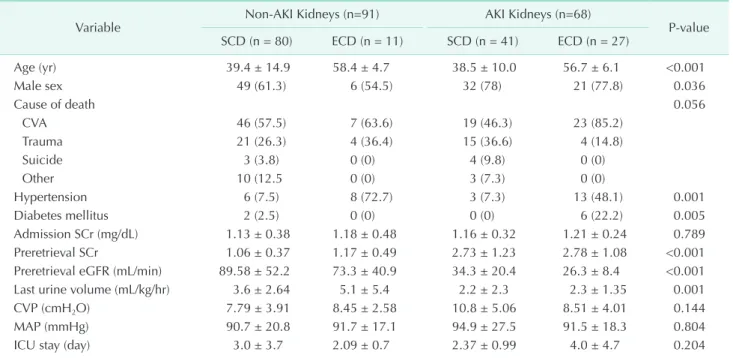
There was insignificant difference in the donor SCr on the admission day across the 4 groups (P = 0.789). However, the preretrieval donor SCr was higher (P < 0.001), while the preretrieval donor eGFR was lower in the AKI groups than they were in the non-AKI groups, respectively (P < 0.001). The last donor urine volume was also smaller in the AKI groups than it was in the non AKI groups (P = 0.001) (Table 1).
Recipient characteristics by group
There were significantly different baseline characteristics
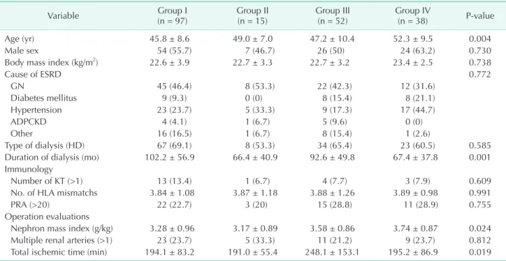
across the 4 groups. According to the group classification, the median patient age in ECD groups (groups II and IV) was older than that in the non-ECD groups (P = 0.004). The duration of dialysis was significantly shorter in the ECD groups than it was in the non-ECD groups (P = 0.001). The nephron mass index (g/
kg) was largest in group IV (P = 0.024). The total ischemic time was longest in group III (Table 2).
Graft renal function, AR episodes, and DGF
The mean MDRD GFR levels among the 4 groups were significantly different for up to 2 years posttransplantation (P
= 0.02). However, 3–5 years after transplantation, the MDRD GFR levels were not significantly different across the groups (P = 0.122, P = 0.229, P = 0.708, respectively) (Table 3, Fig. 2).
Although the incidence of DGF was significantly more frequent in patients in the AKI group than it was in those in non-AKI groups (8.13%, 0%, 25.0%, 23.7%, respectively; P = 0.008), the incidence of biopsy-proven AR was comparable among the groups (P = 0.463).
Graft and patient survival
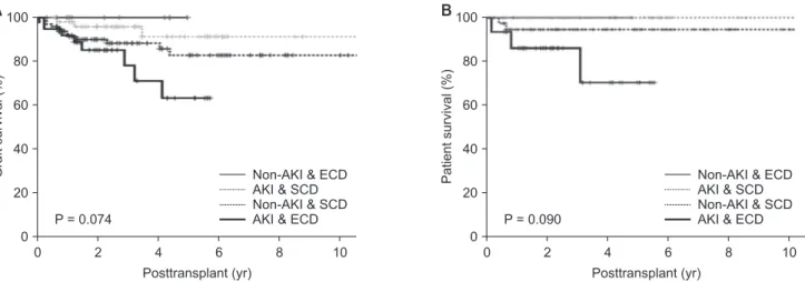
Actual graft and patient survival rates were also similar among the groups, with a mean follow-up period of 40.3 months (P = 0.114, P = 0.094, respectively) (Table 4). Graft survival rates at 1-, 2-, and 5-year posttransplantation were: 91.2%, 89.9%, and 82.7%, respectively, in group I; 100% in group II; 98.0%, 95.8%,
Table 1. Donor characteristics by group (n = 159)
Variable Non-AKI Kidneys (n=91) AKI Kidneys (n=68)
P-value SCD (n = 80) ECD (n = 11) SCD (n = 41) ECD (n = 27)
Age (yr) 39.4 ± 14.9 58.4 ± 4.7 38.5 ± 10.0 56.7 ± 6.1 <0.001
Male sex 49 (61.3) 6 (54.5) 32 (78) 21 (77.8) 0.036
Cause of death 0.056
CVA 46 (57.5) 7 (63.6) 19 (46.3) 23 (85.2)
Trauma 21 (26.3) 4 (36.4) 15 (36.6) 4 (14.8)
Suicide 3 (3.8) 0 (0) 4 (9.8) 0 (0)
Other 10 (12.5 0 (0) 3 (7.3) 0 (0)
Hypertension 6 (7.5) 8 (72.7) 3 (7.3) 13 (48.1) 0.001
Diabetes mellitus 2 (2.5) 0 (0) 0 (0) 6 (22.2) 0.005
Admission SCr (mg/dL) 1.13 ± 0.38 1.18 ± 0.48 1.16 ± 0.32 1.21 ± 0.24 0.789
Preretrieval SCr 1.06 ± 0.37 1.17 ± 0.49 2.73 ± 1.23 2.78 ± 1.08 <0.001
Preretrieval eGFR (mL/min) 89.58 ± 52.2 73.3 ± 40.9 34.3 ± 20.4 26.3 ± 8.4 <0.001
Last urine volume (mL/kg/hr) 3.6 ± 2.64 5.1 ± 5.4 2.2 ± 2.3 2.3 ± 1.35 0.001
CVP (cmH2O) 7.79 ± 3.91 8.45 ± 2.58 10.8 ± 5.06 8.51 ± 4.01 0.144
MAP (mmHg) 90.7 ± 20.8 91.7 ± 17.1 94.9 ± 27.5 91.5 ± 18.3 0.804
ICU stay (day) 3.0 ± 3.7 2.09 ± 0.7 2.37 ± 0.99 4.0 ± 4.7 0.204
Values are presented as mean ± standard deviation or number (%).
AKI, acute kidney injury; SCD, standard criteria donor; ECD, expanded criteria donor; CVA, cerebrovascular accident; SCr, serum creatinine; eGFR, estimated glomerular filtration rate; CVP, central venous pressure; MAP, mean arterial pressure; ICU, intensive care unit.
and 91.2%, respectively, in group III; and 91.8%, 85.0% and 63.0%, respectively, in group IV. Patient survival rates at 1-, 2-, and 5-year posttransplantation were: 97.8%, in group I; 100% in groups II and III; and 94.4%, 94.4% and 88.1%, respectively, in
group IV. There were no significant differences in graft survival rate (P = 0.074) or actual patient survival rate among the 4 groups (P = 0.090) (Fig. 3). However, the long-term allograft survival rate was significantly lower in group IV than it was in Table 2. Recipient characteristics by group (No. of recipients = 20)
Variable Group I
(n = 97) Group II
(n = 15) Group III
(n = 52) Group IV
(n = 38) P-value
Age (yr) 45.8 ± 8.6 49.0 ± 7.0 47.2 ± 10.4 52.3 ± 9.5 0.004
Male sex 54 (55.7) 7 (46.7) 26 (50) 24 (63.2) 0.730
Body mass index (kg/m2) 22.6 ± 3.9 22.7 ± 3.3 22.7 ± 3.2 23.4 ± 2.5 0.738
Cause of ESRD 0.772
GN 45 (46.4) 8 (53.3) 22 (42.3) 12 (31.6)
Diabetes mellitus 9 (9.3) 0 (0) 8 (15.4) 8 (21.1)
Hypertension 23 (23.7) 5 (33.3) 9 (17.3) 17 (44.7)
ADPCKD 4 (4.1) 1 (6.7) 5 (9.6) 0 (0)
Other 16 (16.5) 1 (6.7) 8 (15.4) 1 (2.6)
Type of dialysis (HD) 67 (69.1) 8 (53.3) 34 (65.4) 23 (60.5) 0.585
Duration of dialysis (mo) 102.2 ± 56.9 66.4 ± 40.9 92.6 ± 49.8 67.4 ± 37.8 0.001
Immunology
Number of KT (>1) 13 (13.4) 1 (6.7) 4 (7.7) 3 (7.9) 0.609
No. of HLA mismatchs 3.84 ± 1.08 3.87 ± 1.18 3.88 ± 1.26 3.89 ± 0.98 0.991
PRA (>20) 22 (22.7) 3 (20) 15 (28.8) 11 (28.9) 0.755
Operation evaluations
Nephron mass index (g/kg) 3.28 ± 0.96 3.17 ± 0.89 3.58 ± 0.86 3.74 ± 0.87 0.024
Multiple renal arteries (>1) 23 (23.7) 5 (33.3) 11 (21.2) 9 (23.7) 0.812
Total ischemic time (min) 194.1 ± 83.2 191.0 ± 55.4 248.1 ± 153.1 195.2 ± 86.9 0.019 Values are presented as mean ± standard deviation or number (%).
Group I: Non-AKI & SCD (n = 97); group II: Non-AKI & ECD (n = 15); group III: AKI & SCD (n = 52); group IV: AKI & ECD (n = 38);
AKI, acute kidney injury; SCD, standard criteria donor; ECD, expanded criteria donor; ESRD, end-stage renal disease; GN, glomerulonephritis; ADPCKD, autosomal dominant polycystic kidney disease; HD, hemodialysis; KT, kidney transplantation; HLA, human leukocyte antigen; PRA, panel-reactive antibody.
Table 3. Change in the graft function with time (No. of recipients = 202) eGFR by MDRD (mL/min/1.73 m2) Group I
(n = 97) Group II
(n = 15) Group III
(n = 52) Group IV
(n = 38) P-value
Immediate postoperative 5.82 ± 2.45 4.20 ± 1.26 5.32 ± 1.70 5.54 ± 1.92 0.838
POD#2 days 20.4 ± 17.3 18.5 ± 8.93 10.1 ± 8.75 9.57 ± 4.94 <0.001
POD#5 days 41.4 ± 27.6 40.2 ± 17.3 20.1 ± 21.3 19.6 ± 16.1 <0.001
POD#7 days 52.0 ± 27.6 52.8 ± 18.4 25.0 ± 23.9 29.0 ± 20.5 <0.001
POD#1 month 63.0 ± 19.5 53.3 ± 17.0 53.9 ± 27.3 45.4 ± 18.3 <0.001
POD#3 months 60.3 ± 18.1 48.5 ± 11.7 58.0 ± 19.5 45.1 ± 17.8 0.002
POD#6 months 61.1 ± 19.1 47.1 ± 8.73 59.3 ± 17.2 46.9 ± 17.8 0.010
POD#12 months 63.4 ± 23.6 51.3 ± 11.6 60.1 ± 17.5 46.2 ± 18.9 0.001
POD#18 months 60.3 ± 22.5 48.1 ± 6.82 58.9 ± 19.9 46.2 ± 20.0 0.018
POD#2 years 61.2 ± 22.1 47.1 ± 7.92 62.3 ± 16.6 42.9 ± 23.5 0.020
POD#3 years 55.2 ± 26.2 48.1 ± 7.97 59.4 ± 17.9 41.6 ± 20.2 0.122
POD#4 years 57.8 ± 24.7 46.7 ± 10.8 61.7 ± 21.8 41.8 ± 21.9 0.229
POD#5 years 56.8 ± 21.8 46.0 ± 0.0 62.4 ± 13.2 56.5 ± 7.48 0.708
Values are presented as mean ± standard deviation.
Group I: Non-AKI & SCD (n = 97); group II: Non-AKI & ECD (n = 15); group III: AKI & SCD (n = 52); group IV: AKI & ECD (n = 38);
AKI, acute kidney injury; SCD, standard criteria donor; ECD, expanded criteria donor; eGFR, estimated glomerular filtration rate;
MDRD, modification of diet in renal disease; POD, postoperative day.
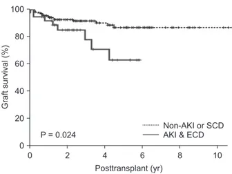
the other groups (P = 0.024) (Fig. 4).
DISCUSSION
AKI has diverse causes. AKI can also be relatively benign, with a minimal elevation in SCr levels, or devastating and lead to anuric renal failure [14]. One third of AKI in hospitalized patients represents transient azotemia. However, transient azotemia is also independently correlated with an increased risk of death [15]. AKI frequently develops in patients who become deceased donors, because these patients are critically ill in ICUs and are subject to both ischemic and nephrotoxic insults [16]. DGF manifests as severe ischemic-reperfusion injury (IRI) in the kidney graft [17]. Kidney grafts with pre- existing AKI prior to transplantation may be more vulnerable to ischemic injury during recovery, and additional injuries as calcineurin inhibitor toxicity [11].
In transplantation, IRI negatively effects kidney function.
There are also several potential etiologies for graft fibrosis,
Posttransplant
Post KT
Posttransplant 0
100
80
60
40
20
2 Days 7 Days 1 Mo 3 Mo 6 Mo 12 Mo 24 Mo 36 Mo 60 Mo Non-AKI & SCD
Non-AKI & ECD AKI & SCD
AKI & ECD * * *
*
*
*
*
*
*P < 0.05 MDRD GFR
(mL/min/1.73 m )2
Fig. 2. Change in graft function. AKI, acute kidney injury;
SCD, standard criteria donor; ECD, expanded criteria donor;
MDRD, modification of diet in renal disease; eGFR, estimated glomerular filtration rate; KT, kidney transplantation.
Table 4. Outcomes by group
Variable Group I
(n = 97) Group II
(n = 15) Group III
(n = 52) Group IV
(n = 38) P-value
Actual patient survivala) 95 (97.9) 15 (100) 52 (100) 35 (92.1) 0.094
Actual graft survivalb) 84 (87.5) 15 (100) 48 (92.3) 30 (78.9) 0.114
Follow-up (mo) 43.7 ± 35.2 27.0 ± 20.0 43.8 ± 29.1 32.1 ± 19.9 0.060
Delayed graft function 8 (8.3) 0 (0) 13 (25.0) 9 (23.7) 0.008
BPAR episode 41 (42.7) 3 (20.0) 17 (32.7) 14 (36.8) 0.463
Values are presented as number (%) or mean ± standard deviation.
Group I: Non-AKI & SCD (n = 97); group II: Non-AKI & ECD (n = 15); group III: AKI & SCD (n = 52); group IV: AKI & ECD (n = 38);
AKI, acute kidney injury; SCD, standard criteria donor; ECD, expanded criteria donor; BPAR, biopsy proven acute rejection.
a, b)During the follow-up period.
Graftsurvival(%)
0 2 4 6 8 10
A
Posttransplant (yr)
Non-AKI & ECD AKI & SCD Non-AKI & SCD AKI & ECD 0
100
80
60
40
20
P = 0.074
Patientsurvival(%)
0 2 4 6 8 10
B
Posttransplant (yr) 0
100
80
60
40
20
P = 0.090
Non-AKI & ECD AKI & SCD Non-AKI & SCD AKI & ECD
Fig. 3. Graft (A, P = 0.074) and patient survival (B, P = 0.090) among the 4 groups. AKI, acute kidney injury; SCD, standard criteria donor; ECD, expanded criteria donor.
including the immune response and nonimmune factors (e.g., drug toxicities, infections, and urinary tract obstruction) [18].
Hypoperfusion due to shock or rhabdomyolysis after brain death can cause warm ischemia, which may develop prerecovery AKI.
Warm ischemic time is associated with irreversible cell damage leading to DGF and primary dysfunction [19]. A preexisting allograft tissue injury may affect the graft’s function until natural repair is completed. In nontransplanted kidneys, the phases of AKI consist of initiation, repair, and recovery; this process may continue for up to 6 months [20,21]. Due to this potentially prolonged recovery time after AKI, the rates of DGF are greater in donors who have elevated creatinine levels than they are in those without [22]. However, recent studies have shown that there are comparable outcomes after KT from donors with and without AKI [8,9]. Hall et al. [23] found that the rates of DGF increased as the AKI stage increased; however, there was no significant difference in the 1-year graft survival between AKI and non-AKI groups. Heilman et al. [24] also found that there was no difference in 1-year graft survival or 1-year eGFR between the AKI and control groups. Although the high incidence of DGF did not suggest a poor outcome in AKI donors, DGF itself remains an important risk factor for AR. DGF in non- AKI kidneys developed frequently from AR, which led to poor allograft outcomes [17,25,26].
With regard to baseline characteristics, the duration of dialysis was longest in group I, although the nephron mass index was larger in AKI groups. These findings may reflect that recipients on the waiting list prefer to receive grafts without AKI; in particular, they prefer SCD kidneys without AKI.
We investigated the clinical outcomes of KT from deceased donors with AKI. AKI was defined using the AKIN criteria and
ECD classification. The incidence of DGF was higher in the AKI groups than it was in the non-AKI groups. The graft function (MDRD GFR levels) among the 4 groups was significantly different for up to 2 years posttransplantation. After 3 years (and persisting until at least 5 years after KT), the MDRD GFR levels did not differ significantly among the groups. In group IV, the graft function may be affected by both AKI and ECD. It seems that after completion of the repair process, preexisting AKI did not affect the long-term outcome of the allograft kidney [11]. In this study, we found that there was no significant difference in graft survival or patient survival among the 4 groups. According to our past report, ECD may have less of an effect on graft survival and patient survival than previously thought in spite of the incidence of DGF was higher than SCD. There are 2 possible reasons for this result. One is the short cold ischemic time (mean, 4.05 ± 2.18 hours), and the other is the ethnic homogeneity of Koreans [27]. However, the long-term allograft survival rate was significantly lower in group IV compared than it was in the other groups. We can assumed that this subgroup analysis showed a statistically significance because of the negative synergic effect from ECD and AKI groups. Therefore, KT from ECD kidneys with terminal AKI must be carefully considered.
This study has several limitations. First, this was a retrospective study, which makes it vulnerable to bias in general. In addition, the subgroup analysis made up between group IV and the other groups could be prone to the statistical bias of random assignment. Second, according to the Korean guidelines for the management of deceased donors, it is recommended that potential deceased donors be transferred to an appointed institution for solid organ procurement. In this study, the final donor SCr levels were typically measured within 48 hours of the day of admission (and therefore measurement of the baseline SCr level). Therefore, the incidence of AKI may have been underestimated. Lastly, we did not include the AKIN staging categorization in this study. In stage 3, the graft discard rate may have been overestimated. Therefore, the DGF rate and follow up GFR levels could be different according to the stage of AKI.
In conclusion, 42.8% of deceased donors were diagnosed with AKI, while 23.9% were defined as ECD. As the waitlist population for kidneys has increased, using ECD organs with AKI has become a more important alternative. There is no significant difference in graft and patient survival rates with and without AKI. So, the utilization of renal grafts from ECDs with terminal AKI is a feasible approach to address the critical organ shortage.
CONFLICTS OF INTEREST
No potential conflict of interest relevant to this article was
Graftsurvival(%)
0 2 4 6 8 10
Posttransplant (yr)
Non-AKI or SCD AKI & ECD 0
100
80
60
40
20
P = 0.024
Fig. 4. Comparing graft survival (P = 0.024) between group IV and the others (groups I, II, and III). Group I: Non-AKI &
SCD (n = 97); group II: Non-AKI & ECD (n = 15); group III:
AKI & SCD (n = 52); group IV: AKI & ECD (n = 38); AKI, acute kidney injury; SCD, standard criteria donor; ECD, expanded criteria donor.
reported.
ACKNOWLEDGEMENTS
This research was supported by Basic Science Research
Program through the National Research Foundation of Korea (NRF) funded by the Ministry of Education (2017R1D1A1B03034715).
REFERENCES
1. Lo CM. Deceased donation in Asia:
challenges and opportunities. Liver Transpl 2012;18 Suppl 2:S5-7.
2. Jung CW, Park KT, Kim SY, Kim SJ, Kim MG, Jo SK, et al. Clinical outcomes in kidney transplantation patients from deceased donors with acute kidney injury.
Transplant Proc 2013;45:2941-5.
3. Korean Network for Ogran Sharing. 2016 Annual data report [Internet]. Seoul:
Korean Network for Ogran Sharing; [cited 2017 Sep 8]. Available from: http://konos.
go.kr.
4. Merion RM, Ashby VB, Wolfe RA, Distant DA, Hulbert-Shearon TE, Metzger RA, et al. Deceased-donor characteristics and the survival benefit of kidney transplantation.
JAMA 2005;294:2726-33.
5. Summers DM, Johnson RJ, Allen J, Fuggle SV, Collett D, Watson CJ, et al. Analysis of factors that affect outcome after transplantation of kidneys donated after cardiac death in the UK: a cohort study.
Lancet 2010;376:1303-11.
6. de Mendonca A, Vincent JL, Suter PM, Moreno R, Dearden NM, Antonelli M, et al. Acute renal failure in the ICU: risk factors and outcome evaluated by the SOFA score. Intensive Care Med 2000;26:
915-21.
7. Bonventre JV, Yang L. Cellular patho- physiology of ischemic acute kidney injury. J Clin Invest 2011;121:4210-21.
8. Anil Kumar MS, Khan SM, Jaglan S, Heifets M, Moritz MJ, Saeed MI, et al. Suc- cess ful transplantation of kidneys from deceased donors with acute renal failure:
Three-year results. Trans plantation 2006;
82:1640-5.
9. Ugarte R, Kraus E, Montgomery RA, Bur-
dick JF, Ratner L, Haas M, et al. Excel- lent outcomes after transplantation of deceased donor kidneys with high ter min al creatinine and mild pathologic lesions. Transplantation 2005;80:794-800.
10. Kellum JA. Diagnostic criteria for acute kidney injury: present and future. Crit Care Clin 2015;31:621-32.
11. Lee MH, Jeong EG, Chang JY, Kim Y, Kim JI, Moon IS, et al. Clinical outcome of kidney transplantation from deceased donors with acute kidney injury by Acute Kidney Injury Network criteria. J Crit Care 2014;29:432-7.
12. Metzger RA, Delmonico FL, Feng S, Port FK, Wynn JJ, Merion RM. Expanded crite- ri a donors for kidney transplantation. Am J Transplant 2003;3 Suppl 4:114-25.
13. Mehta RL, Kellum JA, Shah SV, Molitoris BA, Ronco C, Warnock DG, et al. Acute Kidney Injury Network: report of an ini tia tive to improve outcomes in acute kidney injury. Crit Care 2007;11:R31.
14. Hoste EA, Kellum JA. Acute kidney injury:
epidemiology and diagnostic criteria. Curr Opin Crit Care 2006;12:531-7.
15. Uchino S, Bellomo R, Bagshaw SM, Goldsmith D. Transient azotaemia is as so ci ated with a high risk of death in hospitalized patients. Nephrol Dial Trans- plant 2010;25:1833-9.
16. Kolonko A, Chudek J, Pawlik A, Wilk J, Jałowiecki P, Wiecek A. Acute kidney injury before organ procurement is asso- ciated with worse long-term kidney graft outcome. Transplant Proc 2011;43:2871-4.
17. Siedlecki A, Irish W, Brennan DC. Delayed graft function in the kidney transplant.
Am J Transplant 2011;11:2279-96.
18. Heilman RL, Devarapalli Y, Chakkera HA,
Mekeel KL, Moss AA, Mulligan DC, et al.
Impact of subclinical inflammation on the development of interstitial fibrosis and tubular atrophy in kidney transplant recipients. Am J Transplant 2010;10:563- 70.
19. Perico N, Cattaneo D, Sayegh MH, Remu- zzi G. Delayed graft function in kid ney trans plantation. Lancet 2004;364:1814-27.
20. Chawla LS, Kimmel PL. Acute kidney injury and chronic kidney disease: an integrated clinical syndrome. Kidney Int 2012;82:516-24.
21. Macedo E, Bouchard J, Mehta RL. Renal recovery following acute kidney injury.
Curr Opin Crit Care 2008;14:660-5.
22. Boffa C, van de Leemkolk F, Curnow E, Homan van der Heide J, Gilbert J, Sharples E, et al. Transplantation of kidneys from donors with acute kidney injury: friend or foe? Am J Transplant 2017;17:411-9.
23. Hall IE, Schroppel B, Doshi MD, Ficek J, Weng FL, Hasz RD, et al. Associations of deceased donor kidney injury with kidney discard and function after trans- plantation. Am J Transplant 2015;15:1623- 31.
24. Heilman RL, Smith ML, Kurian SM, Huskey J, Batra RK, Chakkera HA, et al.
Trans planting kidneys from deceased do- nors with severe acute kidney injury. Am J Transplant 2015;15:2143-51
25. Farney AC, Rogers J, Orlando G, al-Geizawi S, Buckley M, Farooq U, et al. Evolving ex perience using kidneys from deceased donors with terminal acute kidney injury.
J Am Coll Surg 2013;216:645-55.
26. Seo CH, Ju JI, Kim MH, Jun KW, Ahn SH, Hwang JK, et al. Risk factors and long- term outcomes of delayed graft function
in deceased donor renal transplantation.
Ann Surg Treat Res 2015;89:208-14.
27. Hwang JK, Park SC, Kwon KH, Choi
BS, Kim JI, Yang CW, et al. Long-term outcomes of kidney transplantation from expanded criteria deceased donors at a
single center: comparison with standard criteria deceased donors. Transplant Proc 2014;46:431-6.