Korean J Hepatobiliary Pancreat Surg 2016;20:93-96
http://dx.doi.org/10.14701/kjhbps.2016.20.2.93
Case Report
Incidental detection of pancreatic hemangioma mimicking a metastatic tumor of renal cell carcinoma
Sung Hyun Kim1, Ji-Ye Kim2, Jin Young Choi3, Young Deuk Choi4, and Kyung Sik Kim1
Departments of 1Hepatobiliary and Pancreatic Surgery, 2Pathology, 3Radiology, and 4Urology, Severance Hospital, Yonsei University College of Medicine, Seoul, Korea
Adult pancreatic hemangioma is a rare disease. We presented a case of a woman with pancreatic tail mass mimicking a distant metastasis from the kidney. A 68-year-old woman was found with a left kidney mass on medical checkup.
Computed tomography scan showed a 4.3 cm-sized mass in the left kidney, suggesting renal cell carcinoma (RCC), and a strongly enhancing tiny nodule in the pancreatic tail. We could not rule the possibility of RCC metastasis, hence, surgical resection of the pancreatic mass simultaneously with radical nephrectomy for RCC was conducted. Gross pathologic examination revealed hemangioma. Immunohistochemistry revealed that the tumor was positive for CD34, CD31 and factor VIII-related antigen. There were no significant postoperative events, and the patient was discharged on postoperative day 7 without any complications. Treatment strategies for pancreatic hemangioma have not been established. To our knowledge, this was the first case report of asymptomatic pancreatic hemangioma. In previous literature, treatment differed on a case-by-case basis, ranging from observation to surgical resection. The most im- portant factor in deciding whether to perform surgery is possibly risk-benefit effectiveness; however, tumor location, patient symptoms, and other factors are also important. (Korean J Hepatobiliary Pancreat Surg 2016;20:93-96) Key Words: Pancreas; Hemangioma; Adult hemangioma; Incidental discovery; CD34
Received: August 31, 2015; Revised: September 16, 2015; Accepted: September 21, 2015 Corresponding author: Kyung Sik Kim
Department of Hepatobiliary and Pancreatic Surgery, Severance Hospital, Yonsei University College of Medicine, 50-1 Yonsei-ro, Seodaemun-gu, Seoul 03722, Korea
Tel: +82-2-2228-2100, Fax: +82-2-313-8289, E-mail: [email protected]
Copyright Ⓒ 2016 by The Korean Association of Hepato-Biliary-Pancreatic Surgery
This is an Open Access article distributed under the terms of the Creative Commons Attribution Non-Commercial License (http://creativecommons.org/
licenses/by-nc/4.0) which permits unrestricted non-commercial use, distribution, and reproduction in any medium, provided the original work is properly cited.
Korean Journal of Hepato-Biliary-Pancreatic Surgery ∙ pISSN: 1738-6349ㆍeISSN: 2288-9213
INTRODUCTION
Hemangiomas are tumors characterized by increased numbers of normal or abnormal vessels filled with blood.
Occasionally, hemangiomas can occur internally, and nearly one-third of these internal lesions are found in the liver. Pancreatic hemangiomas are especially rare; pancre- atic vascular neoplasms collectively account for only 0.1% of all pancreatic tumors.1 These tumors are usually diagnosed fortuitously by laparotomies performed to diag- nose a large, palpable abdominal mass.2-5 We presented a very rare case in which a woman without specific symp- toms was found with a cavernous hemangioma in the pan- creas tail that mimicked metastatic tumor.
CASE
A 68-year-old woman was found with a mass in her left kidney on medical checkup. She had no significant past medical history except hypertension and no symptoms (e.g., hematuria, abdominal pain, or abdominal discomfort).
An axial contrast-enhanced computed tomography (CT) scan showed a heterogeneous solid mass in the left kidney, suggesting the presence of renal cell carcinoma (RCC).
There was a strongly enhancing tiny nodule in the tail of the pancreas that was most likely either a neuroendocrine tumor or a RCC metastasis (Fig. 1A). Because she had no specific symptoms or abnormal laboratory findings, the possibility of RCC metastasizing into the pancreas could not be ruled out. Therefore, surgical resection, including left radical nephrectomy and distal pancreatectomy was conducted. There were no significant postoperative events,
94 Korean J Hepatobiliary Pancreat Surg Vol. 20, No. 2, May 2016
Fig. 1. Imaging study and gross morphology. (A) Axial contrast-enhanced CT scan during the arterial phase shows a strongly enhancing tiny nodule in the pancreatic tail (arrowhead); (B) Gross specimen; pancreatic hemangioma confined to the pancreas (arrow).
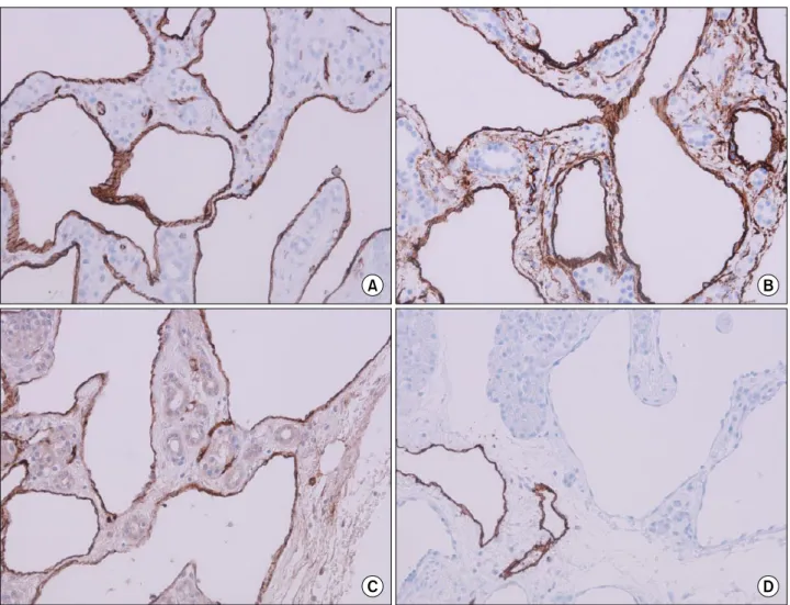
Fig. 2. Immunohistochemical stain shows a cavernous, ectatic endothelial neoplasm arising amid pancreatic parenchymal tissue.
(A) CD31 (100×); (B) CD34 (100×); (C) factor VIII-related antigen (100×); and (D) D2-40 (100×).
and the patient was discharged home on postoperative day 7 without any morbidity or complications.
Gross pathologic examination revealed a 0.6×0.5 cm-sized
hemangioma confined to the pancreas, and the tissue had a tumor-free margin (Fig. 1B). Immunohistochemical anal- ysis showed that the tumor was positive for CD31, CD34,
Sung Hyun Kim, et al. Incidental detection of pancreatic hemangioma 95
and factor VIII-related antigen, and negative for D2-40, RCC, and CD56 (SCLC), supporting the diagnosis of he- mangioma (Fig. 2). The left kidney mass was confirmed as RCC with no evidence of metastasis.
DISCUSSION
Hemangiomas rarely occur in the pancreas and often are not suspected clinically; only 14 patients are reported in the literature since 1939. Review of the previous literature reports on pancreatic hemangioma indicated that most he- mangiomas occur in females (12/15 patients, including our patient) and are symptomatic (9/15 patients had abdominal pain, and one patient each had melena, thrombocytopenia, abdominal distension, and palpitation, suggesting bleed- ing). Only 1 tumor was found incidentally, in 1939 upon autopsy.2-7
The hemangioma was found incidentally at a pre- operative evaluation for RCC. Unlike previous studies, we found no symptoms suggesting pancreatic hemangioma, likely because it was a tiny pancreatic tail mass. Typically, hemangiomas are strongly contrast enhancing in the arterial phase of conventional contrast-enhanced CT imaging;8 however, our case did not present these findings, likely because of the small lesion size. The pancreatic hemangio- ma thus mimicked metastatic cancer originating from the RCC. To our knowledge, this case was the first report of pancreatic hemangioma without a symptomatic event, and the tumor is the smallest of the reported cases. After sur- gery, the pancreatic tail tumor was pathologically con- firmed as a hemangioma.
Microscopic findings revealed a typical feature of he- mangioma i.e., blood-filled spaces separated by fibrous con- nective tissue. For a definite diagnosis, immunohistochemistry is required to assess the presence of the factor VIII-related antigen, a marker for vascular endothelium that was re- ported by Chang and colleagues.9 Subsequently, Mundinger and colleagues reported that neoplastic cells in hemangio- ma also express the endothelial markers CD31 and CD34.5 In our patient, immunohistochemical findings were positive for all 3 markers; whereas, D2-40, a marker for lymphatic endothelium, and CD56, a marker for neural cell, were both negative, further indicating that the tumor mass was a hemangioma.
Because of its rarity, there is no standard treatment for
pancreatic hemangioma. Reviewing the previous literature on pancreatic hemangioma, we found that multiple differ- ent treatments were administered, from observation to sur- gical resection.2-7 Furthermore, the possibility of abdomi- nal pain or hemorrhagic events is typically increased in patients with larger hemangioma masses. Therefore, some clinicians suggested that surgery is the best treatment option. However, other clinicians suggested that if the pa- tient’s symptoms are minimal, observation is a possible treatment option, because pancreatic hemangiomas are benign.
The location of the pancreatic hemangioma is variable, and may be important for determining the best treatment option. Upon reviewing previous literature, we found that pancreatic hemangiomas were located at the head (8/15 patients), neck (1/15 patients), or body/tail (6/15 pa- tients).2-7 When the tumor is located at the body/tail, distal pancreatectomy is an option. However, if the tumor is lo- cated at the proximal site of the pancreas, pylorus-preserv- ing pancreaticoduodenectomy is indicated. Patients who underwent pylorus-preserving pancreaticoduodenectomy had a higher rate of morbidity than patients who under- went distal pancreatectomy (34.7% vs. 27.8%, p<0.05).10 Therefore, if a patient has a pancreas head hemangioma with minimal symptoms that can be controlled, close ob- servation and regular follow-up can be one of the treat- ment options according to risk-benefit analysis. Because our case was confined to a tiny mass at the distal pancreas, and we could not rule out distant metastasis from the RCC tumor, we decided to perform a distal pancreatectomy.
The literature review indicated that treatment decisions require assessment of the severity of symptoms and loca- tion of the tumor. When all the cases were collectively considered, determining the timing of surgery based on comparison of surgical risk-benefit analysis emerged as an important factor. Future reports will provide more data on the optimal treatment strategies for pancreatic hemangioma.
REFERENCES
1. Robbins SL, Kumar V. Robbins and Cotran pathologic basis of disease. 8th ed. Philadelphia, PA: Saunders/Elsevier, 2010.
2. Weidenfeld J, Zakai BB, Faermann R, Barshack I, Aviel-Ronen S. Hemangioma of pancreas: a rare tumor of adulthood. Isr Med Assoc J 2011;13:512-514.
3. Lee J, Raman K, Sachithanandan S. Pancreatic hemangioma
96 Korean J Hepatobiliary Pancreat Surg Vol. 20, No. 2, May 2016
mimicking a malignant pancreatic cyst. Gastrointest Endosc 2011;73:174-176.
4. Jarboui S, Salem A, Gherib BS, Ben Moussa M, Rajhi H, Mnif N, et al. Hemangioma of the pancreas in a 60-year-old woman:
a report of a new case. Gastroenterol Clin Biol 2010;34:569-571.
5. Mundinger GS, Gust S, Micchelli ST, Fishman EK, Hruban RH, Wolfgang CL. Adult pancreatic hemangioma: case report and lit- erature review. Gastroenterol Res Pract 2009;2009:839730.
6. Williamson JM, Finch-Jones M, Pope I. Endoscopic ultra- sonography allowing expectant management of pancreatic haemangioma. Ann R Coll Surg Engl 2014;96:e1-e2.
7. Naito Y, Nishida N, Nakamura Y, Torii Y, Yoshikai H, Kawano H, et al. Adult pancreatic hemangioma: a case report. Oncol Lett 2014;8:642-644.
8. Kobayashi H, Itoh T, Murata R, Tanabe M. Pancreatic cavernous hemangioma: CT, MRI, US, and angiography characteristics.
Gastrointest Radiol 1991;16:307-310.
9. Chang WT, Lee KT, Yang SF. Cavernous hemangioma of the pancreas: report of a case. Pancreas 2003;26:310-312.
10. Kneuertz PJ, Pitt HA, Bilimoria KY, Smiley JP, Cohen ME, Ko CY, et al. Risk of morbidity and mortality following hepato-pan- creato-biliary surgery. J Gastrointest Surg 2012;16:1727-1735.