Learning fiberoptic intubation for awake nasotracheal intubation
Hyuk Kim
1, Eunsun So
2, Myong-Hwan Karm
2, Hyun Jeong Kim
1, Kwang-Suk Seo
11Department of Dental Anesthesiology, School of Dentistry, Seoul National University, Seoul, Korea
2Department of Dental Anesthesiology, Seoul National University Dental Hospital, Seoul, Korea
Background: Fiberoptic nasotracheal intubation (FNI) is performed if it is difficult to open the mouth or if intubation using laryngoscope is expected to be difficult. However, training is necessary because intubation performed by inexperienced operators leads to complications.
Methods: Every resident performed intubation in 40 patients. Success of FNI was evaluated as the time of FNI. First intubation time was restricted to 2 min 30 s. If the second attempt was unsuccessful, it was considered a failed case, and a specialist performed nasotracheal intubation. If the general method of intubation was expected to be difficult, awake intubation was performed. The degree of nasal bleeding during intubation was also evaluated.
Results: The mean age of the operators (11 men, 7 women) was 27.8 years. FNI was performed in a total of 716 patients. The success rate was 88.3% for the first attempt and 94.6% for the second attempt. The failure rate of intubation in anesthetized patients was 4.9%, and 13.6% in awake patients. When intubation was performed in anesthetized patients, the failure rate from the first to fifth trial was 9.6%, which decreased to 0.7% when the number of trials increased to > 30 times. In terms of awake intubation, there was no failed attempt when the resident had performed the FNI > 30 times. The number of FNIs performed and nasal bleeding were important factors influencing the failure rate.
Conclusion: The success rate of FNI increased as the number of FNI performed by residents increased despite the nasal bleeding.
Keywords: Fiberscope; Learning curve; Nasotracheal intubation.
This is an Open Access article distributed under the terms of the Creative Commons Attribution Non-Commercial License (http://creativecommons.org/licenses/by-nc/4.0/) which permits unrestricted non-commercial use, distribution, and reproduction in any medium, provided the original work is properly cited.
Received: December 11, 2017•Revised: December 21, 2017•Accepted: December 26, 2017
Corresponding Author: Kwang-Suk Seo, Department of Dental Anesthesiology, School of Dentistry, Seoul National University, 101 Daehaka-ro Jongno-gu, Seoul 03080, Korea
Tel: +82-2-2072-0622 Fax: +82-2-766-9427 E-mail: [email protected] Copyrightⓒ 2017 Journal of Dental Anesthesia and Pain Medicine
INTRODUCTION
Generally, oral intubation is performed to secure the airway during general anesthesia; however, because oral cavity is operation field during oral and maxillofacial surgery and dental treatment, nasotracheal intubation is performed to maintain the airway [1]. For nasotracheal intubation, generally, a laryngoscope and Magill forceps are used; however, if any difficulty in securing the airway
is expected, for example, cervical fracture and dislocation or airway tumor and abscess, fiberoptic nasotracheal intubation (FNI) is performed. Depending on the situa- tion, if risks, such as difficult facial mask ventilation, restricted mouth opening, or pulmonary aspiration are present, then awake fiberoptic intubation is performed [2].
An anesthesiology resident should be able to perform FNI for the management of difficult airways.
Because of the complex anatomical structure of the nasal cavity, performing nasotracheal intubation can
cause complications such as nasal cavity damage, dental injuries, airway bleeding and bronchospasm. Thus, securing the airway during FNI can be difficult due to increased risk for bleeding and discharge during emergency situations such as damage to the oral cavity, edema, or hematoma after surgery [3]. Therefore, training is required to perform proficient awake fiberoptic intubation.
FNI can be performed in two ways: inserting the tube into the nasal cavity first, then finding the vocal cord using a fiberscope; or inserting the fiberscope first into trachea through the nasal cavity, then inserting the tube into the trachea through the nostril. Although the former method was introduced earlier, and requires fiberscope insertion when it is difficult to secure the airway [4] and the risk for nasal bleeding may increase, the tube can be used as a guide to easily insert a flexible endoscope into the oral cavity. The latter method is generally performed if any difficulty in securing the airway is initially expected when performing FNI. Although interference of the visual field due to bleeding in the nasal cavity can be prevented, damage to the nasal cavity can be more severe when inserting the tube following the fiberscope.
To train intubation using a fiberscope in case of difficulty in securing the airway during nasotracheal intubation, the method of inserting the tube first into the nasal cavity was taught. Furthermore, awake intubation training was also conducted for the same method.
For every nasotracheal intubation using a fiberscope that was performed, success status was evaluated to plot a learning curve and the time it takes to train individuals to perform awake FNI. In addition, factors that influence the level of difficulty in FNI were also investigated.
METHODS
1. Study subjects
Operators: For this study, Institutional Review Board approval was obtained from Seoul National University
Dental Hospital (SNUDH, No. L0605-12). Second-year anesthesiology residents of Seoul National University Hospital who were on 1-month rotation from August 2005 to April 2007 were included. Although the operators had at least 1 year of experience with oral intubation, they had virtually no experience with nasotracheal intubation and FNI.
Patients: All patients who were undergoing surgery at SNUDH during the residents’ rotation period and receiv- ing nasotracheal intubation were considered. However, only those who were educated about the possible complications of general anesthesia and nasotracheal intubation and provided informed written consent were included in the study. Patients with American Society of Anesthesiologists physical class III or lower, children <
8 years of age, and patients with lung or heart disease were excluded. Patients in whom a difficulty with nasotracheal intubation was anticipated due to nasal cavity obstruction, mass or deformity, and those under- going emergency surgery were also excluded. If the use of mask ventilation was expected to be difficult, due to reasons such as severe maxillofacial deformities or mass in the oral cavity, or the visual field with laryngoscope was not satisfactory due to trismus, awake FNI was performed.
Study subjects only included individuals for whom intubation was performed by residents. Each resident performed FNI in at least 40 patients.
2. Protocol
2.1. Resident training
All residents who participated received more than 1 h of FNI practice using an airway management trainer (Laerdal Co., Stavanger, Norway) before performing nasotracheal intubation. Up to the third time performing FNI on patients, specialists provided training using videos, and these attempts were not included in the study analysis.
2.2. Preparation of general anesthesia and naso- tracheal intubation
When a patient was transferred to the operating room, the anatomical condition of the nasal cavity was assessed, and the appropriate nostril was chosen for intubation. An RAE endotracheal tube with internal diameter (ID) 6.5 mm was used in women, while ID 7.0 mm was used in men. For children between 8 and 15 years of age, the (age/4 +3) mm equation was used to select the appropriate RAE tube ID with cuff.
Anticholinergics were not administered as pretreatment for anesthesia. The tube was softened by immersion in water heated to a temperature of approximately 40 ℃, and then coated with surgical gel for insertion into the nasal cavity. While performing intubation, an assistant held the patient’s chin upward.
A flexible endoscope with a diameter of 4.2 mm (LF2, Olympus Co., Japan) was used and if the tube ID was
< 5.5 mm, then a 2.8 mm tube (LF-DP, Olympus Co., Japan) was used. The ocular of the endoscope was equipped with a camera (AR-TF08E; Olympus Co., Japan) that enables viewing through a video system.
While performing FNI, the operator viewed through the ocular, and the observer viewed the monitor. The fiberscope was cleaned with antiseptic solution before and after use to minimize infection risk. Before intubation, the flexible endoscope was coated with surgical lubricant and the lens was wiped with a cotton ball soaked in alcohol to secure the view.
Before injecting the anesthetic, 100% oxygen ventila- tion was administered. Intravenous anesthetic (thiopental or propofol) was injected and the inhalation anesthetic or propofol was continuously administered until complete loss of consciousness. Vecuronium 0.15 mg/kg was injected to induce complete muscle relaxation, then nasotracheal intubation was performed.
If awake intubation was required, the patient was nebulized with 4% lidocaine for 20 min, then fentanyl 25-50 mcg and midazolam 1- 2 mg were administered by intravenous injection before performing FNI.
2.3. Evaluation of FNI success
FNI success was evaluated as the time required for intubation. The endotracheal tube was inserted into the nasal cavity and tip of the tube was positioned at the nasopharynx. Next, the fiberscope was inserted into the endotracheal tube to find the vocal cord, and then the tip of the fiberscope was inserted through the trachea until the carina was seen. An RAE tube was inserted into the trachea following the fiberscope. An assistant held the patient’s chin upward with one hand to facilitate intuba- tion. Another assistant measured the time from the mo- ment the tip of the fiberscope was inserted into the endotracheal tube until the carina was seen on the monitor.
2.4. Time measurement interval and safety main- tenance
Before inserting the endotracheal tube into the nostril, 100% oxygen saturation level was maintained by manual ventilation of oxygen and inhalation anesthetics. Tracheal intubation time was restricted to 2 min 30 s. Once 2 min 30 s passed, the endotracheal tube was removed from the nasal cavity, and 100% oxygen was provided via mask ventilation for 1 min. If significant bleeding and discharge were occurred, suction was performed. Even if the time was within 2 min 30 s, if the oxygen saturation level dropped below 95%, then 100% oxygen manual ventilation was performed. The first two attempts were performed by the same operator. After two failed attempts, a specialist intervened and performed the intubation. If bleeding was severe or too much time had passed, then nasotracheal intubation was performed using laryngoscope and Magill forceps. The resident’s failure after two attempts was evaluated as a failed case [5].
2.5. Measurement of nasal bleeding
The degree of nasal bleeding during nasotracheal intubation with fiberscope was measured by the degree of visibility: grade 1 no bleeding (no interference with securing the visual field); grade 2 mild bleeding (the
Characteristic Status of consciousness at intubation
Total (716)
Anesthetized (n = 672) Awake (n = 44)
M:F, n 356:316 33:11 389:327
Age (yr) 32.8 ± 17.4 (8–84) 39.6 ± 17.7 (18–76) 33.2 ± 17.5 (8–84)
Height (cm) 164.3 ± 10.8 (121–194) 167.2 ± 8.8 (150–184.5) 164.5 ± 10.7 (121–194)
Weight (kg) 61.0 ± 13.3 (19–119) 63.5 ± 10.8 (42–87.5) 61.1 ± 13.1 (19–119)
Data presented as mean ± standard deviation (range) unless otherwise indicated.
Table 1. Patient characteristics
Status of consciousness at intubation
Total Anesthetized (n = 672) Awake (n = 44)*
1st trial success 601 (89.4) 31 (70.5) 632 (88.3)
2nd trial success 38 (5.7) 7 (15.9) 45 (6.3)
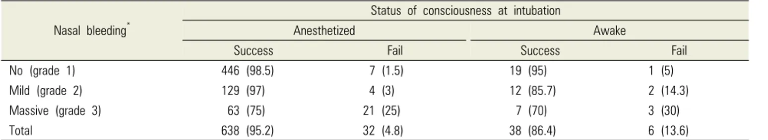
Fail 33 (4.9) 6 (13.6) 39 (5.4)
Total 672 (100) 44 (100) 716 (100)
Data are presented as n (%). *Anesthetized versus awake, P = 0.001 (chi-squared test).
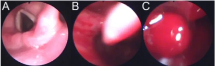
Table 2. Success rate of fiberoptic nasotracheal intubation (divided by awake intubation and postanesthesia intubation) Fig. 1. Grade of intra-oral bleeding. A. No bleeding (grade 1), B. Mild
bleeding: the degree of bleeding which does not influence on fiberoptic nasal intubation (grade 2), C. Massive bleeding: the degree of bleeding which influences on fiberoptic nasal intubation (grade 3).
degree of bleeding that did not influence the visual field);
and grade 3 massive bleeding (the degree of bleeding that influenced the visual field) (Fig. 1). If too much bleeding and discharge were occurred, suction was performed by the assistant.
2.6. Learning curve analysis method
Patient sex, age, weight, height, and the size of the tube were recorded. For each patient, the time required for FNI by the resident and the degree of bleeding observed with endoscope were also recorded.
The times recorded as required time for FNI were depicted as a learning curve, and the ratio of FNI success and failure cases were evaluated. For awake FNI, success was evaluated separately. The success rate was analyzed by dividing into several intervals based on the sum of 5 trials. The effect of nasal bleeding on the degree of difficulty was determined, and logistic regression analysis
was used for comparative analysis of the influence of awake status, degree of nasal bleeding, and increased FNI trials on the success rate of intubation.
Depending on the situation, the chi-squared test, Student’s t-test, and logistic regression analysis were performed for analysis, and a P < 0.05 was considered to be statistically significant.
RESULTS
A total of 18 second-year anesthesiology residents (11 men, 7 women) were included in the present study. The age of the residents were 27 (n = 10), 28 (n = 4), 29 (n = 1), 30 (n = 2), and 31 (n = 1) years.
All residents performed a total of 716 FNIs, in which 15 residents performed the procedure 40 times, 2 performed it 39 times, and 1 performed it 38 times.
Awake FNI was performed a total of 44 times (mean 2.4 times), in which 2 residents performed it 5 times, 3 residents performed it 4 times, 3 residents performed it 3 times, 6 residents performed it 2 times twice, 1 resident performed it once; 3 residents were not able to perform the maneuver at all (Table 1).
The success rate was 88.3% for the first attempt and 94.6% for the second attempt. The failure rate was 4.9%
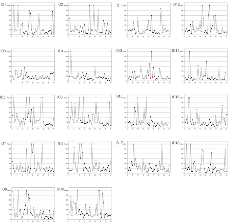
Fig. 2. The learning curve of each of the anesthesiology residents. Up to 150 s, one successful intubation is represented by a dot. Dots between 150 and 300 s represent the success of the 2nd intubation attempt. Dots at 300 s represent failed intubation. Red dots represent awake intubation.
in anesthetized patients and 13.6% in awake patients (Table 2).
Time for each successful intubation for each resident was represented as a learning curve (Fig. 2). Analysis of the learning curves revealed that improvement was observed as the number of attempts increased. The learning curves exhibited diverse patterns, in which some showed frequent failures in the beginning, interim and end, some had a high interim failure rate, and some had
a steady success rate from beginning to end.
In terms of the mean time to successful performance, the mean time was 53.8 ± 47.4 s for anesthetized intubation and 103.2 ± 79.8 s for awake intubation, demonstrating a difference in time to successful intuba- tion depending on awake status (P = 0.001) (Fig. 3).
When analyzing the cumulative success rate by dividing by 5 trial intervals, the failure rate up to the first fifth post-anesthesia intubation was 9.0%, and the failure rate
Fig. 4. Percentage of success and numbers of failure cases in the 5th cumulative interval (post-anesthesia intubation).
Fig. 5. Percentage of success and numbers of cases in the 5th cumulative interval (awake intubation).
Fig. 3. Plot of average time in cumulative trials in successful cases (divided by awake intubation and post-anesthesia intubation).
Factor B (SE) Wald P-value
Awake 1.07 (0.54) 3.82 0.05
Nasal bleeding 1.51 (0.22) 45.58 < 0.0001
Intubation trial count -0.05 (0.017) 9.33 0.002
Table. 4. Logistic analysis of factors affecting success rate of flexion endoscopy Nasal bleeding*
Status of consciousness at intubation
Anesthetized Awake
Success Fail Success Fail
No (grade 1) 446 (98.5) 7 (1.5) 19 (95) 1 (5)
Mild (grade 2) 129 (97) 4 (3) 12 (85.7) 2 (14.3)
Massive (grade 3) 63 (75) 21 (25) 7 (70) 3 (30)
Total 638 (95.2) 32 (4.8) 38 (86.4) 6 (13.6)
Data presented as n (%). *Nasal bleeding grade P < 0.001 (chi-squared analysis).
Table 3. Influence of nasal bleeding on the success rate of flexible endoscopy
decreased to 0.7 % when the total performance number was > 30 (Fig. 4). Even for awake intubation, the failure rate was 0% when performed > 30 times (Fig. 5).
When nasal bleeding was severe, the failure rate increased to 30% when FNI was performed while awake (Table 3) (P < 0.001 chi-squared test). According to logistic regression analysis, awake status, degree of nasal bleeding, and the number of attempts for nasotracheal intubation had an influence on the intubation success rate and, among them, nasal bleeding was the factor with the most influence (Table 4).
DISCUSSION
The results of this study demonstrated that the success rate of intubation and awake FNI increased as the number of attempts increased. However, when bleeding occurred as the endotracheal tube was inserted, the difficulty of FNI increased.
In patients for whom endotracheal intubation is expected to be difficult, awake intubation using fiber- scope has been presented as the standard method of airway management [2]. However, to proficiently perform intubation using fiberscope without complications, significant training and performance experience are needed [6].
For new residents, the FNI learning curve for a normal airway simulator showed that it converged to 51 seconds to reach the carnia from the nostril and that the number of trials required for proficient intubation performance varied from 27 to 58 [7]. For oral intubation, the first attempt required 4 minutes, but it fell to 1.53 minutes after 10 trials, with a success rate > 95% [8]. Another study reported that when fiberscope was first inserted to nasal cavity, followed by nasotracheal intubation, the intubation time was approximately 50 seconds in 12 residents who performed intubation 20 times [5]. In the present study, the mean performance time for anes- thetized FNI was 53.8 ± 47.4 seconds and that of awake FNI was 103.2 ± 79.8 seconds, yielding similar results.
An American Society Anesthesiologists Closed Claims Study collected data on adverse respiratory events from the 1980s and found that improper ventilation accounted for 38%, esophageal intubation for 18%, and difficult endotracheal intubation for 17% of the adverse respiratory events [9]. According to data collected after the 1990s, esophageal intubation has decreased significantly to less than 5%; however, difficult endotracheal intubation remains an important issue, at 27% [10].
Adverse respiratory events mainly occur during anes- thesia induction, [11] and two failed attempts at intubation into the airway has been associated with
hypoxia, arrhythmia, cardiac arrest, brain damage and death. Therefore, the competence of the operator is related to the incidence of adverse respiratory events [12].
The use of a fiberscope is a highly successful method in patients for whom securing the airway is difficult. Even if the laryngeal view of laryngoscope is poor, a fiberscope inserted up to the carina can be a guide for the endotracheal tube to appropriately position within the trachea, and the position can be confirmed while the fiberscope is removed. A previous study reported that among patients for whom intubation was impossible with the usual method, more than 90% showed success with fiberscope [13].
Many methods of local anesthesia for the airway are available for awake FNI, and the authors have chosen a highly stable and noninvasive lidocaine nebulizer [14,15]. In addition, an appropriate amount of midazolam and fentanyl were administered when inserting the fiberscope to reduce airway reflexes and to safely maintain natural ventilation [16].
Endotracheal intubation generally uses a laryngoscope, and the degree of difficulty is evaluated based on the laryngeal view grade established by Cormack and Lehane [17]. In addition to cases with a high Cormack and Lehane’s laryngeal view grade, a flexible endoscope can be useful in cases with difficulty in securing the airway due to poor cervical extension, short thyromental distance, and modified Mallampati test [18,19].
The correlation analysis between the degree of difficulty of flexible laryngoscope intubation and the laryngeal view grade revealed that laryngeal view grade had no effect on the degree of difficulty [20]. Results of this study demonstrate that excessive bleeding and discharge are the most significant disadvantages inter- fering with the visual field of laryngoscope, making intubation difficult [21]. Therefore, to increase residents’
success rate in FNI, reducing intra-oral bleeding and discharge are important.
Intra-oral and nasal cavity discharge can be reduced using antisialagogues such as glycopyrrolate [22]. Nasal bleeding occurs commonly during nasotracheal intubation
when endotracheal tubes with large diameters are used, when too much force is applied during tube insertion, or when multiple attempts at intubation are made [23].
Many studies have investigated methods to reduce nasal bleeding, such as using a siphon as a guide at the tip of the tube [24,25], softening of the tube by immersing it in warm water [25], and applying epinephrine [26]. In addition, the presence of systemic diseases associated with major bleeding disorders should be screened through patient history taking, and the appropriate nostril for intubation should be selected through physical examina- tion [1,21]. Insertion of a flexible laryngoscope into the nasal cavity first was one method to reduce the frequency of nasal bleeding when performing FNI [27]. In this study, however, the purpose was to train residents for FNI in cases of unexpected difficult airway during naso- tracheal intubation. Thus, methods of inserting the tube into the nasal cavity first were pursued.
In conclusion, even with some nasal bleeding, the success rate of FNI was found to increase with residents’
FNI proficiency.
AUTHOR ORCIDs
Hyuk Kim: https://orcid.org/0000-0003-3352-9536 Eunsun So: https://orcid.org/0000-0003-3335-1563 Myong-Hwan Karm: https://orcid.org/0000-0002-7494-4747 Hyun Jeong Kim: https://orcid.org/0000-0002-9265-7549 Kwang-Suk Seo: https://orcid.org/0000-0001-5906-0639
CONFLICTS OF INTEREST: The authors have no conflicts of interest to declare.
REFERENCES
1. Mahmood S, Lowe T. Management of epistaxis in the oral and maxillofacial surgery setting: An update on current practice. Oral Surg Oral Med Oral Pathol Oral Radiol Endod 2003; 95: 23-9.
2. Apfelbaum JL, Hagberg CA, Caplan RA, Blitt CD, Connis RT, Nickinovich DG, et al. Practice guidelines for
management of the difficult airway: An updated report by the american society of anesthesiologists task force on management of the difficult airway. Anesthesiology 2013;
118: 251-70.
3. Delaney KA, Hessler R. Emergency flexible fiberoptic nasotracheal intubation: A report of 60 cases. Ann Emerg Med 1988; 17: 919-26.
4. Ovassapian A, Yelich SJ, Dykes MH, Brunner EE.
Fiberoptic nasotracheal intubation--incidence and causes of failure. Anesth Analg 1983; 62: 692-5.
5. Smith JE, Jackson AP, Hurdley J, Clifton PJ. Learning curves for fibreoptic nasotracheal intubation when using the endoscopic video camera. Anaesthesia 1997; 52: 101-6.
6. Ovassapian A, Yelich SJ, Dykes MH, Golman ME.
Learning fibreoptic intubation: Use of simulators v.
Traditional teaching. Br J Anaesth 1988; 61: 217-20.
7. Dalal PG, Dalal GB, Pott L, Bezinover D, Prozesky J, Bosseau Murray W. Learning curves of novice anes- thesiology residents performing simulated fibreoptic upper airway endoscopy. Can J Anaesth 2011; 58: 802-9.
8. Johnson C, Roberts JT. Clinical competence in the performance of fiberoptic laryngoscopy and endotracheal intubation: A study of resident instruction. J Clin Anesth 1989; 1: 344-9.
9. Caplan RA, Posner KL, Ward RJ, Cheney FW. Adverse respiratory events in anesthesia: A closed claims analysis.
Anesthesiology 1990; 72: 828-33.
10. Bailie R, Posner KL. New trends in adverse respiratory events. ASA Newsletter 2011; 75: 28-9.
11. Huitink JM, Lie PP, Heideman I, Jansma P, Greif R, Schagen Nv, et al. A prospective, cohort evaluation of major and minor airway management complications during routine anaesthetic care at an academic medical centre.
Anaesthesia 2017; 72: 42-8.
12. Buis ML, Maissan IM, Hoeks SE, Klimek M, Stolker RJ.
Defining the learning curve for endotracheal intubation using direct laryngoscopy: A systematic review.
Resuscitation 2016; 99: 63-71.
13. Ovassapian A. Fiberoptic Airway Endoscopy in Anesthesia and Critical Care In: Fiberoptic tracheal intubation. New York, Raven Press. 1990, pp57-79.
14. Korttila K, Tarkkanen J, Tarkkanen L. Comparison of laryngotracheal and ultrasonic nebulizer administration of lidocaine in local anaesthesia for bronchoscopy. Acta Anaesthesiol Scand 1981; 25: 161-5.
15. Somerson SJ, Sicilia MR. AANA journal course: update for nurse anesthetists--beyond the laryngoscope: advanced techniques for difficult airway management. AANA J 1993;
61: 64-71.
16. Machata AM, Gonano C, Holzer A, Andel D, Spiss CK, Zimpfer M, et al. Awake nasotracheal fiberoptic intubation:
Patient comfort, intubating conditions, and hemodynamic stability during conscious sedation with remifentanil.
Anesth Analg 2003; 97: 904-8.
17. Cormack RS, Lehane J. Difficult tracheal intubation in obstetrics. Anaesthesia 1984; 39: 1105-11.
18. Collins SR, Blank RS. Fiberoptic intubation: An overview and update. Respir Care 2014; 59: 865-78.
19. Nasa VK, Kamath SS. Risk factors assessment of the difficult intubation using intubation difficulty scale (IDS).
J Clin Diagn Res 2014; 8: GC01-3.
20. Kim HW, Seo KS, Shin TJ, Kim HJ. The effect of laryngeal view grade and intraoral bleeding on intubation difficulty during fiberoptic nasotracheal intubation. J Korean Dent Soc Anesthesiol 2009; 9: 91-7.
21. Sanuki T, Hirokane M, Kotani J. Epistaxis during
nasotracheal intubation: A comparison of nostril sides. J Oral Maxillofac Surg 2010; 68: 618-21.
22. Lai HC, Chang YH, Huang RC, Hung NK, Lu CH, Chen JH, et al. Efficacy of sevoflurane as an adjuvant to propofol-based total intravenous anesthesia for attenuating secretions in ocular surgery. Medicine 2017; 96: e6729.
23. O’Connell JE, Stevenson DS, Stokes MA. Pathological changes associated with short-term nasal intubation.
Anaesthesia 1996; 51: 347-50.
24. Morimoto Y, Sugimura M, Hirose Y, Taki K, Niwa H.
Nasotracheal intubation under curve-tipped suction catheter guidance reduces epistaxis. Can J Anaesth 2006;
53: 295-8.
25. Elwood T, Stillions DM, Woo DW, Bradford HM, Ramamoorthy C. Nasotracheal intubation: A randomized trial of two methods. Anesthesiology 2002; 96: 51-3.
26. Kameyama K, Watanabe S, Kano T, Kusukawa J. Effects of nasal application of an epinephrine and lidocaine mixture on the hemodynamics and nasal mucosa in oral and maxillofacial surgery. J Oral Maxillofac Surg 2008; 66:
2226-32.
27. Kwon MA, Song J, Kim S, Ji SM, Bae J. Inspection of the nasopharynx prior to fiberoptic-guided nasotracheal intubation reduces the risk epistaxis. J Clin Anesth 2016;
32: 7-11.