Copyright © 2019 The Korean Society for Bone and Mineral Research
This is an Open Access article distributed under the terms of the Creative Commons Attribution Non-Commercial Li- cense (http://creativecommons.org/licenses/by-nc/4.0/) which permits unrestricted non-commercial use, distribu- tion, and reproduction in any medium, provided the original work is properly cited.
Influence of Thyroid-stimulating Hormone
Suppression Therapy on Bone Mineral Density in Patients with Differentiated Thyroid Cancer: A Meta- analysis
Byung-Ho Yoon1*, Youjin Lee2*, Hyun Jin Oh3, Sung Han Kim4, Young-Kyun Lee5
1Department of Orthopaedic Surgery, Inje University College of Medicine, Seoul Paik Hospital, Seoul;
2Department of Internal Medicine, Center for Thyroid Cancer, National Cancer Center, Goyang;
3Division of Gastroenterology, Department of Internal Medicine, Center for Cancer Prevention and Detection, National Cancer Center, Goyang;
4Department of Urology, Urological Cancer Center, National Cancer Center, Goyang;
5Department of Orthopaedic Surgery, Seoul National University Bundang Hospital, Seongnam, Korea
Background: The effects of subclinical hyperthyroidism on bone mineral density (BMD) induced by thyroid-stimulating hormone (TSH) suppression therapy in patients with dif- ferentiated thyroid cancer (DTC) remains unclear. We conducted a meta-analysis to de- termine the influence of TSH suppression therapy on BMD. Methods: We performed a systematic search to identify studies which included BMD measurement of femoral neck, total hip or lumbar spine in patients on TSH suppression therapy for DTC. Main outcome measures were difference of BMD of femoral neck, total hip or lumbar spine measured by dual energy X-ray absorptiometry between patients and controls. Results:
A systematic search yielded a total of 11 published controlled cross-sectional studies (in- cluding about 571 patients and 836 controls). TSH suppression therapy was associated with the lower BMD of total hip (weighted mean difference [WMD], -0.023; 95% confi- dence interval [CI], -0.047 to 0.000; P=0.050) and spine (WMD, -0.041; 95% CI, -0.057 to -0.026; P<0.001) in postmenopausal women with DTC, while it was not associated with that in premenopausal women and men with DTC. Conclusions: Although the included studies were limited by small numbers, results suggested possible association between chronic TSH suppression therapy and the lower BMD of spine and total hip in postmeno- pausal women (but not in premenopausal women and men) with DTC. A large, well-de- signed study with long-term follow-up would provide further insight into the influence of TSH suppression therapy and loss of BMD.
Key Words: Bone density · Meta-analysis · Osteoporosis · Thyroid neoplasms
INTRODUCTION
Thyroid cancer is one of the most common endocrine malignancy.[1] Majority of thyroid cancer is differentiated thyroid cancer (DTC) raised from thyroid follicu- lar epithelial cells, which are considered well-differentiated tumors, and have an overall excellent prognosis, with reported 10-year survival rates reaching 90%.[1- 3] The excellent prognosis of DTC is because of a combination of the favorable bi- Corresponding author
Young-Kyun Lee
Department of Orthopaedic Surgery, Seoul National University Bundang Hospital, 82 Gumi-ro, 173 beon-gil, Bundang-gu, Seongnam 13620, Korea
Tel: +82-31-787-7204 Fax: +82-31-787-4056 E-mail: [email protected] Received: February 4, 2019 Revised: February 11, 2019 Accepted: February 14, 2019
No potential conflict of interest relevant to this article was reported.
* Byung-Ho Yoon and Youjin Lee equally contributed to this work, and should be considered co-first authors.
ologic behavior of tumor cell and effective therapeutic modalities. The ‘standard’ treatment strategy for DTC in- cludes surgery (near total/total thyroidectomy) followed by radioactive iodine (131-I) ablation of the surgical rem- nant and metastatic lesion, and long-term thyrotropin (TSH) suppression therapy.[3,4] DTC expresses thyroid- stimulating hormone (TSH) receptor on the cell membrane and TSH stimulates cell growth rate.[5] Thus the long-term suppression of TSH by supra-physiologic doses of L-thyrox- ine is used to treat patients with DTC with the purpose of decreasing the risk of cancer recurrence.[6-10]
However, it has been suggested that the long-term TSH suppression may be associated with potential undesired adverse effects of thyroxine on bone metabolism [11,12]
as well as the major cardiovascular events [13-18] and atri- al fibrillation,[19,20] because this represents in effect a state of chronic subclinical hyperthyroidism. Although normal euthyroid status during childhood and adolescence is re- quired for acquisition of peak bone mass, overt hyperthy- roidism is associated with an increased risk for osteoporo- sis.[21-23] The elevated level of thyroid hormone can ex- cessively stimulate a bone turnover,[24] and shorten the bone remodeling cycle [25] which lead to consequent bone loss and decrease of bone mineral density (BMD). There- fore, patients who underwent TSH suppression therapy af- ter thyroidectomy could be vulnerable to osteoporosis and decreased BMD.[19,26-28]
However, there is no consensus about the influence of long-term TSH suppression following thyroidectomy on BMD in patients with DTC, because of different study de- sign (cross-sectional and longitudinal study), included pa- tient groups (premenopausal and postmenopausal wom- en, and men), methodology measuring BMD, area of inter- est for BMD (femoral neck and lumbar spine), and choice of outcome parameters (T-score, Z-score, and absolute val- ue of BMD).[29-32]
Therefore, the purpose of this study was to determine whether TSH suppression therapy in patients with DTC in- fluence BMD from the literature review and meta-analysis.
METHODS
This study was exempted from Institutional Review Board review because it did not involve any human subjects.
1. Search strategy
This meta-analysis was conducted according to the up- dated Preferred Reporting Items for Systematic review and Meta-Analysis Protocols (PRISMA-P) guidelines.[33] Search- es of PubMed-Medline, EMBASE, and Cochrane Library were conducted by using key terms (“thyroid cancer or thyroid- ectomy” and “osteoporosis or osteoporotic fracture”) (Sup- plementary Appendix 1). The last search was conducted on September 26, 2018. Two authors (YJL and YKL) inde- pendently screened the titles and abstracts to identify stud- ies on BMD in thyroid cancer. They also checked the refer- ence lists of all potentially eligible studies and review pa- pers to find out additional relevant publications.
2. Selection criteria
Studies were screened and selected by all investigators on the basis of a priori criteria.
The inclusion criteria were (1) published as an original article in English; (2) included TSH suppression therapy in patients with DTC; (3) controlled cross-sectional studies (pa- tients compared to a normal control group more or less carefully matched for age, sex, and menopausal status at least); (4) evaluated the BMD as primary outcome by using dual energy X-ray absorptiometry (DXA) in femoral neck, total hip or lumbar spine; and (5) available numerical data for both patients and controls (number of patients, mean and standard deviation of BMD according to the meno- pausal status).
Exclusion criteria were (1) cannot evaluate numerical data for patients with DTC; if the study included other conditions such as medullary cancer and toxic goiter; (2) not included TSH suppression therapy; (3) not available menopausal status; (4) measured BMD not by using DXA; (5) not report- ed value of BMD of femoral neck, total hip or lumbar spine;
(6) not have normal control group; and (7) reviews and col- lection of abstracts for conference meeting.
Two authors (YJL and YKL) reviewed the retrieved full manuscripts to determine whether value of BMD after TSH suppression therapy in patients with DTC in femoral neck or lumbar spine had been reported.
3. Outcome measure and data extraction The primary outcomes for the meta-analysis was the dif- ference of BMD between patients with TSH suppression ther- apy after surgery for DTC and control group.
The studies were categorized according to gender and menopausal state and subgroup analysis undertaken ac- cordingly (premenopausal women, postmenopausal wom- en, and men).
For every eligible study, the following data were extract- ed and entered in a spread sheet by the 2 reviewers: the family name of the first author, year of publication, coun- try, number of patients, mean duration of TSH suppression therapy, sample characteristics (age and gender), the mean value of BMD (g/cm2) in femoral neck or lumbar spine.
4. Quality assessment and publication bias Two of the authors (YJL and YKL) independently evaluat- ed the quality of all the studies, using Newcastle-Ottawa Scales.[34] This tool comprises three parameters: selection, comparability, and outcome. Each parameter consists of
subcategorized questions: selection (a maximum of 4 stars), comparability (a maximum of 2 stars), and exposure or out- come (a maximum of 3 stars). We assessed the publication bias with Begg’s funnel plot [35] and Egger’s test [36].
5. Statistical analysis
We calculated the weighted mean difference (WMD) rep- resenting the magnitude of the difference between the comparative groups for each outcome, because all studies used the same outcome and unit of measurement (g/cm2).
[37] WMD were computed separately for all available treat- ment and control groups for each study. We had used a fixed-effects or random-effects model depending on the results of heterogeneity to quantify the pooled effect size of the included studies (Values of P-value of less than 0.1 or an I2 value higher than 50% meant significant heteroge-
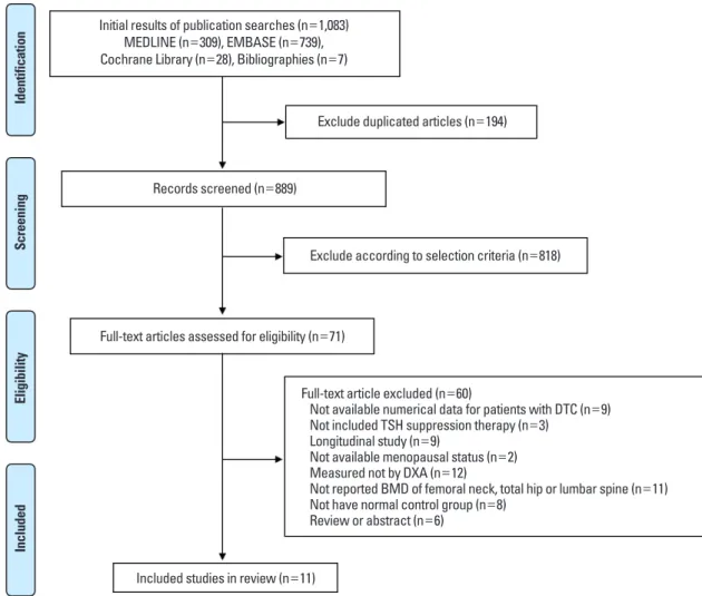
Fig. 1. Preferred Reporting Items for Systematic review and Meta-analysis flow diagram details the process of relevant study selection. DTC, dif- ferentiated thyroid cancer; TSH, thyroid-stimulating hormone; DXA, dual-energy X-ray absorptiometry; BMD, bone mineral density.
Initial results of publication searches (n=1,083) MEDLINE (n=309), EMBASE (n=739), Cochrane Library (n=28), Bibliographies (n=7)
Identification
Exclude duplicated articles (n=194)
Records screened (n=889)
Exclude according to selection criteria (n=818)
Full-text articles assessed for eligibility (n=71)
Full-text article excluded (n=60)
Not available numerical data for patients with DTC (n=9) Not included TSH suppression therapy (n=3)
Longitudinal study (n=9)
Not available menopausal status (n=2) Measured not by DXA (n=12)
Not reported BMD of femoral neck, total hip or lumbar spine (n=11) Not have normal control group (n=8)
Review or abstract (n=6) Included studies in review (n=11)
ScreeningEligibilityIncluded
neity and a random-effects model should be applied). All analyses were performed using STATA (version 14.0; Stata Corp., College Station, TX, USA).
RESULTS
A primary search from the PubMed-Medline, EMBASE, and Cochrane Library, yielded 1,083 published articles. Af- ter duplicates removed, 889 articles were primarily screened by title and abstract. As a result, 71 articles were selected
and reviewed for eligibility by full-text papers and a total of 11 cross-sectional studies fulfilling all inclusion criteria were included in the final analysis (Fig. 1).[11,38-47]
The results of the subgroup analyses according to gen- der and menopausal state were as follows.
1. Premenopausal women
The effect of TSH suppression therapy on BMD in pre- menopausal women is described in 8 studies involving a total of 183 patients and 227 controls (Table 1). Femoral Table 1. Bone mineral density of thyroid-stimulating hormone suppression therapy group and control group in premenopausal women
References Patients Controls Femoral neck Total hip Lumbar spine
Patients Controls Patients Controls Patients Controls
Franklyn et al.[11] 18 18 1.000±0.110 0.970±0.130 0.760±0.140 0.780±0.150
Giannini et al.[46] 12 10 NA NA 1.100±0.300 1.000±0.030
Goerres et al.[45] 7 7 0.892±0.141 0.861±0.094 1.006±0.143 0.903±0.128
Toivonen et al.[39] 15 22 1.032±0.124 1.017±0.125 NA NA
Reverter et al.[40] 44 44 NA NA 1.229±0.167 1.223±0.155
Eftekhari et al.[47] 22 22 NA NA 1.080±0.180 1.050±0.090
Tournis et al.[38] 40 29 0.940±0.100 0.900±0.100 0.970±0.100 0.930±0.100 1.200±0.100 1.100±0.100 Moon et al.[42] 25 75 0.930±0.100 0.900±0.090 0.980±0.100 0.960±0.080 1.210±0.110 1.180±0.120 The data is presented as number or mean±standard deviation.
NA, not applicable.
Fig. 2. Forest plot of effect of thyroid-stimulating hormone suppression therapy on femoral neck, total hip, and lumbar spine bone mineral density in premenopausal women with differentiated thyroid cancer determined by fixed effects meta-analysis. Effect sizes are indicated as Hedges’ g standardized mean differences and 95% confidence interval (CI). WMD, weighted mean difference.
neck BMD (pooled WMD, 0.029; 95% confidence interval [CI], 0.005-0.054; P=0.020), and spine BMD (pooled WMD, 0.049; 95% CI, 0.022-0.076; P<0.001) were significantly higher in patients with TSH suppression therapy than con- trol group, while total hip BMD (pooled WMD, 0.029; 95%
CI, -0.003 to 0.061; P=0.076) did not differ significantly in premenopausal women (Fig. 2).
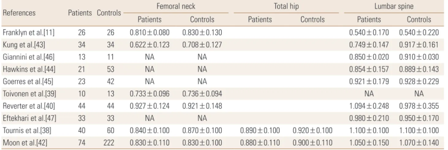
2. Postmenopausal women
The effect of TSH suppression therapy in postmenopaus- al women was investigated in 10 studies involving a total 318 patients and 538 controls (Table 2). Total hip BMD (pooled WMD, -0.023; 95% CI, -0.047 to 0.000; P=0.050), and spine BMD (pooled WMD, -0.041; 95% CI, -0.057 to -0.026; P<0.001) and were significantly lower in patients with TSH suppres-
Table 2. Bone mineral density of thyroid-stimulating hormone suppression therapy group and control group in postmenopausal women
References Patients Controls Femoral neck Total hip Lumbar spine
Patients Controls Patients Controls Patients Controls
Franklyn et al.[11] 26 26 0.810±0.080 0.830±0.130 0.540±0.170 0.540±0.220
Kung et al.[43] 34 34 0.622±0.123 0.708±0.127 0.749±0.147 0.917±0.161
Giannini et al.[46] 13 11 NA NA 0.850±0.020 0.910±0.030
Hawkins et al.[44] 21 53 NA NA 0.854±0.157 0.889±0.143
Goerres et al.[45] 23 42 NA NA 0.921±0.179 0.928±0.229
Toivonen et al.[39] 10 13 0.733±0.096 0.736±0.094 NA NA
Reverter et al.[40] 44 44 0.927±0.124 0.921±0.148 1.094±0.248 0.978±0.355
Eftekhari et al.[47] 33 33 NA NA 0.980±0.210 0.950±0.170
Tournis et al.[38] 40 60 0.840±0.100 0.870±0.100 0.890±0.100 0.920±0.100 1.100±0.100 1.100±0.100 Moon et al.[42] 74 222 0.830±0.110 0.830±0.100 0.880±0.110 0.900±0.110 1.050±0.150 1.070±0.140 The data is presented as number or mean±standard deviation.
NA, not applicable.
Fig. 3. Forest plot of effect of thyroid-stimulating hormone suppression therapy on femoral neck, total hip, and lumbar spine bone mineral density in postmenopausal women with differentiated thyroid cancer determined by fixed effects meta-analysis. Effect sizes are indicated as Hedges’ g standardized mean differences and 95% confidence interval (CI). WMD, weighted mean difference.
sion therapy than control group, while femoral neck BMD (pooled WMD, -0.016; 95% CI -0.035 to 0.002; P=0.084) did not differ significantly (Fig. 3).
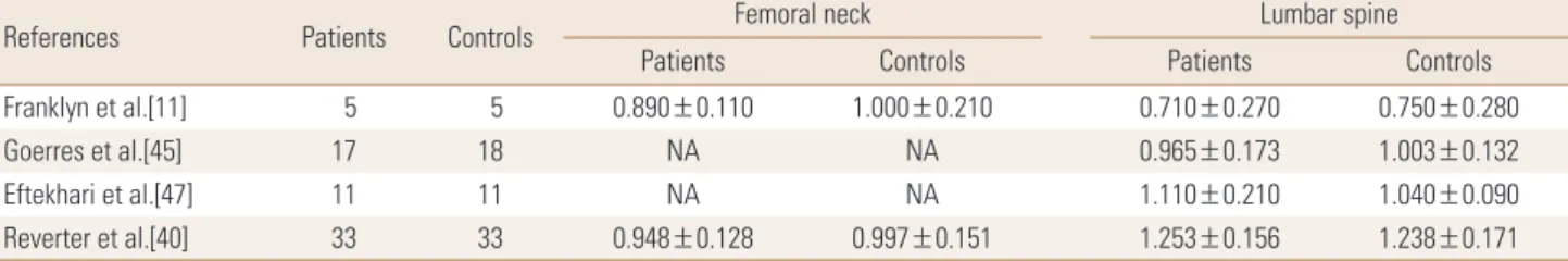
3. Men
Four studies involving a total 66 patients and 67 controls were identified for analysis addressed the effects of TSH suppression therapy on bone metabolism in men in a cross- sectional study design (Table 3). No study showed a signifi- cant difference of BMD between patients and controls. Fem- oral neck BMD (pooled WMD, -0.055; 95% CI, -0.119 to 0.009;
P=0.094), and spine BMD (pooled WMD, 0.007; 95% CI, -0.049 to 0.063; P=0.803) did not differ significantly in men (Fig. 4).
4. Quality assessment and publication bias In terms of the methodological quality, the mean value of the awarded star was 5.3 (5 stars [2 studies], 6 stars [9 studies]; Supplementary Table 1). The Begg’s funnel plot was not asymmetrical, and P-value for bias were not signif- icant in all outcomes (Fig. 5).
DISCUSSION
The clinical implications of long-term TSH suppression therapy on bone are critical, largely because of the favorable prognosis of DTC and long-term survival of patients with DTC.[10] Subclinical thyroid dysfunction has been known to be associated with increased risk of hip fracture,[48] but the influence of chronic subclinical hyperthyroidism, TSH Table 3. Bone mineral density of thyroid-stimulating hormone suppression therapy group and control group in men
References Patients Controls Femoral neck Lumbar spine
Patients Controls Patients Controls
Franklyn et al.[11] 5 5 0.890±0.110 1.000±0.210 0.710±0.270 0.750±0.280
Goerres et al.[45] 17 18 NA NA 0.965±0.173 1.003±0.132
Eftekhari et al.[47] 11 11 NA NA 1.110±0.210 1.040±0.090
Reverter et al.[40] 33 33 0.948±0.128 0.997±0.151 1.253±0.156 1.238±0.171
The data is presented as number or mean±standard deviation.
NA, not applicable.
Fig. 4. Forest plot of effect of thyroid-stimulating hormone suppression therapy on femoral neck and lumbar spine bone mineral density in men with differentiated thyroid cancer determined by fixed effects meta-analysis. Effect sizes are indicated as Hedges’ g standardized mean differ- ences and 95% confidence interval (CI). WMD, weighted mean difference.
Fig. 5. The Begg’s funnel plot and P-value by Egger’s test shows publication bias of femoral neck, total hip, and lumbar spine bone mineral density in each group. (A-C) Premenopausal women, (D-F) postmenopausal women, and (G, H) men. WMD, weighted mean difference.
0 0.01
0.02 0.03
0.04
se (WMD)
-0.05 0 0.05 0.10 WMD
Funnel plot with pseudo 95% confidence limits
P=0.917
0
0.02
0.04
0.06
0.08
se (WMD)
-0.2 -0.1 0 0.1 WMD
Funnel plot with pseudo 95% confidence limits
P=0.251
0
0.005
0.010
0.015
0.020
se (WMD)
-0.06 -0.04 -0.02 0 0.02 WMD
Funnel plot with pseudo 95% confidence limits P value non-available
0
0.01
0.02
0.03
0.04
se (WMD)
-0.1 -0.05 0 0.05 WMD
Funnel plot with pseudo 95% confidence limits
P=0.463
0 0.02 0.04 0.06 0.08 0.10
se (WMD)
-0.3 -0.2 -0.1 0 0.1 0.2 WMD
Funnel plot with pseudo 95% confidence limits
P value non-available
0
0.05
0.10
0.15
0.20
se (WMD)
-0.4 -0.2 0 0.2 0.4 WMD
Funnel plot with pseudo 95% confidence limits
P=0.976 0 0.02 0.04 0.06 0.08
se (WMD)
-0.1 0 0.1 0.2
WMD
Funnel plot with pseudo 95% confidence limits
P=0.799
0 0.005 0.010 0.015 0.020 0.025
se (WMD)
-0.02 0 0.02 0.04 0.06 0.08 WMD
Funnel plot with pseudo 95% confidence limits P value non-available
A B C
D E F
G H
suppression therapy, on decreased BMD in patients who underwent thyroidectomy for DTC remain controversial.
Our aim was to review the literature on the effects of TSH suppression therapy on BMD in patients with DTC.
Although there have been many studies on this issue, each study had a different outcome as well as different tools for measurement of BMD. Many previous studies have used single or dual photon absorptiometry, not DXA.[49-54]
The majority of studies reported no effect of TSH suppres- sion therapy on BMD in men and premenopausal women.
[40-42] Our meta-analysis also showed no influence of TSH suppression therapy on BMD in men and premenopausal women. On the other hands, the influence in postmeno- pausal women remain unclear. Our meta-analysis showed the TSH suppression therapy was associated with lower BMD of total hip and spine in postmenopausal women. We
could not determine conclusively this issue, because of too small number of the included studies and pooled patients, although we performed a meta-analysis.
However, some large population-based cohort studies showed the increased risk of osteoporosis and osteoporot- ic fracture such as hip fracture and vertebral fracture.[55,56]
Lin et al.[56] compared the risk of osteoporosis and osteo- porotic fracture among 9,398 thyroid cancer patients with levothyroxine use (n=538), those (n=8,860) without levo- thyroxine use and propensity-score-matched controls (n=
9,398). They showed that the incidence of osteoporosis and osteoporotic fracture in the thyroid cancer patients (8.69/
1,000 person-years) was higher than that in the non-thy- roid-cancer cohort (6.60/1,000 person-years) (adjusted haz- ard ratio, 1.39; 95% CI, 1.22-1.58). They also presented that long duration of levothyroxine use, and high cumulative
dose of levothyroxine were significantly associated with an increased risk of osteoporosis in thyroid cancer patients following thyroidectomy.[56]
Based on our meta-analysis of available data, we identi- fied postmenopausal women with DTC receiving TSH sup- pression therapy as a risk group for bone loss. Considering menopause as the most important risk factor of osteopo- rosis,[57] the discrepancy in results between premenopaus- al and postmenopausal women might be explained by a different susceptibility according to menopausal status.
The present study has a limitation. The cumulative sam- ple size was not very large because most of the studies in- cluded relatively few patients. Thus the results of subgroup analysis are also very limited due to small sample size. But we used the weighted effect sizes by including studies only had used DXA, the effect size is easily interpreted from a clinical point of view.
CONCLUSIONS
Overall, although studies were limited by small numbers, results suggested possible association between chronic TSH suppression therapy and the higher risk of low BMD in postmenopausal women with TSH suppression therapy.
And, it is clear that larger-scale, better-designed studies that report effects of TSH suppression therapy on BMD are needed in the future to determine the influence of TSH sup- pression therapy on risk of osteoporotic fracture in DTC.
ACKNOWLEDGMENTS
This research was supported by a grant of the Korea Health Technology R&D Project through the Korea Health Indus- try Development Institute (KHIDI), funded by the Ministry of Health & Welfare, Republic of Korea (grant no. HI18C0284).
REFERENCES
1. Lim H, Devesa SS, Sosa JA, et al. Trends in thyroid cancer incidence and mortality in the United States, 1974-2013.
JAMA 2017;317:1338-48.
2. Mirian C, Grønhøj C, Jensen DH, et al. Trends in thyroid can- cer: retrospective analysis of incidence and survival in Den- mark 1980-2014. Cancer Epidemiol 2018;55:81-7.
3. Tam S, Boonsripitayanon M, Amit M, et al. Survival in dif-
ferentiated thyroid cancer: comparing the AJCC cancer staging seventh and eighth editions. Thyroid 2018;28:1301- 10.
4. Mazzaferri EL, Kloos RT. Clinical review 128: current appro- aches to primary therapy for papillary and follicular thy- roid cancer. J Clin Endocrinol Metab 2001;86:1447-63.
5. Williams GR, Bassett JHD. Thyroid diseases and bone health.
J Endocrinol Invest 2018;41:99-109.
6. Haugen BR. 2015 American thyroid association manage- ment guidelines for adult patients with thyroid nodules and differentiated thyroid cancer: what is new and what has changed? Cancer 2017;123:372-81.
7. Mazzaferri EL, Jhiang SM. Long-term impact of initial sur- gical and medical therapy on papillary and follicular thy- roid cancer. Am J Med 1994;97:418-28.
8. Goretzki PE, Frilling A, Simon D, et al. Growth regulation of normal thyroids and thyroid tumors in man. Recent Re- sults Cancer Res 1990;118:48-63.
9. Cooper DS, Specker B, Ho M, et al. Thyrotropin suppres- sion and disease progression in patients with differentiat- ed thyroid cancer: results from the National Thyroid Can- cer Treatment Cooperative Registry. Thyroid 1998;8:737- 44.
10. McGriff NJ, Csako G, Gourgiotis L, et al. Effects of thyroid hormone suppression therapy on adverse clinical outcomes in thyroid cancer. Ann Med 2002;34:554-64.
11. Franklyn JA, Betteridge J, Daykin J, et al. Long-term thy- roxine treatment and bone mineral density. Lancet 1992;
340:9-13.
12. Quan ML, Pasieka JL, Rorstad O. Bone mineral density in well-differentiated thyroid cancer patients treated with suppressive thyroxine: a systematic overview of the litera- ture. J Surg Oncol 2002;79:62-9.
13. Biondi B, Palmieri EA, Fazio S, et al. Endogenous subclini- cal hyperthyroidism affects quality of life and cardiac mor- phology and function in young and middle-aged patients.
J Clin Endocrinol Metab 2000;85:4701-5.
14. Biondi B, Palmieri EA, Lombardi G, et al. Effects of thyroid hormone on cardiac function: the relative importance of heart rate, loading conditions, and myocardial contractili- ty in the regulation of cardiac performance in human hy- perthyroidism. J Clin Endocrinol Metab 2002;87:968-74.
15. Napoli R, Biondi B, Guardasole V, et al. Impact of hyperthy- roidism and its correction on vascular reactivity in humans.
Circulation 2001;104:3076-80.
16. Sgarbi JA, Villaça FG, Garbeline B, et al. The effects of early antithyroid therapy for endogenous subclinical hyperthy- roidism in clinical and heart abnormalities. J Clin Endocri- nol Metab 2003;88:1672-7.
17. Smit JW, Eustatia-Rutten CF, Corssmit EP, et al. Reversible diastolic dysfunction after long-term exogenous subclini- cal hyperthyroidism: a randomized, placebo-controlled study. J Clin Endocrinol Metab 2005;90:6041-7.
18. Schlumberger M, Pacini F, Wiersinga WM, et al. Follow-up and management of differentiated thyroid carcinoma: a European perspective in clinical practice. Eur J Endocrinol 2004;151:539-48.
19. Rosario PW, Carvalho M, Calsolari MR. Symptoms of thyro- toxicosis, bone metabolism and occult atrial fibrillation in older women with mild endogenous subclinical hyper- thyroidism. Clin Endocrinol (Oxf) 2016;85:132-6.
20. Karunakaran P, Maharajan C, Chockalingam R, et al. The effect of total thyroidectomy on the recovery of bone min- eral density in subjects with hyperthyroidism. Surgery 2019;
165:80-4.
21. Greenspan SL, Greenspan FS. The effect of thyroid hormone on skeletal integrity. Ann Intern Med 1999;130:750-8.
22. Tuchendler D, Bolanowski M. The influence of thyroid dys- function on bone metabolism. Thyroid Res 2014;7:12.
23. Williams GR. Actions of thyroid hormones in bone. Endo- krynol Pol 2009;60:380-8.
24. Eriksen EF, Mosekilde L, Melsen F. Trabecular bone remod- eling and bone balance in hyperthyroidism. Bone 1985;6:
421-8.
25. Mosekilde L, Melsen F, Bagger JP, et al. Bone changes in hyperthyroidism: interrelationships between bone mor- phometry, thyroid function and calcium-phosphorus me- tabolism. Acta Endocrinol (Copenh) 1977;85:515-25.
26. Abrahamsen B, Jørgensen HL, Laulund AS, et al. The ex- cess risk of major osteoporotic fractures in hypothyroid- ism is driven by cumulative hyperthyroid as opposed to hypothyroid time: an observational register-based time- resolved cohort analysis. J Bone Miner Res 2015;30:898- 905.
27. Gürlek A, Gedik O. Effect of endogenous subclinical hyper- thyroidism on bone metabolism and bone mineral densi- ty in premenopausal women. Thyroid 1999;9:539-43.
28. Kim CW, Hong S, Oh SH, et al. Change of bone mineral den- sity and biochemical markers of bone turnover in patients on suppressive levothyroxine therapy for differentiated
thyroid carcinoma. J Bone Metab 2015;22:135-41.
29. Stĕpán JJ, Límanová Z. Biochemical assessment of bone loss in patients on long-term thyroid hormone treatment.
Bone Miner 1992;17:377-88.
30. Jódar E, Martínez-Díaz-Guerra G, Azriel S, et al. Bone min- eral density in male patients with L-thyroxine suppressive therapy and Graves disease. Calcif Tissue Int 2001;69:84-7.
31. Heijckmann AC, Huijberts MS, Geusens P, et al. Hip bone mineral density, bone turnover and risk of fracture in pa- tients on long-term suppressive L-thyroxine therapy for differentiated thyroid carcinoma. Eur J Endocrinol 2005;
153:23-9.
32. Jódar E, Begoña López M, García L, et al. Bone changes in pre- and postmenopausal women with thyroid cancer on levothyroxine therapy: evolution of axial and appendicu- lar bone mass. Osteoporos Int 1998;8:311-6.
33. Shamseer L, Moher D, Clarke M, et al. Preferred reporting items for systematic review and meta-analysis protocols (PRISMA-P) 2015: elaboration and explanation. BMJ 2015;
350:g7647.
34. Stang A. Critical evaluation of the Newcastle-Ottawa scale for the assessment of the quality of nonrandomized stud- ies in meta-analyses. Eur J Epidemiol 2010;25:603-5.
35. Begg CB, Mazumdar M. Operating characteristics of a rank correlation test for publication bias. Biometrics 1994;50:
1088-101.
36. Egger M, Davey Smith G, Schneider M, et al. Bias in meta- analysis detected by a simple, graphical test. BMJ 1997;
315:629-34.
37. Becker BJ. Synthesizing standardized mean-change mea- sures. Br J Math Stat Psychol 1988;41:257-78.
38. Tournis S, Antoniou JD, Liakou CG, et al. Volumetric bone mineral density and bone geometry assessed by periph- eral quantitative computed tomography in women with differentiated thyroid cancer under TSH suppression. Clin Endocrinol (Oxf) 2015;82:197-204.
39. Toivonen J, Tähtelä R, Laitinen K, et al. Markers of bone turnover in patients with differentiated thyroid cancer with and following withdrawal of thyroxine suppressive therapy. Eur J Endocrinol 1998;138:667-73.
40. Reverter JL, Holgado S, Alonso N, et al. Lack of deleterious effect on bone mineral density of long-term thyroxine sup- pressive therapy for differentiated thyroid carcinoma. En- docr Relat Cancer 2005;12:973-81.
41. Reverter JL, Colomé E, Holgado S, et al. Bone mineral den-
sity and bone fracture in male patients receiving long-term suppressive levothyroxine treatment for differentiated thy- roid carcinoma. Endocrine 2010;37:467-72.
42. Moon JH, Jung KY, Kim KM, et al. The effect of thyroid stim- ulating hormone suppressive therapy on bone geometry in the hip area of patients with differentiated thyroid car- cinoma. Bone 2016;83:104-10.
43. Kung AW, Lorentz T, Tam SC. Thyroxine suppressive thera- py decreases bone mineral density in post-menopausal women. Clin Endocrinol (Oxf) 1993;39:535-40.
44. Hawkins F, Rigopoulou D, Papapietro K, et al. Spinal bone mass after long-term treatment with L-thyroxine in post- menopausal women with thyroid cancer and chronic lym- phocytic thyroiditis. Calcif Tissue Int 1994;54:16-9.
45. Goerres G, Theiler R, Müller-Brand J. Interfemur variation of bone mineral density in patients receiving high-dose thyroxin therapy. Calcif Tissue Int 1998;63:98-101.
46. Giannini S, Nobile M, Sartori L, et al. Bone density and min- eral metabolism in thyroidectomized patients treated with long-term L-thyroxine. Clin Sci (Lond) 1994;87:593-7.
47. Eftekhari M, Asadollahi A, Beiki D, et al. The long term ef- fect of levothyroxine on bone mineral density in patients with well differentiated thyroid carcinoma after treatment.
Hell J Nucl Med 2008;11:160-3.
48. Lee JS, Buzková P, Fink HA, et al. Subclinical thyroid dys- function and incident hip fracture in older adults. Arch In- tern Med 2010;170:1876-83.
49. Stall GM, Harris S, Sokoll LJ, et al. Accelerated bone loss in
hypothyroid patients overtreated with L-thyroxine. Ann Intern Med 1990;113:265-9.
50. Adlin EV, Maurer AH, Marks AD, et al. Bone mineral density in postmenopausal women treated with L-thyroxine. Am J Med 1991;90:360-6.
51. Greenspan SL, Greenspan FS, Resnick NM, et al. Skeletal integrity in premenopausal and postmenopausal women receiving long-term L-thyroxine therapy. Am J Med 1991;
91:5-14.
52. Paul TL, Kerrigan J, Kelly AM, et al. Long-term L-thyroxine therapy is associated with decreased hip bone density in premenopausal women. JAMA 1988;259:3137-41.
53. Recker DP, Shapiro B. The effect of thyroidectomy on bone mineral content in perimenopausal women. Thyroidology 1989;1:59-65.
54. Ross DS, Neer RM, Ridgway EC, et al. Subclinical hyperthy- roidism and reduced bone density as a possible result of prolonged suppression of the pituitary-thyroid axis with L-thyroxine. Am J Med 1987;82:1167-70.
55. Melton LJ 3rd, Ardila E, Crowson CS, et al. Fractures fol- lowing thyroidectomy in women: a population-based co- hort study. Bone 2000;27:695-700.
56. Lin SY, Lin CL, Chen HT, et al. Risk of osteoporosis in thyroid cancer patients using levothyroxine: a population-based study. Curr Med Res Opin 2018;34:805-12.
57. Diab DL, Watts NB. Postmenopausal osteoporosis. Curr Opin Endocrinol Diabetes Obes 2013;20:501-9.