Background and Purpose McDonald criteria for multiple sclerosis diagnosis have been re- vised over the years, diagnostic procedures have been simplified and earlier diagnosis facilitat- ed. The new 2017 revision introduces other important changes, with a further simplification for the diagnosis. Oligoclonal bands reassume a more relevant role in the workup.
Methods We describe 3 typical cases of patients admitted for clinically isolated syndrome and illustrate how the application of the new criteria can change the diagnostic approach with respect to the previous criteria.
Results In two of the three cases a diagnosis of multiple sclerosis is now possible.
Conclusions The new 2017 Multiple Sclerosis criteria may have an important impact in clini- cal practice with an earlier treatment to avoid the risk of disease dissemination. Their application requires a careful assessment to avoid misdiagnosis and mistreatments.
Key Words multiple sclerosis, clinically isolated syndrome, criteria, McDonald criteria, oligoclonal bands.
Clinical Application of 2017 McDonald Diagnostic Criteria for Multiple Sclerosis
INTRODUCTION
Clinically isolated syndrome (CIS) describes the first clinical neurological episode when a patient has symptoms and signs suggestive of multiple sclerosis (MS). It usually occurs in young adults involving optic nerve, brainstem or spinal cord. CIS is often the first manifes- tation of MS.1 About two thirds of patients presenting with CIS will have further clinical epi- sodes or new lesions at MRI and convert to relapsing-remitting MS.2
McDonald criteria for MS diagnosis have been revised over the years-McDonald’s 2001,3 2005,4 20105-and the new update was formulated in 2017.6 With these amendments, diag- nostic procedures have been simplified and earlier diagnosis facilitated.
The 2010 revisions of the McDonald criteria for MS5 have introduced important changes in the clinical approach, with possibility of diagnosis at the first observation. In fact, for the first time, a single MRI examination allowed the diagnosis of MS, if dissemination of le- sions in space (DIS) and time (DIT) were demonstrated without a no better explanation.
The criteria for DIS were defined by 1 or more lesions in at least 2 of 4 typical areas of the CNS (periventricular, juxtacortical, infratentorial, and spinal cord). DIT was defined by presence of an asymptomatic gadolinium enhancing lesion and a T2 lesion or a new T2 lesion at a follow-up MRI.
The new 2017 revision of McDonald criteria 6 introduces other important changes, with a further simplification for the diagnosis. Aim of the revision is to facilitate earlier diagno- sis and to preserve the specificity of the 2010 McDonald criteria reducing the frequency of Vittorio Manteroa
Lucia Abateb Roberto Balgeraa Loredana La Mantiac Andrea Salmaggia
a Neurological Unit, MS Centre, A. Manzoni Hospital-ASST Lecco, Lecco, Italy
b Neurological Unit,
ASST Valtellina Alto Lario, Sondrio, Italy
c Unit of Neurorehabilitation, Multiple Sclerosis Center, I.R.C.C.S. Santa Maria Nascente- Fondazione Don Gnocchi, Milano, Italy
pISSN 1738-6586 / eISSN 2005-5013 / J Clin Neurol 2018;14(3):387-392 / https://doi.org/10.3988/jcn.2018.14.3.387
Received January 22, 2018 Revised March 25, 2018 Accepted March 27, 2018 Correspondence Vittorio Mantero, MD Neurological Unit, MS Centre, A. Manzoni Hospital-ASST Lecco, Via dell’Eremo 9/11,
23900 Lecco, Italy Tel +390341489332 Fax +390341489801
E-mail vittorio.mantero@hotmail.com
cc This is an Open Access article distributed under the terms of the Creative Commons Attribution Non-Com- mercial License (http://creativecommons.org/licenses/by-nc/4.0) which permits unrestricted non-commercial use, distribution, and reproduction in any medium, provided the original work is properly cited.
JCN
Open Access ORIGINAL ARTICLEClinical Application of 2017 McDonald Criteria
JCN
misdiagnosis. The first important change is the introduction of the presence of oligoclonal bands (OCBs) in cerebrospinal fluid (CSF) to make the diagnosis of MS in a patient with ev- idence of DIS, allowing for substitution of DIT. The second major change is that either symptomatic or asymptomatic gadolinium enhancing lesions can be considered in deter- mining DIS and DIT. Moreover, cortical lesions can be used in addiction to juxtacortical ones to support DIS.
METHODS
The aim of the paper is to show, through the discussion of typical CIS patients, how the application of the new criteria can change the diagnostic approach with respect to the previ- ous criteria. The patients gave their consent for research.
RESUlT
Case 1
A 23-year-old woman came to our observation complaining of left side hypoesthesias from the mammillar line and par- esthesias in the hands, which lasted about one week. She had no medical history and assumed estroprogestinic therapy.
At entry, neurological examination revealed left hypoes- thesia with D5–D6 level, reduction of superficial abdominal reflexes to the left side, brisk deep tendon reflexes, no weak-
ness, no Lhermitte sign and no sphincter dysfunction. Dis- ability was 2, 5 Expanded Disability Status Scale (EDSS) points according to Kurtzke’s scale.7
Multimodal evoked potentials were normal. We excluded systemic pathologies and other CNS diseases. Anti-aquapo- rin-4 (anti-AQP4) and anti-myelin oligodendrocyte glyco- protein (anti-MOG) antibodies resulted negative. CSF exami- nation revealed normal cell count and the presence of OCBs.
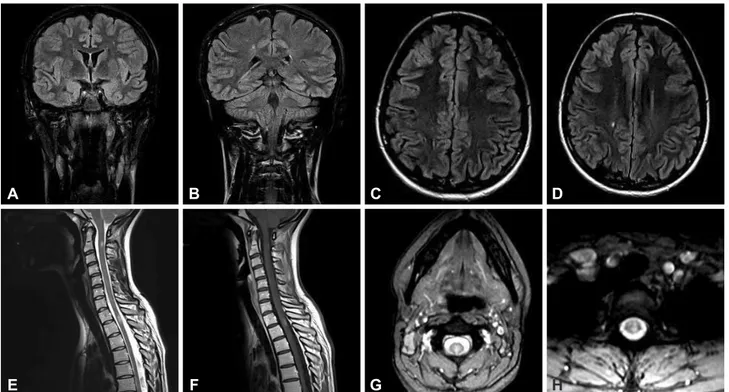
Brain MRI showed T2-weighted lesions involving right centrum semiovale and left frontal lobe (Fig. 1A-D). Spinal MRI showed two enhancing lesions at C2 and D1–D2 level (Fig. 1E-H).
Diagnosis of MS can be made with the new criteria, but not with the 2010 criteria. The criteria for DIS and DIT are now respected, including the enhancing lesions of the spinal cord, although both are symptomatic (C2 for hands pares- thesias and D1–D2 for left side hypoesthesia). In fact either symptomatic or asymptomatic gadolinium enhancing lesions can be considered in determining DIS and DIT.
Case 2
A 27-year-old man came to our observation complaining from one week of blurred vision in the left eye with pain dur- ing ocular movements. No significant medical event had oc- curred previously.
At entry, neurological examination showed impaired vi-
Fig. 1. Brain coronal plane FLAIR-weighted and axial plane T2-weighted scan showing hyperintense lesions in the left frontal lobe (A and C) and in right centrum semiovale (B and D), spinal cord sagittal and axial plane T2-weighted showing hyperintense lesions at C2 (E and G), and D1-D2 level (E and H), with enhancement on T1 post-Gadolinium scan (F).
A
E
B
F
C
G
D
H
Mantero V et al.
JCN
sion in the left eye with normal pupillary light reflex, brisk deep tendon reflexes. At ophthalmologic investigation, visual acuity was 1/10 in left eye with normal fundus oculi. Disabil- ity was 4, 5 EDSS points.
Visual evoked potentials (VEP) showed increased latency (134 ms) and decreased amplitude of the left P100 wave; mo- tor evoked potentials proved increased latency and decreased amplitude with wave dispersion and increased central con- duction time at right lower limb; sensory and auditory evoked potentials were normal. Other CNS diseases were excluded, in particular neuromyelitis optica spectrum disorders (NMOSD) with negativity of anti-AQP4 and anti-MOG antibodies. OCBs were detected in the CSF that presented normal cell count.
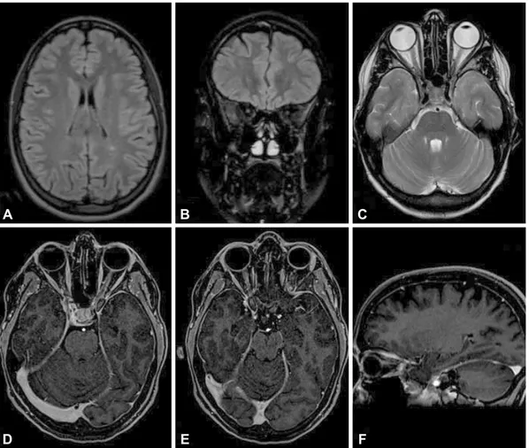
Brain MRI showed multiple focal T2-weighted periven- tricular, cortical-juxtacortical, and infratentorial lesions and in left optic nerve (Fig. 2A-C and E). Spinal cord MRI showed multiple T2-weighted hyperintensity (Fig. 2D). None of these lesions presented enhancement (Fig. 2F-H).
Also in this case, diagnosis of MS can be made with the new criteria, but not with the 2010 criteria. Other diseases were excluded and the criteria for DIS but not for DIT are respected. The diagnosis of MS is possible because the pres- ence of OCBs allows for substitution of DIT.
Case 3
A 24-year-old-woman came to our observation about ten days after acute onset of blurred vision in the right eye associated with slight pain with ocular movements. Her past medical history was silent.
At entry, neurological examination was normal, except for impaired vision in the right eye with normal pupillary light reflex. Ophthalmologic investigation revealed visual acuity 2/10 in the right eye and optic disc with shallow nasal mar- gin. Disability was 3 EDSS points.
VEP showed increased latency (140 ms) and decreased amplitude of the right P100 wave. Optical coherence tomog- raphy (OCT) proved a slight thinning of the right retinal nerve. We excluded other CNS, autoimmune diseases and acute ischemic optic neuropathy. Anti-AQP4 and anti-MOG antibodies resulted negative. CSF examination revealed nor- mal cell count and the presence of OCBs.
Brain MRI showed a T2-weighted lesion in the left pe- ritrigonal white matter and slight hyperintensity in the right optic nerve (Fig. 3A and B). No enhancing lesions were de- tected. Spinal cord MRI showed normal findings.
Following high dose methylprednisolone treatment (1g dai- ly iv for 5 days), visual acuity returned to normal limits over two weeks, with a residual relative afferent pupillar defect.
Fig. 2. Brain coronal plane FLAIR-weighted scan showing hyperintense juxtacortical (A), periventricular and infratentorial lesions (B and C) with- out enhancement on T1 post-Gadolinium scan (F and G), axial plane T2-weighted scan showing slight signal hyperintensity in left optic nerve (E), spinal cord sagittal plane T2-weighted showing hyperintense lesions at multiple levels (D) without enhancement on T1 post-Gadolinium scan (H).
A
E
B
F
C
G
D
H
Clinical Application of 2017 McDonald Criteria
JCN
In this case MRI data were inconsistent with DIS. In fact optic nerve is not a typical area for MS. Despite the pres- ence of OCBs, diagnosis of MS cannot be allowed.
DISCUSSION
We describe 3 typical cases of patients admitted to our hos- pital for CIS. We underline the differences between 2010 and 2017 criteria for MS diagnosis and discuss the advan- tages of this further revision in the clinical practice. The first implication is the anticipation of MS diagnosis. As a matter of fact, in the first two cases herein presented, the diagnosis of MS was achieved at clinical onset according to the new criteria, while with the previous criteria the diagnosis would have remained as CIS. In all three patients NMOSD diagno- sis was carefully excluded.
The first patient presented an acute transverse myelitis.
Brain MRI showed three T2 lesions, while spinal MRI showed two symptomatic T1-weighted gadolinium-enhancing le- sions. According to the new criteria, diagnosis of MS could
be performed, with demonstration of DIS and DIT. Based on the previous criteria, symptomatic enhancing lesion(s) did not satisfy DIT criteria. The MRI in MS (MAGNIMS) group has recently suggested no distinction should be made be- tween symptomatic and asymptomatic MRI lesions.8 In fact, some studies reported that the presence of a symptomatic le- sion identified patients with MS with a high sensitivity and accuracy and without compromising specificity.9-11
The second report describes a CIS patient in whom MRI showed numerous T2 lesions suggestive of DIS. The diag- nosis of MS would not have been possible with the 2010 cri- teria, in the absence of at least one gadolinium-enhancing lesion. With the new criteria, even if in absence of DIT, MS diagnosis was possible for the presence of OCBs in CSF.
OCBs reassume now a more relevant role in the MS work- up. In 2010 criteria “the Panel reaffirmed that positive CSF findings (elevated immunoglobulin G index or 2 or more OCBs) can be important to support the inflammatory de- myelinating nature of the underlying condition, to evaluate alternative diagnoses, and to predict clinically definite MS.”
Fig. 3. Brain axial plane FLAIR-weighted scan showing hyperintensity of the left peritrigonal white matter (A) and coronal and axial plane FLAIR- weighted scan showing slight hyperintensity in the right optic nerve (B and C) without clear enhancement on T1 post-Gadolinium axial and sagit- tal scan (D, E, and F).
A
D
B
E
C
F
Mantero V et al.
JCN
In the 2001 and 2005 a positive CSF finding could be used for reaching diagnosis in case of incomplete MRI findings.
Now OBCs can be viewed as substitution for the DIT re- quirement. Its prognostic value remains undiscussed12: CIS patients with positive OCBs are more than twice as likely to having a second attack as OCB-negative ones, independent- ly from MRI findings.13-15 Patients both with OCBs and sev- eral T2 lesions present a risk at almost 90% of developing MS within five years.13 The presence of OCBs is the best biologi- cal marker to predict conversion to MS and it is associated with a shorter time conversion.14
The third case is a typical optic neuritis (ON) with hyper- intense lesion on the right optic nerve. More than one third of CIS are ON. It is important to note that optic nerve is not in- cluded as a typical area and should not be considered either for DIS or for DIT. In this case, diagnosis of MS cannot be performed with the new criteria. This patient should there- fore be considered as CIS.
The role of optic nerve lesions is debated. In fact, in the MAGNIMS consensus guidelines, the expert panel proposed that lesions in the optic nerve should be added to the crite- ria for DIS as an additional CNS area.8 It is known that pa- tients presenting with ON as first attack have a lower risk for conversion to MS than patients with other presentations, even if the crucial issue at MS presentation is not CIS topography but MRI at baseline. In fact if a patient with ON has abnor- mal baseline MRI, his prognosis does not differ from that of other different CIS.16 The 2017 revision of McDonald criteria states that studies to validate MRI, VEP, or OCT in fulfilling DIS or DIT in support of a MS diagnosis are identified as high priority. OCT detects thinning of the retinal nerve fiber layer in the eyes of patients with MS and is important to iden- tify the presence of a unilateral ON in MS.17 In addition, there are other possible future OCT markers of MS disease activity:
macular ganglion cell layer+inner plexiform layer for brain atrophy and inner nuclear layer thickness for inflammatory activity.18
An anticipated diagnosis of MS may avoid further clinical and MRI monitoring to support the clinical conclusion, and allow a preventive discussion and counseling for the patients.
However, more simplified criteria do not imply an easier di- agnosis for clinicians. A correct interpretation of symptoms and signs, atypical symptoms and red flags, to obtain no better explanation,19,20 remains mandatory. To avoid misdi- agnosis, during evaluation of patients with suspected MS, a careful assessment is necessary, in particular for NMOSD, in fact in some cases uncertainty can occur, particularly with AQP4-seronegative patients.6
Finally, the access to disease modifying therapies to pre- vent further clinical or radiological dissemination could be
anticipated and extended to more patients.
In conclusion, the new 2017 MS criteria may have an im- portant impact in clinical practice: leading to an earlier diag- nosis, to an increase in MS cases being diagnosed, anticipat- ing patients’ counselling and treatment to avoid the risk of disease dissemination. However, their application requires a more careful assessment of the patients (including CSF anal- ysis) to avoid misdiagnosis and mistreatments, taking into account that many of them are characterized by non-negli- gible and partly still unknown long-term systemic effects.
Conflicts of Interest
The authors have no financial conflicts of interest.
REFERENCES
1. Miller DH, Chard DT, Ciccarelli O. Clinically isolated syndromes.
Lancet Neurol 2012;11:157-169.
2. Brownlee WJ, Miller DH. Clinically isolated syndromes and the rela- tionship to multiple sclerosis. J Clin Neurosci 2014;21:2065-2071.
3. McDonald WI, Compston A, Edan G, Goodkin D, Hartung HP, Lub- lin FD, et al. Recommended diagnostic criteria for multiple sclerosis:
guidelines from the International Panel on the diagnosis of multiple sclerosis. Ann Neurol 2001;50:121-127.
4. Polman CH, Reingold SC, Edan G, Filippi M, Hartung HP, Kappos L, et al. Diagnostic criteria for multiple sclerosis: 2005 revisions to the
“McDonald criteria”. Ann Neurol 2005;58:840-846.
5. Polman CH, Reingold SC, Banwell B, Clanet M, Cohen JA, Filippi M, et al. Diagnostic criteria for multiple sclerosis: 2010 revisions to the
‘‘McDonald criteria”. Ann Neurol 2011;69:292-302.
6. Thompson AJ, Banwell BL, Barkhof F, Carroll WM, Coetzee T, Comi G, et al. Diagnosis of multiple sclerosis: 2017 revisions of the McDon- ald criteria. Lancet Neurol 2018;17:162-173.
7. Kurtzke JF. Rating neurologic impairment in multiple sclerosis: an ex- panded disability status scale (EDSS). Neurology 1983;33:1444-1452.
8. Filippi M, Rocca MA, Ciccarelli O, De Stefano N, Evangelou N, Kap- pos L, et al. MRI criteria for the diagnosis of multiple sclerosis: MAG- NIMS consensus guidelines. Lancet Neurol 2016;15:292-303.
9. Caucheteux N, Maarouf A, Genevray M, Leray E, Deschamps R, Chaunu MP, et al. Criteria improving multiple sclerosis diagnosis at the first MRI. J Neurol 2015;262:979-987.
10. Brownlee WJ, Swanton JK, Miszkiel KA, Miller DH, Ciccarelli O.
Should we include lesions in the symptomatic site in dissemination in space in patients with clinically isolated syndromes? Mult Scler 2015;
23:65.
11. Tintore M, Otero-Romero S, Río J, Arrambide G, Pujal B, Tur C, et al.
Contribution of the symptomatic lesion in establishing MS diagnosis and prognosis. Neurology 2016;87:1368-1374.
12. Tintore M, Rovira À, Río J, Otero-Romero S, Arrambide G, Tur C, et al. Defining high, medium and low impact prognostic factors for de- veloping multiple sclerosis. Brain 2015;138:1863-1874.
13. Kuhle J, Disanto G, Dobson R, Adiutori R, Bianchi L, Topping J, et al.
Conversion from clinically isolated syndrome to multiple sclerosis: a large multicentre study. Mult Scler 2015;8:1013-1024.
14. Tintoré M, Rovira A, Río J, Tur C, Pelayo R, Nos C, et al. Do oligoclo- nal bands add information to MRI in first attacks of multiple sclero- sis? Neurology 2008;70:1079-1083.
15. Martinelli V, Dalla Costa G, Messina MJ, Di Maggio G, Sangalli F, Moiola L, et al. Multiple biomarkers improve prediction of multiple sclerosis in clinically isolated syndromes. Acta Neurol Scand 2017;136:
454-461.
16. Tintoré M, Rovira A, Rio J, Nos C, Grivé E, Téllez N, et al. Is optic neu-
Clinical Application of 2017 McDonald Criteria
JCN
ritis more benign than other first attacks in multiple sclerosis? Ann Neurol 2005;57:210-215.
17. Nolan RC, Galetta SL, Frohman TC, Frohman EM, Calabresi PA, Cas- trillo-Viguera C, et al. Optimal intereye difference thresholds in retinal nerve fiber layer thickness for predicting a unilateral optic nerve lesion in multiple sclerosis. J Neuroophthalmol 2018 Jan 29 [Epub] available from: https://doi.org/10.1097/WNO.0000000000000629.
18. Bischof A, Caverzasi E, Cordano C, Hauser SL, Henry RG. Advances
in imaging multiple sclerosis. Semin Neurol 2017;37:538-545.
19. Kelly SB, Chaila E, Kinsella K, Duggan M, Walsh C, Tubridy N, et al.
Using atypical symptoms and red flags to identify non-demyelinating disease. J Neurol Neurosurg Psychiatry 2012;83:44-48.
20. Charil A, Yousry TA, Rovaris M, Barkhof F, De Stefano N, Fazekas F, et al. MRI and the diagnosis of multiple sclerosis: expanding the concept of “no better explanation”. Lancet Neurol 2006;5:841-852.