http://dx.doi.org/10.4174/astr.2016.90.3.131 Annals of Surgical Treatment and Research
Incidental gallbladder cancer after routine cholecystectomy: when should we suspect it
preoperatively and what are predictors of patient survival?
Yongchel Ahn, Cheon-Soo Park1, Shin Hwang2, Hyuk-Jai Jang1, Kun-Moo Choi1, Sung-Gyu Lee2
Departments of Hematology-Oncology and 1Surgery, GangNeung Asan Hospital, University of Ulsan College of Medicine, Gangneung, 2Division of Hepatobiliary Surgery and Liver Transplantation, Department of Surgery, Asan Medical Center, University of Ulsan College of Medicine, Seoul, Korea
INTRODUCTION
Gallbladder (GB) cancer is the fifth most common malignancy of the gastrointestinal tract, with an incidence of 0.8%–1.2%, and is the most common malignancy of the biliary tract [1].
Occasionally, GB malignancy is found on pathologic reports after routine cholecystectomy. Recent studies have reported
an increase in incidental GB cancer (iGBC), with approximately 50%–58% of all new GBC cases [2,3]. Some authors have reported incidence of iGBC after laparoscopic cholecystectomy (LC) was approximately 0.2%–2.1% [4-6].
When iGBC is detected after cholecystectomy, additional procedures such as liver resection, lymph node dissection, and/or bile duct resection (BDR) may be performed depending Purpose: In about 1% of cases, incidental gallbladder cancers (iGBC) are found after routine cholecystectomy. The aim of this study is to compare clinical features of iGBC with benign GB disease and to evaluate factors affecting recurrence and survival.
Methods: Between January 1998 and March 2014, 4,629 patients received cholecystectomy and 73 iGBC patients (1.6%) were identified. We compared clinical features of 4,556 benign GB disease patients with 73 iGBC patients, and evaluated operative outcomes and prognostic factors in 56 eligible patients.
Results: The iGBC patients were older and concomitant diseases such as hypertension and anemia were more common than benign ones. And an age of more than 65 years was the only risk factor of iGBC. Adverse prognostic factors affecting patients’ survival were age over 65, advanced histology, lymph node metastasis, and lymphovascular invasion on multivariate analysis. Age over 65 years, lymph node involvement, and lymphovascular invasion were identified as unfavorable factors affecting survival in subgroup analysis of extended cholecystectomy with bile duct resection (EC with BDR, n = 22).
Conclusion: Prior to routine cholecystectomy, incidental GB cancer should be suspected especially in elderly patients.
And advanced age, lymph node metastasis, and lymphovascular invasion are important prognostic factors in EC with BDR cohorts.
[Ann Surg Treat Res 2016;90(3):131-138]
Key Words: Gallbladder neoplasms, Cholecystectomy, Prognosis
Reviewed January February March April May June July August September October November December
Received August 27, 2015, Revised October 5, 2015, Accepted November 4, 2015
Corresponding Author: Cheon-Soo Park
Department of Surgery, GangNeung Asan Hospital, University of Ulsan College of Medicine, 38 Bangdong-gil, Gangneung 25440, Korea
Tel: +82-33-610-3226, Fax: +82-33-610-4960 E-mail: [email protected]
Copyright ⓒ 2016, the Korean Surgical Society
cc Annals of Surgical Treatment and Research is an Open Access Journal. All articles are distributed under the terms of the Creative Commons Attribution Non- Commercial License (http://creativecommons.org/licenses/by-nc/4.0/) which permits unrestricted non-commercial use, distribution, and reproduction in any medium, provided the original work is properly cited.
on pathologic and imaging findings. Glenn and Hays [7] first introduced ‘radical cholecystectomy’ (Glenn operation), a radical resection technique with regional lymphadenectomy.
Pack et al. [8] and Fahim et al. [9] have also reported combined hepatectomy and portal lymph node dissection in pT1b or more advanced GBC.
In general, it is difficult to anticipate GB malignancy in most routine cholecystectomies for preoperative diagnosis of benign GB diseases. Additionally, surgeons often encounter the concerns of tumor spread in perioperative GB perforation and of reoperation extent. To date, there have been few reports on the predictive factors of iGBC. A recently published article described old age, female gender, Asian or African American, elevated serum ALP and conversion to open cholecystectomy as risk factors in LC patients [10].
In this retrospective study, we reviewed the clinicopatho- logical characteristics of iGBC compared with benign GB diseases and sought to find predictive risk factors. We also analyzed prognostic factors affecting recurrence and survival in iGBC patients.
METHODS
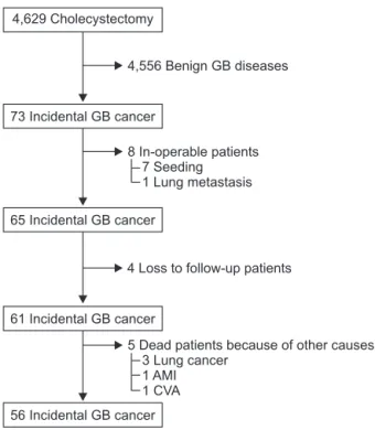
In our institution, 4,629 patients received cholecystectomy between January 1998 and March 2014. Seventy-three patients (1.6%) were confirmed to have iGBC on final pathology.
Demographics and clinical characteristics of these patients were retrospectively retrieved and were compared to those of 4,556 benign patients. For continuous variables, Student t-test was used for comparisons. Categorical variables were analyzed with the chi-square test or Fisher exact test. And multiple logistic regression models for a dichotomous outcome were developed, and odds ratios were utilized to evaluate preoperative iGBC risk factors.
Seventeen patients were excluded from further survival analysis: 8 due to confirmed metastasis or seeding during operation; 4 were lost to follow up; and 5 as a result of death by other diseases (3 cases of primary lung cancer, 1 acute myocardial infarction, 1 brain infarction) (Fig. 1). Disease-free and overall survival (OS) was calculated by the Kaplan-Meier method. Prognostic factors were analyzed by the univariate Kaplan-Meier method and compared by the log-rank test to identify the predictors for survival. Multivariate regression analysis was performed using the Cox proportional hazards model to identify the independent prognostic factors for survival. A P-value less than 0.05 was considered statistically significant. All Statistical calculations were performed with IBM SPSS Statistics ver. 19.0 (IBM Co., Armonk, NY, USA).
RESULTS
Clinical features and predictive risk factors
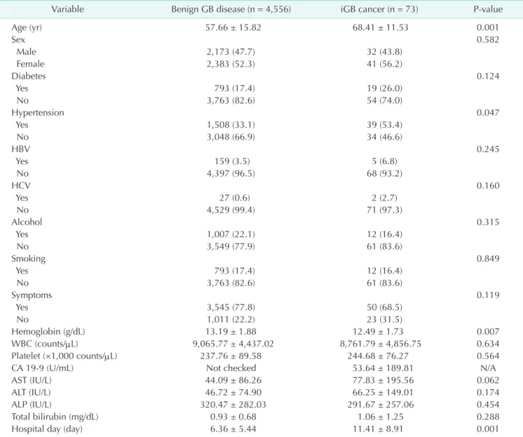
A total of 73 patients were identified as iGBC cases and the other 4,556 were benign GB diseases. The mean age of iGBC patients was older than that of benign ones (68.4 ± 11.5 years vs. 57.7 ± 15.8 years, P = 0.001). The hemoglobin levels differed significantly between the two groups (iGBC vs. benign, 12.5
± 1.7 g/dL vs. 13.2 ± 1.9 g/dL; P = 0.007). But levels of total bilirubin, AST, ALT, ALP, WBC, and platelet counts were not sig- nificantly different. In addition, more iGBC patients were on antihypertensive medications (P = 0.047) (Table 1).
Among the 73 cancer patients, 32 (43.8%) were men and 50 (68.5%) had preceding gastrointestinal symptoms. Preoperative diagnoses were diverse: 22 symptomatic GB stones (30.1%), 23 acute cholecystitis (31.5%), 17 GB polyps (23.3%), 6 GB empyema (8.2%), 3 GB polypoid mass (4.1%), 1 combined GB stone and polyp (1.4%), and 1 choledochal cyst (1.4%). Multiple logistic regression analysis identified that only an age more than 65 years was significantly associated with iGBC (odds ratio, 2.542;
95% confidence interval [CI], 1.352–4.777; P = 0.004).
Recurrence and survival outcomes of iGBC
Fifty-six eligible patients underwent different operations for iGBC according to pathologic and imaging findings (Table 2). The most common curative reoperation for advanced GBC was EC with BDR. We defined EC as cholecystectomy in-
4,556 Benign GB diseases
4 Loss to follow-up patients
5 Dead patients because of other causes 3 Lung cancer
7 Seeding 1 Lung metastasis 8 In-operable patients
1 AMI 1 CVA 4,629 Cholecystectomy
73 Incidental GB cancer
65 Incidental GB cancer
61 Incidental GB cancer
56 Incidental GB cancer
Fig. 1. Selection process of patients with incidentally con
firmed gallbladder cancer (n = 56). GB, gallbladder; AMI, acute myocardial infarction; CVA, cerebral vascular accident.
cluding wedge resection of the GB fossa with a rim of normal hepatic tissue (approximately 2 cm in thickness or more), or segmentectomy of liver segments (IVb and V) with regional lymph node dissection. EC after primary cholecystectomy was usually performed for (1) pT2 or pT3, (2) suspicious lymph node
enlargement on postoperative CT scan, and (3) positive cystic duct resection margin. And BDR was considered in situations of (2) and (3) in our institution. In fact, additional surgical proce- dures were not strictly protocol-driven and eighteen patients received only cholecystectomy because of patient refusal, comorbid medical conditions and so on.
Univariate analysis revealed that older age (≥65 years), higher serum level of CA 19-9 (≥50 U/mL), acute cholecystitis, and GB empyema on preoperative diagnosis were adverse clinical factors of recurrence and survival. Also, in perioperative GB perforation subset, early recurrence was significantly greater and their 6-month, 1-year, and 3-year DFS rates compared with nonperforation group were 58% vs. 79%; 44% vs. 65%;
and 36% vs. 59%, respectively (P = 0.04). However, OS was not significantly different between perforation and nonperforation Table 1. Clinical features of incidental gallbladder (GB) cancer and benign gallbladder diseases
Variable Benign GB disease (n = 4,556) iGB cancer (n = 73) Pvalue
Age (yr) 57.66 ± 15.82 68.41 ± 11.53 0.001
Sex 0.582
Male 2,173 (47.7) 32 (43.8)
Female 2,383 (52.3) 41 (56.2)
Diabetes 0.124
Yes 793 (17.4) 19 (26.0)
No 3,763 (82.6) 54 (74.0)
Hypertension 0.047
Yes 1,508 (33.1) 39 (53.4)
No 3,048 (66.9) 34 (46.6)
HBV 0.245
Yes 159 (3.5) 5 (6.8)
No 4,397 (96.5) 68 (93.2)
HCV 0.160
Yes 27 (0.6) 2 (2.7)
No 4,529 (99.4) 71 (97.3)
Alcohol 0.315
Yes 1,007 (22.1) 12 (16.4)
No 3,549 (77.9) 61 (83.6)
Smoking 0.849
Yes 793 (17.4) 12 (16.4)
No 3,763 (82.6) 61 (83.6)
Symptoms 0.119
Yes 3,545 (77.8) 50 (68.5)
No 1,011 (22.2) 23 (31.5)
Hemoglobin (g/dL) 13.19 ± 1.88 12.49 ± 1.73 0.007
WBC (counts/µL) 9,065.77 ± 4,437.02 8,761.79 ± 4,856.75 0.634
Platelet (×1,000 counts/µL) 237.76 ± 89.58 244.68 ± 76.27 0.564
CA 199 (U/mL) Not checked 53.64 ± 189.81 N/A
AST (IU/L) 44.09 ± 86.26 77.83 ± 195.56 0.062
ALT (IU/L) 46.72 ± 74.90 66.25 ± 149.01 0.174
ALP (IU/L) 320.47 ± 282.03 291.67 ± 257.06 0.454
Total bilirubin (mg/dL) 0.93 ± 0.68 1.06 ± 1.25 0.288
Hospital day (day) 6.36 ± 5.44 11.41 ± 8.91 0.001
Values are presented as mean ± standard deviation or number (%).
Table 2. Operations according to the pathologic Tstage
Variable pT1a pT1b pT2 pT3 Pvalue
Cholecystectomy 6 (75.0) 2 (50.0) 17 (45.9) 1 (14.3) 0.319 EC+ BDR 2 (25.0) 1 (25.0) 14 (37.8) 5 (71.4) Other 0 (0) 1 (25.0) 6 (16.2) 1 (14.3)
Total 8 4 37 7
Values are presented as number (%).
EC, extended cholecystectomy; BDR, bile duct resection.
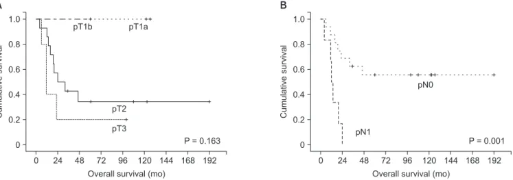
subgroups (Table 3). In a subgroup analysis of EC with BDR (n = 22), GB perforation was not statistically significant in DFS (P = 0.127) and OS (P = 0.113). And advanced pT stage, advanced pN stage, positive resection margin, moderate or poor differentiation, positive perineural invasion, and positive lymphovascular invasion were also risk factors of recurrence and survival on univariate analysis. However, tumor size and types of operation did not affect patients’ outcome (Table 4, Fig.
2). In subgroup analysis for those who received EC with BDR (n
= 22), cumulative OS curves based on pathologic T staging and
pathologic N staging are respectively shown (Fig. 3A, B).
On a multivariate analysis of all iGBC patients, age more than 65 years, positive lymph node, moderately or poorly dif- ferentiated tumor, and presence of lymphovascular invasion were statistically significant predictors of poor prognosis.
But in EC with BDR subgroup analysis, elderly age (hazard ratio [HR], 107.7; 95% CI, 2.812–4,132.1; P = 0.012), lymph node involvement (HR, 10.88; 95% CI, 1.004–117.9; P = 0.049), lymphovascular invasion (HR, 33.62; 95% CI, 2.444–462.401; P = 0.009) were identified as adverse prognostic factors. Depth of Table 3. Clinical prognostic factors of recurrence and survival in patients with incidental gallbladder cancer (n = 56)
Variable DFS (%) OS (%)
6 mo 1 yr 3 yr Pvalue 6 mo 1 yr 3 yr Pvalue
Age (yr) 0.030 0.004
<65 90 74 68 100 100 74
≥65 63 50 39 72 60 37
Sex 0.729 0.858
Male 82 62 56 78 78 52
Female 67 57 50 85 72 50
Diabetes 0.787 0.967
Yes 68 61 54 81 74 52
No 75 58 52 82 74 50
Hypertension 0.822 0.836
Yes 67 63 57 76 63 53
No 77 56 48 87 83 49
Hepatitis B virus 0.411 0.273
Yes 80 27 27 80 80 27
No 72 61 54 82 74 53
Hepatitis C virus 0.961 0.827
Yes 50 50 50 100 50 50
No 74 59 52 81 75 51
Alcohol 0.319 0.649
Yes 75 45 45 75 75 45
No 73 61 53 83 74 52
Smoking 0.872 0.653
Yes 69 46 46 69 69 46
No 73 60 53 84 75 52
Symptoms 0.508 0.228
Yes 67 55 51 75 64 48
No 84 67 56 95 95 57
CA 199 (U/mL) 0.003 0.032
<50 80 67 59 85 78 58
≥50 43 26 26 65 51 17
Preoperative Dx 0.011 0.027
Stone 76 63 55 82 82 50
Polyp 93 93 85 93 93 93
Stone & polyp 100 100 100 100 100 100
Cholecystitis 61 38 30 81 54 27
Other 59 39 31 77 56 25
GB perforation 0.041 0.074
Yes 58 44 36 65 52 36
No 79 65 59 90 84 58
DFS, disease free survival; OS, overall survival; SR, survival rate; GB, gallbladder; Dx, diagnosis.
tumor (T-stage), however, was not a significant prognostic factor in both all-iGBC patients group and EC with BDR subgroup (Table 5).
DISCUSSION
Cholecystectomy is one of the most commonly performed surgical treatments around the world. As the safety and feasibility of LC have been demonstrated, it is being performed with increasing frequency even in elderly patients [11]. And technical advances in ultrasonography and computed tomo- graphy have contributed to earlier detection of GB cancer, preoperatively. Still, incidental GB malignancy is reported in 0.2%–2.1% of all routine cholecystectomy for benign GB diseases [4-6]. In our cohort, seventy-three (1.57%) iGBC cases were verified.
The importance of preoperative suspicion of malignancy Table 4. Pathological and surgical prognostic factors of recurrence and survival in patients with incidental gallbladder cancer (n = 56)
Variable DFS OS
Median ± SD Pvalue Median ± SD Pvalue
T stage 0.001 0.001
T1a Not reached Not reached
T1b Not reached Not reached
T2 18.30 ± 5.47 24.50 ± 6.58
T3 9.10 ± 0.39 22.70 ± 6.61
N stage <0.001 <0.001
N0 Not reached Not reached
N1 7.70 ± 1.59 18.50 ± 5.13
Resection margin 0.003 <0.001
Positive (R1, R2) 7.70 ± 3.33 11.30 ± 2.29
Negative (R0) Not reached Not reached
Tumor size (cm) 0.078 0.300
<2 Not reached Not reached
≥2 19.10 ± 6.69 25.30 ± 13.50
Differentiation 0.034 0.001
Well Not reached Not reached
Moderate 14.00 ± 2.27 22.70 ± 3.38
Poor 2.30 ± 1.14 4.00 ± 0.33
Other Not reached 6.80
Perineural invasion <0.001 <0.001
Yes 3.90 ± 1.57 9.10 ± 5.37
No Not reached Not reached
Lymphovascular invasion 0.001 <0.001
Yes 9.10 ± 3.88 11.10 ± 2.86
No Not reached Not reached
Operation 0.242 0.591
Cholecystectomy Not reached Not reached
EC + BDR 16.70 24.30 ± 15.17
Other 16.60 ± 23.09 25.10
DFS, disease free survival; OS, overall survival; SD, standard deviation; R0, no residual tumor; R1, microscopic residual tumor; R2, macroscopic residual tumor; EC, extended cholecystectomy; BDR, bile duct resection.
Cumulativesurvival
0 0.8 0.6
0.2 0
Overall survival (mo) 0.4
24 48 72 168
1.0
96 120 144 192
P = 0.591 Other operations
Cholecystectomy EC + BDR
Fig. 2. KaplanMeier survival curves according to types of operation in all cancer patients (n = 56). EC, extended cholecystectomy; BDR, bile duct resection.
cannot be emphasized enough. Prior to surgery, patients at an increased risk for iGBC need to be identifiable in clinical practice. Even though several GBC risk factors have been proposed, many of them are based on epidemiology, image findings, or small size of cohorts in advanced tumors.
Unfortunately, few reports on predictors of iGBC are available in the English literature until now. Some investigators suggested iGBC was more likely found in elderly patients, dilated bile duct, and thickened GB wall [12]. Another retrospective study revealed that advanced age, female sex, Asian or African American ethnicity, an elevated ALP, and converted open cholecystectomy as risk factors [10]. We found that iGBC patients tend to be older, anemic, and on hypertensive medication compared with benign GB diseases. But an age 65 years or older was the only independent predictor in our study.
In the literature review, an advanced age has been consistently a major risk for iGBC but there is no consensus on the other factors. This is because incidence of iGBC is relatively rare, thus most studies are retrospective in study design. Also, sensitivity and specificity of imaging modalities can be inaccurate to forecast tumorous conditions, especially in severe cholecystitis or empyema accompanied by GB wall thickening.
The prognosis of GB cancer is poor and large studies have
shown only 2.7%–15% 5-year OS rate in advanced stages [13,14].
Many prognostic factors have been reported and the depth of tumor and lymph node metastasis remain the best among GBC experts [13-16]. Recently, a Japanese multicenter study determined that age ≥70 years, female sex, tumor stage, and operative procedures were independent prognostic factors [16].
Nevertheless, little evidence is available regarding survival outcome of unsuspected GB malignancy. And it is not clear whether iGBC patients have a better or similar prognosis when compared with the same stage of nonincidental cases.
In a meta-analysis, conversion to radical surgery warranted a survival benefit in pT2 or more advanced iGBC, although additional surgical procedures were not uniform [17].
A comprehensive decision of subsequent radical surgery is the most important clinical issue for both surgeons and patients. In our study, reoperations were not always protocol- driven for various reasons. Seventeen pT2 patients and one pT3 received only cholecystectomy. Because of advanced age (median, 75 years) and concomitant comorbidities, 16 of them refused reoperations. Another 2 patients were on palliative chemotherapy for other primary malignancies. Since data from these exceptive patients can cause a selection bias, we did further subgroup analysis on patients who received following EC with BDR. In addition, 2 pT1a patients were included; 1 patient with lymph node enlargement on postoperative CT scan and the other with positive cystic duct resection margin.
The types of operation did not influence the all-iGBC patients’
survival. This finding was probably confounded by nodal status.
First, reoperations for pT2 or more advanced were not routinely performed as mentioned above, so patients with positive lymph node could have been included in cholecystectomy only group. Second, even in patients who received EC with BDR, survival outcomes were strongly affected by the presence of node metastasis. Our study demonstrated age ≥65 years, Table 5. Multivariate prognostic factors of survival in exten
ded cholecystectomy with bile duct resection subgroup (n = 22)
Variable Hazard ratio 95% CI Pvalue
Age ≥ 65 yr 107.7 2.812–4,132.1 0.012
Nstage 10.9 1.004–117.9 0.049
Lymphovascular invasion 33.6 2.444–462.4 0.009
Tstage 2.4 0.451–12.5 0.307
CI, confidence interval.
Cumulativesurvival
0 0.8 0.6
0.2 0
Overall survival (mo) 0.4
24 48 72 168
1.0
96 120 144 192
P = 0.163
A
Cumulativesurvival
0 0.8 0.6
0.2 0
Overall survival (mo) 0.4
24 48 72 168
1.0
96 120 144 192
P = 0.001
B
pT1b pT1a
pT2 pT3
pN0
pN1
Fig. 3. KaplanMeier survival curves according to pathologic T (A) and pathologic N (B) stages in extended cholecystectomy with bile duct resection subgroup (n = 22).
positive lymph node, and lymphovascular invasion were independent prognostic factors for survival in EC with BDR subgroup. Differentiation of tumor played a prognostic role in our preliminary analysis and in other studies [18,19], but was not statistically significant in EC with BDR patients. Unlike a Chinese study [4], tumor depth was not correlated with survival in our cohorts. These incoherent results from previous reports and our study may originate from the inevitable limitations of retrospective analysis for the relatively small and heterogeneous population. Even though our result is based on a small number of patients, findings from the homogeneous EC with BDR subgroup can provide clinicians with background information for future clinical trials.
Many authors reported that perforated GB during surgery is a prognostic factor for recurrence or survival [20-24]. Ouchi et al.
[22] mentioned that GB perforation during LC was up to 20% of patients, and the incidence was irrespective of depth of cancer invasion. In our study, intraoperative perforation was noted in 17 out of 56 (30.4%). Perforation was frequently observed in preoperative diagnoses of cholecystitis (6 of 16, 37.5%) and empyema (4 of 5, 80%); and 10 perforated patients (58.8%) received only cholecystectomy because of patient refusal
and other reasons. To control a selection bias, we performed subgroup (EC with BDR) analysis wherein perforation (n = 5) did not affect recurrence (P = 0.127) and OS (P = 0.113) in our study. However, in cases of preoperatively assumed severe cholecystitis or GB empyema, surgical procedures need to be performed with caution, if possible, to avoid GB perforation.
Our study has some limitations. First, the number of the iGBC patients was relatively small, hence this study was designed in a retrospective manner. Second, surgical procedures were not always protocol-driven for various clinical reasons.
Therefore, a large multicenter study should be conducted to overcome the limitations of our findings.
In summary, iGBC should be preoperatively suspected, especially in old-aged patients. An age older than 65 years, lymph node metastasis, and lymphovascular invasion are important prognostic factors in iGBC patients who received subsequent EC with BDR.
CONFLICTS OF INTEREST
No potential conflict of interest relevant to this article was reported.
1. Jemal A, Siegel R, Ward E, Murray T, Xu J, Smigal C, et al. Cancer statistics, 2006. CA Cancer J Clin 2006;56:106-30.
2. Butte JM, Matsuo K, Gonen M, D'Angelica MI, Waugh E, Allen PJ, et al. Gallbladder cancer: differences in presentation, surgi- cal treatment, and survival in patients treated at centers in three countries. J Am Coll Surg 2011;212:50-61.
3. Shih SP, Schulick RD, Cameron JL, Lillemoe KD, Pitt HA, Choti MA, et al.
Gallbladder cancer: the role of laparos- copy and radical resection. Ann Surg 2007;245:893-901.
4. Zhang WJ, Xu GF, Zou XP, Wang WB, Yu JC, Wu GZ, et al. Incidental gallbladder carcinoma diagnosed during or after laparoscopic cholecystectomy. World J Surg 2009;33:2651-6.
5. Yamamoto H, Hayakawa N, Kitagawa Y, Katohno Y, Sasaya T, Takara D, et al.
Unsuspected gallbladder carcinoma after laparoscopic cholecystectomy. J Hepato-
biliary Pancreat Surg 2005;12:391-8.
6. Kwon A H, Imamura A , K it ade H, Kamiyama Y. Unsuspected gallbladder cancer diagnosed during or after laparos- copic cholecystectomy. J Surg Oncol 2008;
97:241-5.
7. Glenn F, Hays DM. The scope of radical surgery in the treatment of malignant tumors of the extrahepatic biliary tract.
Surg Gynecol Obstet 1954;99:529-41.
8. Pack GT, Miller TR, BrasfielD RD. Total right hepatic lobectomy for cancer of the gallbladder; report of three cases. Ann Surg 1955;142:6-16.
9. Fahim RB, McDonald JR, Richards JC, Ferris DO. Carcinoma of the gallbladder:
a study of its modes of spread. Ann Surg 1962;156:114-24.
10. Pitt SC, Jin LX, Hall BL, Strasberg SM, Pitt HA. Incidental gallbladder cancer at cholecystectomy: when should the surgeon be suspicious? Ann Surg 2014;
260:128-33.
11. Lee SI, Na BG, Yoo YS, Mun SP, Choi NK.
Clinical outcome for laparoscopic chole- cystectomy in extremely elderly patients.
Ann Surg Treat Res 2015;88:145-51.
12. Koshenkov VP, Koru-Sengul T, Franceschi D, Dipasco PJ, Rodgers SE. Predictors of incidental gallbladder cancer in pa tients undergoing cholecystectomy for benign gallbladder disease. J Surg Oncol 2013;
107:118-23.
13. Manfredi S, Benhamiche AM, Isambert N, Prost P, Jouve JL, Faivre J. Trends in incidence and management of gallbladder carcinoma: a population-based study in France. Cancer 2000;89:757-62.
14. Donohue JH. Present status of the diag- nosis and treatment of gallbladder carci- noma. J Hepatobiliary Pancreat Surg 2001;
8:530-4.
15. Donohue JH, Stewart AK, Menck HR.
The National Cancer Data Base report on carcinoma of the gallbladder, 1989-1995.
Cancer 1998;83:2618-28.
REFERENCES
16. Kayahara M, Nagakawa T, Nakagawara H, Kitagawa H, Ohta T. Prognostic factors for gallbladder cancer in Japan. Ann Surg 2008;248:807-14.
17. Choi KS, Choi SB, Park P, Kim WB, Choi SY. Clinical characteristics of inci dental or unsuspected gallbladder can cers diagnosed during or after cholecystec- tomy: a systematic review and meta- analysis. World J Gastroenterol 2015;21:
1315-23.
18. Mazer LM, Losada HF, Chaudhry RM, Velazquez-Ramirez GA, Donohue JH, Kooby DA, et al. Tumor characteristics and survival analysis of incidental versus suspected gallbladder carcinoma. J Gas-
trointest Surg 2012;16:1311-7.
19. Choi SB, Han HJ, Kim CY, Kim WB, Song TJ, Suh SO, et al. Incidental gallbladder cancer diagnosed following laparoscopic cholecystectomy. World J Surg 2009;33:
2657-63.
20. Z'graggen K, Birrer S, Maurer CA, Wehrli H, Klaiber C, Baer HU. Incidence of port site recurrence after laparoscopic chole- cystectomy for preoperatively unsus- pected gallbladder carcinoma. Surgery 1998;124:831-8.
21. Lundberg O. Port site metastases after laparoscopic cholecystectomy. Eur J Surg Suppl 2000;(585):27-30.
22. Ouchi K, Mikuni J, Kakugawa Y; Organi-
zing Committee, The 30th Annual Cong- ress of the Japanese Society of Biliary Surgery. Laparoscopic cholecystectomy for gallbladder carcinoma: results of a Japanese survey of 498 patients. J Hepato- biliary Pancreat Surg 2002;9:256-60.
23. Suzuki K, Kimura T, Ogawa H. Is laparos- copic cholecystectomy hazardous for gallbladder cancer? Surgery 1998;123:311-4.
24. Yamaguchi K, Chijiiwa K, Ichimiya H, Sada M, Kawakami K, Nishikata F, et al.
Gallbladder carcinoma in the era of lapar- oscopic cholecystectomy. Arch Surg 1996;
131:981-4.