Expression of Intercellular Adhesion Molecule-1 and E-Selectin in Gastric Cancer and Their Clinical Significance
Woo-Chul Jung, You-Jin Jang, Jong-Han Kim, Sung-Soo Park, Seong-Heum Park, Seung-Joo Kim, Young-Jae Mok, and Chong-Suk Kim
Department of Surgery, Korea University College of Medicine, Seoul, Korea
Purpose: Among cell adhesion molecules, serum levels of intercellular adhesion molecule-1 and E-selectin are known to be correlated with the metastatic potential of gastric cancer. In the present study, the authors investigated the expression of intercellular adhesion mol- ecule-1 and E-selectin in gastric cancer tissues and cultured gastric cancer cells, and examined their clinical value in gastric cancer.
Materials and Methods: The protein was extracted from gastric cancer tissues and cultured gastric cancer cells (MKN-28 and Kato-III) and the expression of intercellular adhesion molecule-1 and E-selectin was examined by western blotting. The clinical significance of in- tercellular adhesion molecule-1 and E-selectin was explored, using immunohistochemical staining of specimens from 157 gastric cancer patients.
Results: In western blot analysis, the expressions of intercellular adhesion molecule-1 in gastric cancer tissues and cultured gastric can- cer cells were increased, however, E-selectin in gastric cancer tissues and cells were not increased. Among 157 gastric cancer patients, 79 patients (50%) were intercellular adhesion molecule-1 positive and had larger tumor size, an increased depth of tumor invasion, lymph node metastasis and perineural invasion. The intercellular adhesion molecule-1 positive group showed a higher incidence of tu- mor recurrence (40.5%), and a poorer 3-year survival than the negative group (54.9 vs. 85.9%, respectively).
Conclusions: Intercellular adhesion molecule-1 is overexpressed in gastric cancer tissues and cultured gastric cancer cells, whereas E- selectin is not overexpressed. Increased expression of intercellular adhesion molecule-1 in gastric cancer could be related to the aggres- sive nature of the tumor, and has a poor prognostic effect on gastric cancer.
Key Words: Stomach neoplasms; Intercellular adhesion molecule-1; E-selectin
Correspondence to: Jong-Han Kim
Department of Surgery, Korea University Ansan Hospital, 123, Jeokgeum-ro, Danwon-gu, Ansan 425-707, Korea
Tel: +82-31-412-4833, Fax: +82-31-413-4829 E-mail: [email protected]
Received May 16, 2012 Revised August 9, 2012 Accepted August 10, 2012
Copyrights © 2012 by The Korean Gastric Cancer Association www.jgc-online.org
This is an open-access article distributed under the terms of the Creative Commons Attribution Non-Commercial License (http://creativecommons.org/
licenses/by-nc/3.0) which permits unrestricted noncommercial use, distribution, and reproduction in any medium, provided the original work is properly cited.
Introduction
The adhesion between endothelial cells and cancer cells is essential for cancer invasion and this process is mediated by in- tercellular adhesion molecules including intercellular adhesion molecule-1 (ICAM-1) and E-selectin.(1,2) There are three distinct steps in the process of cancer cell metastasis, including detachment
of the cancer cells from the primary lesion, penetration through the tissue basement membrane, invasion of blood vessels and migration to the target organ.(3) Concerning the detachment of cancer cells, an increase in metastatic potential has been reported along with the reduction of cell adhesion molecules.(4) Therefore, some research- ers have reported a negative relationship between ICAM-1 expres- sion in cancer cells and tumor progression or metastasis.(5,6)
Generally, host defense mechanisms of immune cells are con- sidered to play an important role in suppression of cancer metas- tasis. ICAM-1, which suppresses the function of immune cells by binding to lymphocytes, may act as a kind of immunosuppressive substance.(7-9) Therefore, with respect to the relationship between cancer cells and ICAM-1, other researchers have reported that el- evation of ICAM-1 expression in cancer cells and increased release
of serum ICAM-1 from cancer cells are related with cancer pro- gression or metastasis.(10-12)
Selectins are adhesion molecules that mediate the initial binding of leukocytes to microvascular endothelium by lectin-type interac- tions with carbohydrate ligands on corresponding target cells.(13,14) E-selectin is detected on the surface of endothelial cells upon ac- tivation by cytokines and binds to target cell surfaces by oligosac- charide recognition.(15) The binding of cancer cells to endothelial cells by E-selectin is reportedly related to their metastatic poten- tial.(16)
In gastric cancer, some researchers have reported that a negative relationship between ICAM-1 expression in gastric cancer cells and tissues and cancer progression or metastatic potential which has a better prognostic effect.(17,18) Otherwise, others have reported a positive relationship between serum levels of circulating ICAM-1, E-selectin and their expression in gastric cancer cells and tumor progression and metastatic potential, which has a poor prognostic effect.(19-22)
In the present study, the authors investigated the expression of ICAM-1 and E-selectin in gastric cancer tissues and cultured gastric cancer cells, and examined their prognostic value in gastric cancer patients.
Materials and Methods
1. Gastric cancer cell culture
Gastric cancer cell lines (MKN28, KATOIII) were cultured in RPMI 1640 medium supplemented with 10% fetal bovine serum, 100 mg/ml streptomycin and 100 unit/ml penicillin and incubated at 37oC in a humidified atmosphere containing 5% CO2 in air (referred to as the normoxic condition).
2. Protein extraction and western blot analysis To obtain the intestinal proteins, cells and tissues were homog- enized in RIPA buffer (20 mM Tris, 1 mM EDTA, 255 mM sucrose, pH 7.1). The supernatants were used. The protein concentrations were determined using a PIERCE Protein Assay Regent (Pierce, Rockford, IL, USA). Each protein sample was mixed with Laemmli sample buffer (Bio-Rad, Hercules, CA, USA). Western blots were performed using 20 mg of total protein. Protein samples were ana- lyzed by sodium dodecyl sulfate-polyacrylamide gel electrophoresis in 10% acrylamide gels. The proteins were transferred onto nitro- cellulose membranes (Protran, Whatman, Dassel, Germany), which were blocked in 5% non-fat dry milk in TBS buffer for 1 hour.
Then, the nitrocellulose membrane was incubated with a polyclonal antibody against E-selectin (Biovision, Inc., Milpitas, CA, USA, 1 : 1,000) and ICAM-1 (SANTACRUZ, Santa Cruz Biotechnology, Inc., Santa Cruz, CA, USA, 1 : 100) diluted in blocking solution overnight at 4oC. Afterwards, the membranes were rinsed three times with TBS, 0.1% Tween-20 for 10 minutes, and re-incubated for 1 hour at room temperature in blocking buffer with 1 : 2,500 diluted horse- radish peroxidase-conjugated secondary antibody, and then washed three times for 10 minutes. Protein detection was performed using the electrochemiluminescence Plus system (Amersham Biosciences, GE Healthcare, Pittsburgh, CT, USA). Optical density analysis of proteins was performed using Image J software (download direc- tory at “imagej.nih.gov/ij/download/.”). Image J is a public domain Java image processing program produced by National Institute of Health. The negative control was the zero signal density, which was the amount of E-selectin protein in KATOIII cells. The reference level was an amount of protein expression in normal stomach tissue determined as one. The amount of protein expression levels in other gastric cancer tissue and cells were measured by the proportion with the amount of density in normal stomach tissue level.
3. Subjects
We studied 157 patients with gastric cancer who underwent gastrectomy at Korea University Hospital from 2003 to 2006. The mean follow-up period was 35.8 months (range, 15~69 months).
The clinical data, including gender, age, and cancer recurrence including distant metastasis were obtained from medical records and the data on tumor size, depth of tumor invasion, lymph node metastasis, Lauren classification, lympho-vascular invasion and perineural invasion were obtained from pathology reports. Before designing this study, we received the approval of the Institutional Review Board in Korea University Hospital during the process of specimen and data collection (Approval number: AS0825-001).
4. Immunohistochemistry
Tissue micro-array was made for the primary tumors of all pa- tients. H&E stained slides of primary tumor tissue were reviewed and central portion and invasion front of tumor was selected. Tumor tissue at the marked portion was aspirated from the paraffin block by Quik-RayTM SYSTEM (Unitma Co., Ltd., Seoul, Korea, Core needle biopsy). Two cores were cut from each donor block for the tissue microarray. Five tissue array blocks were prepared and each contained about 60 tumor cores. Sections of 5 mm thickness were cut and processed for ICAM-1 and E-selectin immunostaining.
Sections were stained for ICAM-1 and E-selectin antigen using the avidin-biotin-peroxidase complex method. We used ICAM-1 rabbit monoclonal antibody (Epitomics, Inc., Burlingame, CA, USA) and E-selectin polyclonal antibody (Biovision, Inc.) as primary antibod- ies. After deparaffinization in xylene and washing in ethanol, sections were heated in 0.01 M sodium citrate buffer (pH 6) at 95oC for 20 minutes to unmask hidden epitopes.
After cooling to room temperature and wash in 0.01 M phosphate-buffered saline, the slides were processed in DAKO EnVsionTM+/HRP kit according to the manufacturer’s instructions (Copenhagen, Denmark). The ICAM-1 monoclonal antibody was diluted 1 : 100 in phosphate-buffered saline and E-selectin poly- clonal antibody was diluted 1 : 1,000, and then they were reacted with tissue specimens at 37oC for 30 minutes. Diaminobenzidine was used as a chromogen, followed by hematoxylin counterstain- ing. Normal rabbit IgG was substituted for primary antibody as the negative control. For positive controls, staining patterns of endothelial cells or leukocytes were checked. The specimens were examined under the microscope. When the tumor specimens were reacted with anti-ICAM-1 and E-selectin antibody, they showed
various staining patterns. The degree of positive staining of tumor cells was represented as an approximate percentage of positive cells. As Yashiro et al.(18) indicated, for analyses, specimens were considered positive if more than 50% of the cancer cells expressed ICAM-1 and E-selectin. Specimens with less than 50% staining of cancer cells were classified into the ICAM-1 and E-selectin- negative groups.
5. Statistical analysis
The statistical significance of the correlation between ICAM-1 expression and clinicopathologic features was evaluated by chi- square test and independent t-test. Survival curves were constructed according to the Kaplan-Meier method. For statistical differences between curves, a P-value was calculated using the log-rank test.
A Cox regression model was used to examine independent prog- nostic factors in patients. All statistical analyses were performed using SPSS 13.0 for Windows (SPSS Inc., Chicago, IL, USA) and a P-value<0.05 was considered statistically significant.
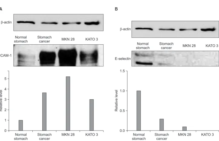
Fig. 1. Intercellular adhesion molecule-1 (ICAM-1), E-selectin expression level in gastric cancer tissues and cell lines (MKN28, KATO3). (A) CAM-1 expression was noted in normal stomach tissues but it was increased in gastric cancer tissues and cultured gastric cancer cells. (B) E-selectin expres- sion was more prominent in normal stomach tissues but it was decreased in gastric cancer tissues and cultured gastric cancer cells.
Results
1. Western blot analysis of ICAM-1 and E-selectin in gastric cancer tissues and cell lines (MKN28, KATO-III)
Western blotting was performed using gastric cancer tissues as well as corresponding tissues from non-cancerous regions and two gastric cancer cell lines, MKN28 and KATO-III. ICAM-1 expres- sion was noted in normal stomach tissues and the level of expres- sion was increased as much as four times in gastric cancer tissues compared with normal stomach tissues. However, E-selectin ex- pression was more prominent in normal stomach tissues and it was decreased in gastric cancer tissues. In cultured gastric cancer cell lines, MKN28 and KATO-III, the level of expression of ICAM-1
was increased, especially in MKN28, but the level of expression of E-selectin was not increased (Fig. 1).
2. Immunohistochemical staining of ICAM-1 and E-selectin in tissue micro array of gastric cancers From 157 gastric cancer tissues, nearly 50% (n=79) tissues were positive for ICAM-1 and 54% (n=85) were positive for E-selectin by immunohistochemical staining (Fig. 2). There was a reverse re- lationship between the positivity of ICAM-1 expression and posi- tivity of E-selectin expression (Table 1).
In our analysis, there was no statistically significant relationship between the expression of E-selectin and clinicopathologic factors and prognosis in these gastric cancer patients.
Fig. 2. Immunohistochemical staining of intercellular adhesion molecule-1 (ICAM-1), E-selectin in tissue microarray slides of gastric cancer. (A) Expression of ICAM-1 was noted in the endothelial cells of gastric cancer tissues. More than 50% of cancer cells were stained (×400). (B) Expres- sion of E-selectin was noted in the diffuse type signet ring gastric cancer cells. More than 50% were stained (×400).
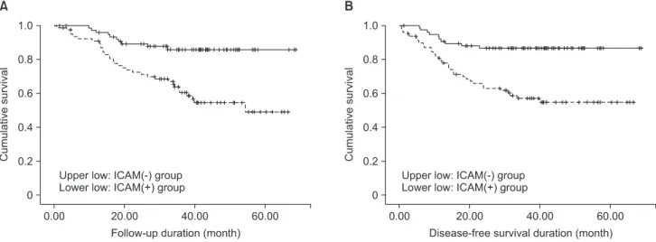
Fig. 3. (A) Overall 3-year survival of gastric cancer patients according to the intercellular adhesion molecule-1 (ICAM-1) immune reactivity. (B) Disease-free 3-year survival of gastric cancer patients according to the ICAM-1 immune reactivity.
3. Immunohistochemical staining of ICAM-1 and its correlation with clinicopathologic factors
Statistical analysis showed that the ICAM-1 positive group had a larger tumor size, advanced depth of tumor invasion, positive lymph node involvement and distant metastasis, namely advanced cancer stage. And hence, the expression of ICAM-1 was signifi- cantly related with the positivity of perineural invasion (Table 2).
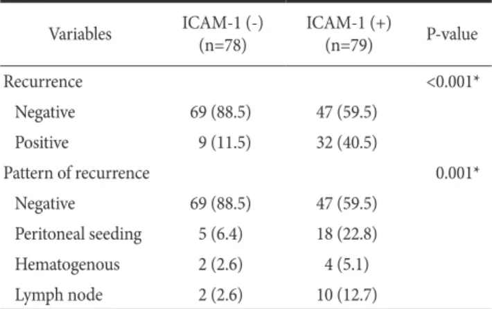
In the relationship between ICAM-1 and cancer recurrence, ICAM-1 was significantly correlated with cancer recurrence and when divided according to the recurrence pattern, it was mostly as- sociated with peritoneal seeding (56.3%, 18/32 cases) (Table 3).
4. ICAM-1 expression and its correlation with prognosis in gastric cancer patients
In survival analysis, ICAM-1-positive group showed a poor 3-year survival than the ICAM-1-negative group. (54.9% vs.
85.9%) and the disease-free survival result was also the same (Fig.
3).
In analysis of prognostic factors in these patients, ICAM-1 positive expression, diffuse type, depth of tumor invasion, lymph node involvement, distant metastasis, lymphovascular invasion and perineural invasion were proved to be negative prognostic factors in univariate analysis. However, in multivariate analysis, depth of tu- mor invasion and distant metastasis were proved to be independent prognostic factors (Table 4).
Discussion
In western blot assay, we could identify the presence of two adhesion molecules (ICAM-1 and E-selectin) in normal stom- Table 1. The relationship between ICAM-1 and E-selectin expression
by immunohistochemical staining Variables ICAM-1 (-)
(n=78) ICAM-1 (+)
(n=79) P-value E-selectin expression
Weak (≤50%) 49 (62.8) 23 (29.1) <0.001*
Strong (>50%) 29 (37.2) 56 (70.9)
Values are presented as number (%). ICAM-1 = intercellular adhesion molecule-1. *P<0.05; statistically significant.
Table 2. The relationship between ICAM-1 expression and clinicopathologic factors in gastric cancer patients
Variables ICAM-1 (-)
(n=78) ICAM-1 (+)
(n=79) P-value
Age (yr) 55.4±14.1 56.6±16.0 0.581
Sex 0.114
Male 42 (53.8) 51 (64.6)
Female 36 (46.2) 28 (35.4)
Tumor size (cm) 4.1±2.8 6.2±3.9 <0.001*
Lauren classification 0.058
Intestinal type 32 (41.0) 22 (27.8)
Diffuse type 46 (59.0) 57 (72.2)
Invasion of tumor depth <0.001*
pT1 (mucosa/submucosa) 58 (74.4) 21 (26.6) pT2 (muscle/subserosa) 8 (10.3) 9 (11.4) pT3/4 (serosa/organ inv.) 12 (15.4) 49 (62.1)
Lymph node involement <0.001*
Negative 47 (60.3) 26 (32.9)
Positive 31 (39.7) 53 (67.1)
Distant metastasis 0.009*
Negative 77 (98.7) 70 (88.6)
Positive 1 (1.3) 9 (11.4)
AJCC stage <0.001*
I 58 (74.4) 26 (32.9)
II 7 (9.0) 4 (5.1)
III 5 (6.4) 29 (36.7)
IV 8 (10.3) 20 (25.3)
Lymphovascular inv. 0.184
Negative 48 (61.5) 42 (53.2)
Positive 30 (38.5) 37 (46.8)
Perineural inv. <0.001*
Negative 69 (88.5) 51 (64.6)
Positive 9 (11.5) 28 (35.4)
Values are presented as mean±standard deviation or number (%).
ICAM-1 = intercellular adhesion molecule-1; inv. = invasion; AJCC = American Joint Committee on Cancer. *P<0.05; statistically significant.
Table 3. The relationship between ICAM-1 expression and recurrence in gastric cancer patients
Variables ICAM-1 (-)
(n=78) ICAM-1 (+)
(n=79) P-value
Recurrence <0.001*
Negative 69 (88.5) 47 (59.5)
Positive 9 (11.5) 32 (40.5)
Pattern of recurrence 0.001*
Negative 69 (88.5) 47 (59.5)
Peritoneal seeding 5 (6.4) 18 (22.8)
Hematogenous 2 (2.6) 4 (5.1)
Lymph node 2 (2.6) 10 (12.7)
Values are presented as number (%). ICAM-1 = intercellular adhesion molecule-1. *P<0.05; statistically significant.
ach tissues and gastric cancer tissues. Interestingly, the expres- sion of ICAM-1 was increased in gastric cancer tissues compared with normal stomach tissues, but the E-selectin expression was decreased in gastric cancer tissues. Furthermore, in the cultured gastric cancer cells, ICAM-1 protein expression was noted but E- selectin protein expression was not noted.
These results can be explained by the hypothesis that E-selec- tins are located on the surface of gastric endothelial cells as adhe- sion molecules in normal gastric tissue, and they maybe lost their
binding effect on the cell to cell interaction during the carcinogen- esis. Therefore they could not show any expression in gastric can- cer cells and decreased in gastric cancer tissue. Otherwise, we can suppose that ICAM-1 plays a more crucial role in carcinogenesis and tumor progression in gastric cancer compared to E-selectin.
ICAM-1 (CD54), a member of immunoglobulin superfamily, is a transmembrane glycoprotein with an extracellular region that has an immunoglobulin-like structure.(23) ICAM-1 is found on the surface of numerous cells including vascular endothelial cells, fibro- Table 4. Uni- and multivariate survival analysis in gastric cancer patients
Variables 3-yr (%) P-value Hazard ratio (95% CI) P-value
ICAM-1 <0.001* 2.012 (0.797~5.081) 0.139
(-) 85.9
(+) 54.5
Age group 0.651
<60 71
≥60 75.3
Sex 0.656
Male 73.1
Female 71.7
Tumor size <0.001* 0.731 (0.313~1.706) 0.469
<5 cm 84
≥5 cm 56.8
Lauren type 0.042* 0.934 (0.399~2.184) 0.875
Intestinal 84.6
Diffuse 66.7
Invasion of tumor depth <0.001* 0.144 (0.030~0.698) 0.016*
pT1 (mucosa/submucosa) 100
pT2 (muscle/subserosa) 83.7
pT3/4 (serosa/organ inv.) 34.7
Lymph node involement <0.001* 0.760 (0.230~2.510) 0.652
Negative 94.4
Positive 54.8
Distant metastasis <0.001* 0.260 (0.110~0.616) 0.002*
Negative 77
Positive 10
Lymphovascular inv. 0.002* 1.257 (0.617~2.560) 0.529
Negative 81.5
Positive 61
Perineural inv. <0.001* 0.714 (0.352~1.450) 0.352
Negative 86.1
Positive 30.6
Values are presented as % or mean (range). CI = confidence interval; ICAM-1 = intercellular adhesion molecule-1; inv. = invasion. *P<0.05;
statistically significant.
blasts, and epithelial cells.(24) After binding to lymphocyte function antigen-1 (LFA-1) on the surface of T lymphocytes, it plays a role in their activation that increases the susceptibility of such tumor cells to lymphocyte-mediated tumor cytotoxicity.(25) ICAM-1 on cancer cells plays an important role in adhering to natural killer (NK) cells or lymphocyte-activated killer (LAK) cells via LFA-1.(26) Mi- gration of granulocytes from blood vessels is mediated by adhesion between ICAM-1 and LFA-1(27) and ICAM-1 is also involved in adhesion to target cells before destruction by killer cells (NK cells, LAK cells).(28) Therefore, ICAM-1 is important for the local im- mune response, i.e. the activity of NK and LAK cells is suppressed by administration of anti-ICAM-1 antibody(8,9) and serum cir- culating ICAM-1 suppresses the adhesion of T cells by binding to LFA-1 and inhibits NK and LAK cell activity.(7,29) Based on these reports, we can hypothesize that ICAM-1 has an important immunosuppressive effect and is involved in carcinogenesis and aggravates cancer progression and this can be applied to our analy- sis.
In the past, there have been several reports that demonstrated the protective effect of ICAM-1 protein in gastric carcinogenesis and cancer progression.(17,18) Fujihara et al.(17) documented lymph node metastasis as a significant risk factor for gastric cancer and proposed that ICAM-1 promotes immunosurveillance system between cancer cells and cytotoxic lymphocytes which are associ- ated with blocking the lymph node metastatic process. Also, in the mechanism of immunologic tolerance, the escape of cancer cells from immune system can be responsible for progression of cancer.
Yashiro et al.(18) suggested that ICAM-1 could be a costimulatory
molecule which plays an important role in cancer cell adhesion to immune cells for this immune tolerance, and hence decreased expression of ICAM-1 on cancer cells might be associated with decreased cytotoxicity of immune cells, which may result in lymph node metastasis and tumor progression in gastric cancer.
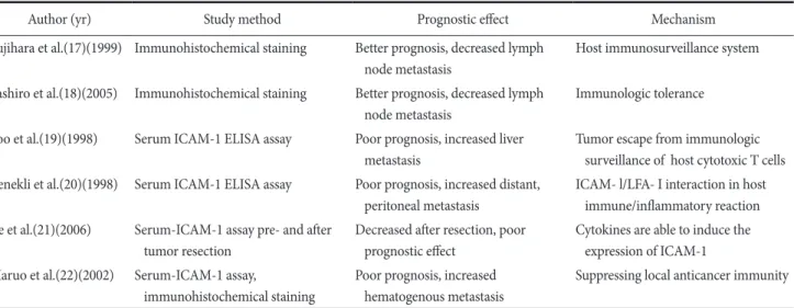
However, most other reports regarding the importance of se- rum circulating ICAM-1 in gastric cancer suggested that increased serum levels of ICAM-1 in gastric cancer may reflect cancer pro- gression and metastasis.(19,20,22) Benekli et al.(20) demonstrated that concentrations of serum circulating ICAM-1 are increased in gastric cancer patients with distant and peritoneal metastasis and they suggested that ICAM-1 could be a reflective metastatic marker for gastric cancer. Also, Maruo et al.(22) proposed that ICAM-1 was over-expressed in cancer cells and tissues and it could be re- leased as a serum circulating ICAM-1, which would promote he- matogenous metastasis by suppressing local antitumor immunity in gastric cancer. Considering the above two conflicting opinions, the role of ICAM-1 as a prognostic marker in gastric cancer has not yet been established because there is a reciprocal effect of ICAM-1 on immune surveillance system which is like two sides of the same coin. These two conflicting results of previous reports are summa- rized in Table 5.
Although we could not obtain the serum levels of circulat- ing ICAM-1 because we used a retrospective study design, we could identify the presence of ICAM-1 in gastric cancer tissues comprised of different types of cells including endothelial cells, lymphocytes, monocytes and other immune cells and they could be a source of serum circulating ICAM-1. And in our analysis,
Table 5. The summary of previous study results about prognostic effect of ICAM-1 on gastric cancer
Author (yr) Study method Prognostic effect Mechanism
Fujihara et al.(17)(1999) Immunohistochemical staining Better prognosis, decreased lymph node metastasis
Host immunosurveillance system Yashiro et al.(18)(2005) Immunohistochemical staining Better prognosis, decreased lymph
node metastasis
Immunologic tolerance Yoo et al.(19)(1998) Serum ICAM-1 ELISA assay Poor prognosis, increased liver
metastasis
Tumor escape from immunologic surveillance of host cytotoxic T cells Benekli et al.(20)(1998) Serum ICAM-1 ELISA assay Poor prognosis, increased distant,
peritoneal metastasis
ICAM- l/LFA- I interaction in host immune/inflammatory reaction Ke et al.(21)(2006) Serum-ICAM-1 assay pre- and after
tumor resection
Decreased after resection, poor prognostic effect
Cytokines are able to induce the expression of ICAM-1 Maruo et al.(22)(2002) Serum-ICAM-1 assay,
immunohistochemical staining
Poor prognosis, increased hematogenous metastasis
Suppressing local anticancer immunity ICAM-1 = intercellular adhesion molecule-1; ELISA = enzyme-linked immunospecific assay; LFA-1 = lymphocyte function antigen-1.
patients with increased expression of ICAM-1 showed an advanced tumor stage and a more frequent incidence of cancer recurrence and peritoneal seeding was the most common form of metastasis.
Therefore, these results reconfirm that ICAM-1 in cancer tissues may have an immunosuppressive effect due to the escape of cancer cells from immune surveillance system which is the similar action of serum circulating ICAM-1 in previous other studies.
Although the negative prognostic effect of ICAM-1 on hema- togenous metastasis in gastric cancer has been emphasized in the previous report,(22) considering that peritoneal seeding is the most common recurrence pattern in gastric cancer, our result can be explained by the general effect of ICAM-1 protein on tumor pro- gression and metastasis in gastric cancer patients.
In our immunohistochemical staining analysis, we could not demonstrate any meaningful relationship between the expression of E-selectin and tumor progression. However, interestingly, a reverse correlation between ICAM-1 and E-selectin protein expression was noted and this was similar to the finding of western blot anal- ysis. A further study regarding the carcinogenetic and prognostic role of E-selectin protein in gastric cancer is needed. Several basic researches about the function and effect of ICAM-1 in immunol- ogy and carcinogenesis were described,(23-28) however, in gastric cancer, the basic research for investigating the role of ICAM-1 on gastric carcinogenesis and prognosis could not yet be performed.
So, we think this basic fundamental study should be needed. Also, more detailed invasion and migration study or proliferation study of gastric cancer cells with or without ICAM-1 inhibitor should be needed for clarification of mechanism in gastric carcinogenesis and cancer progression.
In our gastric cancer patients, we could not demonstrate ICAM- 1 as an independent prognostic factor in multivariate analysis due to the powerful impact of cancer stage on survival. Although subgroup analysis was not possible because the sample size of our study was small, we think that there is a significant possibility of ICAM-1 as the independent prognostic factor in gastric cancer, especially in the advanced stage, when a large- scale, multicenter research study will be undertaken.
As significant progress has been made recently in molecu- lar tumor biology, new therapeutic approaches such as immune- based and targeted therapies in gastric cancer have been intensively investigated.(30) Considering the importance of immune system affecting tumor progression and metastasis, immune therapeutic effects of ICAM-1 and/or anti-ICAM-1 antibody in gastric cancer are worthy of careful study in the future.
In conclusion, the present study demonstrated that ICAM-1 is over-expressed in the gastric cancer tissues and cultured gastric cancer cells compared with normal stomach tissues, and gas- tric cancer patients with high expression of ICAM-1 showed an advanced tumor stage and metastasis. Therefore, the increased expression of ICAM-1 protein in tumor tissues from pathology specimens could be an additional prognostic indicator to monitor for cancer recurrence along with increased serum levels of circulat- ing ICAM-1 during the postoperative follow-up in gastric cancer patients.
Acknowledgments
This work was supported by Korea University Grant.
This manuscript was presented by poster on March, 2012 at the 33rd Annual Spring Congress of Korean Gastric Cancer Associa- tion in Kimdaejung Convention Center, Kwangju, Korea.
References
1. Ohene-Abuakwa Y, Pignatelli M. Adhesion molecules as diag- nostic tools in tumor pathology. Int J Surg Pathol 2000;8:191- 200.
2. Fidler IJ. Critical determinants of metastasis. Semin Cancer Biol 2002;12:89-96.
3. Picker LJ, Butcher EC. Physiological and molecular mecha- nisms of lymphocyte homing. Annu Rev Immunol 1992;10:
561-591.
4. Sommers CL, Thompson EW, Torri JA, Kemler R, Gelmann EP, Byers SW. Cell adhesion molecule uvomorulin expression in human breast cancer cell lines: relationship to morphology and invasive capacities. Cell Growth Differ 1991;2:365-372.
5. Maeda K, Kang SM, Sawada T, Nishiguchi Y, Yashiro M, Oga- wa Y, et al. Expression of intercellular adhesion molecule-1 and prognosis in colorectal cancer. Oncol Rep 2002;9:511-514.
6. Ogawa Y, Hirakawa K, Nakata B, Fujihara T, Sawada T, Kato Y, et al. Expression of intercellular adhesion molecule-1 in invasive breast cancer reflects low growth potential, negative lymph node involvement, and good prognosis. Clin Cancer Res 1998;4:31-36.
7. Rothlein R, Mainolfi EA, Czajkowski M, Marlin SD. A form of circulating ICAM-1 in human serum. J Immunol 1991;147:3788-3793.
8. Wang P, Vánky F, Li SL, Patarroyo M, Klein E. Functional
characteristics of the intercellular adhesion molecule-1 (CD54) expressed on cytotoxic human blood lymphocytes. Cell Immu- nol 1990;131:366-380.
9. Sánchez-Rovira P, Jimenez E, Carracedo J, Barneto IC, Ramirez R, Aranda E. Serum levels of intercellular adhesion molecule 1 (ICAM-1) in patients with colorectal cancer: inhibitory effect on cytotoxicity. Eur J Cancer 1998;34:394-398.
10. Tsujisaki M, Imai K, Hirata H, Hanzawa Y, Masuya J, Nakano T, et al. Detection of circulating intercellular adhesion molecule-1 antigen in malignant diseases. Clin Exp Immunol 1991;85:3-8.
11. Natali P, Nicotra MR, Cavaliere R, Bigotti A, Romano G, Tem- poni M, et al. Differential expression of intercellular adhesion molecule 1 in primary and metastatic melanoma lesions. Can- cer Res 1990;50:1271-1278.
12. Koyama S, Ebihara T, Fukao K. Expression of intercellular ad- hesion molecule 1 (ICAM-1) during the development of inva- sion and/or metastasis of gastric carcinoma. J Cancer Res Clin Oncol 1992;118:609-614.
13. Bevilacqua M, Butcher E, Furie B, Furie B, Gallatin M, Gim- brone M, et al. Selectins: a family of adhesion receptors. Cell 1991;67:233.
14. Bevilacqua MP, Nelson RM. Selectins. J Clin Invest 1993;91:
379-387.
15. Majuri ML, Mattila P, Renkonen R. Recombinant E-selectin- protein mediates tumor cell adhesion via sialyl-Le(a) and sialyl-Le(x). Biochem Biophys Res Commun 1992;182:1376- 1382.
16. Ramphal JY, Hiroshige M, Lou B, Gaudino JJ, Hayashi M, Chen SM, et al. Ligand interactions with E-selectin. Identifica- tion of a new binding site for recognition of N-acyl aromatic glucosamine substituents of sialyl Lewis X. J Med Chem 1996;39:1357-1360.
17. Fujihara T, Yashiro M, Inoue T, Sawada T, Kato Y, Ohira M, et al. Decrease in ICAM-1 expression on gastric cancer cells is correlated with lymph node metastasis. Gastric Cancer 1999;2:221-225.
18. Yashiro M, Sunami T, Hirakawa K. CD54 expression is predic- tive for lymphatic spread in human gastric carcinoma. Dig Dis Sci 2005;50:2224-2230.
19. Yoo NC, Chung HC, Chung HC, Park JO, Rha SY, Kim JH, et al. Synchronous elevation of soluble intercellular adhesion molecule-1 (ICAM-1) and vascular cell adhesion molecule-1 (VCAM-1) correlates with gastric cancer progression. Yonsei
Med J 1998;39:27-36.
20. Benekli M, Güllü IH, Tekuzman G, Savaş MC, Hayran M, Hasçelik G, et al. Circulating intercellular adhesion molecule-1 and E-selectin levels in gastric cancer. Br J Cancer 1998;78:267- 271.
21. Ke JJ, Shao QS, Ling ZQ. Expression of E-selectin, integrin beta1 and immunoglobulin superfamily member in human gastric carcinoma cells and its clinicopathologic significance.
World J Gastroenterol 2006;12:3609-3611.
22. Maruo Y, Gochi A, Kaihara A, Shimamura H, Yamada T, Tanaka N, et al. ICAM-1 expression and the soluble ICAM- 1 level for evaluating the metastatic potential of gastric cancer.
Int J Cancer 2002;100:486-490.
23. Staunton DE, Marlin SD, Stratowa C, Dustin ML, Springer TA. Primary structure of ICAM-1 demonstrates interaction between members of the immunoglobulin and integrin super- gene families. Cell 1988;52:925-933.
24. Dustin ML, Rothlein R, Bhan AK, Dinarello CA, Springer TA.
Induction by IL 1 and interferon-gamma: tissue distribution, biochemistry, and function of a natural adherence molecule (ICAM-1). J Immunol 1986;137:245-254.
25. Staunton DE, Dustin ML, Erickson HP, Springer TA. The ar- rangement of the immunoglobulin-like domains of ICAM- 1 and the binding sites for LFA-1 and rhinovirus. Cell 1990;61:243-254.
26. Schmidt RE, Bartley G, Levine H, Schlossman SF, Ritz J. Func- tional characterization of LFA-1 antigens in the interaction of human NK clones and target cells. J Immunol 1985;135:1020- 1025.
27. Dustin ML, Springer TA. Role of lymphocyte adhesion recep- tors in transient interactions and cell locomotion. Annu Rev Immunol 1991;9:27-66.
28. Vánky F, Wang P, Patarroyo M, Klein E. Expression of the adhesion molecule ICAM-1 and major histocompatibility complex class I antigens on human tumor cells is required for their interaction with autologous lymphocytes in vitro. Cancer Immunol Immunother 1990;31:19-27.
29. Santarosa M, Favaro D, Quaia M, Spada A, Sacco C, Talamini R, et al. Expression and release of intercellular adhesion mol- ecule-1 in renal-cancer patients. Int J Cancer 1995;62:271-275.
30. Amedei A, Benagiano M, della Bella C, Niccolai E, D'Elios MM. Novel immunotherapeutic strategies of gastric cancer treatment. J Biomed Biotechnol 2011;2011:437348.