J Korean Ophthalmol Soc 2014;55(10):1567-1572 pISSN: 0378-6471⋅eISSN: 2092-9374
http://dx.doi.org/10.3341/jkos.2014.55.10.1567
Case Report
혈액학적 이상과 동반된 급성 구후시신경염
Acute Retrobulbar Optic Neuritis with Hematologic Abnormalities
박시윤⋅강은민⋅이윤하⋅김찬윤⋅성공제⋅홍사민
Si Yoon Park, MD, Eun Min Kang, MD, Yun Ha Lee, MD, Chan Yun Kim, MD, PhD, Gong Je Seong, MD, PhD, Samin Hong, MD, PhD
연세대학교 의과대학 안과학교실 및 시기능개발연구소
Institute of Vision Research, Department of Ophthalmology, Yonsei University College of Medicine, Seoul, Korea
Purpose: We present a case of a patient with optic neuritis who had underlying suspicious idiopathic thrombocytopenic purpura.
Case summary: A 35-year-old female with no other systemic disease visited our clinic due to acutely decreased visual acuity in her left eye 10 days in duration. Relative afferent pupillary defect was observed, but without definite papilledema. Based on brain magnetic resonance imaging (MRI), optic neuritis was suspected. Laboratory tests showed increased red blood cells, hemoglo- bin and, hematocrit levels and decreased platelets. Peripheral blood smear test showed decreased platelets, relative lymphocy- tosis and atypical lymphocytes. Specific antibodies for autoimmune disease were not present. High-dose steroid pulse therapy (methyl prednisolone 1.0 g/d, 3 days) was started. One month after treatment her visual acuity and platelet count recovered and her visual field defect improved.
J Korean Ophthalmol Soc 2014;55(10):1567-1572
Key Words: Autoimmune disease, Idiopathic thrombocytopenic purpura, Multiple sclerosis, Neuromyelitis optica, Optic neuritis
■Received: 2014. 6. 21. ■ Revised: 2014. 7. 22.
■Accepted: 2014. 9. 17.
■Address reprint requests to Samin Hong, MD, PhD Department of Ophthalmology, Severance Hospital, #50-1 Yonsei-ro, Seodaemun-gu, Seoul 120-752, Korea Tel: 82-2-2228-3750, Fax: 82-2-312-0541 E-mail: [email protected]
* This work was supported by the Basic Science Research Program through the National Research Foundation of Korea (NRF) fund- ed by the Ministry of Education, Science, and Technology (No.
2011-0013288).
ⓒ2014 The Korean Ophthalmological Society
This is an Open Access article distributed under the terms of the Creative Commons Attribution Non-Commercial License (http://creativecommons.org/licenses/by-nc/3.0/) which permits unrestricted non-commercial use, distribution, and reproduction in any medium, provided the original work is properly cited.
시신경염은 시신경의 염증에 의한 것으로, 다양한 원인이 있고 그에 따라 치료 및 예후가 다르다. 서구에서는 많은 경 우 다발성 경화증(Multiple sclerosis)과 동반된다고 알려졌으 나, 일본 등과 같은 다발성 경화증이 흔하지 않은 지역에서 는 시신경척수염(Neuromyeliris optica)과 동반된 경우가 많 다고 알려졌다.1-3 최근 연구에 따르면 다발성 경화증 및
시신경척수염은 자가면역질환의 일종으로 여겨지고 있으 며, 다른 자가면역질환들과 동반될 수 있다고 보고되고 있 다. 특히 특발성 혈소판 감소성 자반증(Idiopathic thrombo- cytopenic purpura)과 동반된 다발성경화증이 증례보고된 바 있다.4-6 한국에서도 혈액학적 이상을 동반한 환자에게서 발생한 급성 시신경병증을 경험하여 보고하고자 한다.
증례보고
35세 여성이 10일 전 갑자기 발생한 좌안 시력저하를 주 소로 내원하였다. 특이 전신 질환은 없었으나 하루 1갑 이 상의 흡연자였다. 증상 발생 전일 심한 과음 후 응급 경구 피임약을 복용하였고, 다음 날 기상 후 심한 어지러움을 동 반한 좌안 시력저하를 느꼈으며, 이후 증상 호전되지 않아 본원에 내원하였다. 두통 및 안통은 전혀 동반되지 않았다.
내원 시 나안시력은 우안 1.0, 좌안 안전수동 감별이었으
A B
Figure 1. At the first visit, disc photography revealed no definite disc swelling in her both eyes. (A) Right eye, (B) left eye.
A B
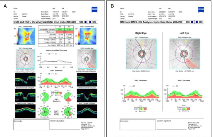
Figure 2. Decreased retinal nerve fiber layer (RNFL) thickness at the inferotemporal area of her left eye in optical coherence
tomography. (A) RNFL quadrants and clock hours maps, (B) RNFL deviation map and thickness. OD = right eye; ONH = optic nerve head; OS = left eye; OU = both eyes; TEMP = temporal; SUP = superior, NAS = nasal, INF = inferior, C/D = cup to disc ratio.며, 좌안 시력은 교정되지 않았다. 안압은 우, 좌안 각각 13, 11 mmHg였다. 양안 모두 세극등 검사에서 전안부 이상소 견은 관찰되지 않았으며, 좌안은 상대적 구심성 동공반사
장애 소견과 이시하라(Ishihara)색각 검사에서 전색맹 소견 을 보였으나 우안은 특이 소견을 보이지 않았다. 안저 검사 에서도 뚜렷한 이상 소견이 나타나지 않았다(Fig. 1). 빛간
A B
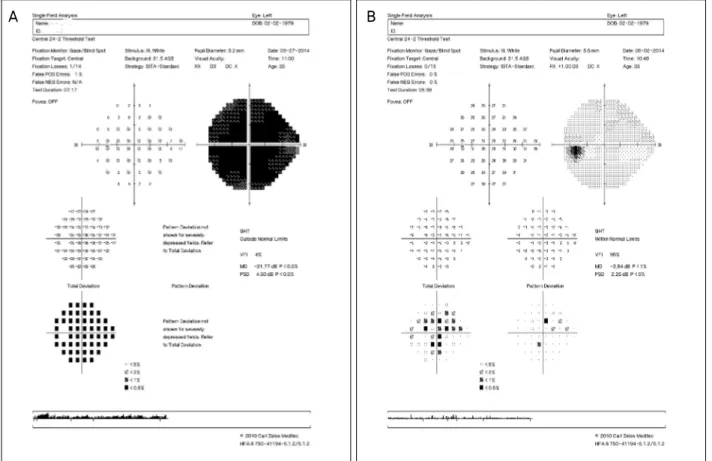
Figure 3. (A) Humphrey visual field test shows almost total scotoma in her left eye before high dose methylprednisolone treatment.
(B) After treatment, improvement of visual field test in her left eye was noted. POS = positive; NEG = negative; GHT = glaucoma hemifield test; VFI = visual field index; MD = mean deviation; PSD = pattern standard deviation.
A B
Figure 4. Brain magentic resonance
image (MRI) with enhance T1 weighted MRI. Diffuse enhance- ment of the left optic nerve, cister- nal segment, suspicious of optic neuritis or neuropathy. No abnor- mal signal intensity or abnormal mass lesion in the brain paren- chyma (A: axial view; B: coronal view).섭단층촬영(Cirrus optical coherence tomography, Carl Zeiss Meditec, Dublin, CA, USA)에서 좌안의 하이측 망막신경섬유 층의 위축 소견을 보였다(Fig. 2). 표준자동시야검사(Humphrey Instruments, Central 24-2 threshold: Carl Zeiss Meditec, Dublin, CA, USA)에서 우안은 정상, 좌안은 전암점소견을 보였다(Fig. 3A). 상기 증상을 바탕으로 좌안 급성 허혈성 시신경병증이 의심되어 전신 검사를 시행하였다. 혈액검사
에서 적혈구(5.55×10⁶/μL), 헤모글로빈(16.1 g/dL), 헤마토크 릿(50.0%)으로 증가해있었고, 혈소판(58,000/μL)수치는 감소 해있었다. 적혈구침강속도 및 C 반응단백정량화 수치는 정 상범위였다. 뇌자기공명영상의 T1 강조, 조영증강영상에서 좌측 시신경의 뇌 수조부분에 조영 증강소견이 관찰되어 구 후시신경염의 가능성이 높다고 판단되어 고용량 스테로이드 정맥주사(Intravenous methylprednisolone, 1 g/d (250 mg×4
회/d), 3 days)를 시작하였다(Fig. 4). 항핵항체(Anti-nuclear antibody) 등 다른 자가면역 질환을 나타내는 표지자에서 는 음성소견을 보였다. 혈액학적 이상에 대해 혈액종양내 과 협진을 시행하였고, 말초혈액도말검사(Peripheral Blood Smear)상, 적혈구의 모양 및 색은 정상범위였으며 혈소판 의 감소, 상대적 림프구 증가 및 비정형 림프구가 관찰되었 다. 진성 적혈구 증가증(Polycythemia vera) 또는 만성 특발 성 혈소판 감소성 자반증(Chronic idiopathic thrombocyto- penic purpura)으로 생각되어 추가 혈액 검사를 시행한 결 과 진성 적혈구 증가증에 상대적으로 특이적인 JAK2 V617F, JAK2 Exon127의 돌연변이는 발견되지 않았으며, 이차적으로 혈소판 감소를 유발할 수 있는 전신질환 소견 은 보이지 않았다. 그러나 허혈성 원인을 완전히 배제할 수 없어 추가적으로 아세틸살리실산(Acetyl salicylic acid) 100 mg/day, 니세르골린(Nicergoline) 20 mg/day을 투약하였고, 브로모디닌 점안액(Brimonidine tartrate 0.15% 5 m, 2회 /day)을 점안하였다. 스테로이드 제제는 경구약으로 전환 후 한 달에 걸쳐 점차적으로 줄여나갔다(Prednisolone ace- tate 60 mg/d × 14 days, 30 mg/d × 14 days).
고용량 스테로이드치료 2주 후 시야검사상 호전소견을 보 였고(Fig. 3B). 치료 한 달 후 최대 교정시력이 좌안 0.9로 호전되었다. 혈액검사에서도 혈소판 수치가 250,000/μL로 회복 소견 보였으며, 헤마토크릿은 47.2%로 감소하였다.
스테로이드치료를 중단 후 다시 혈소판 수치 감소의 우려 가 있고, 이 경우 특발성 혈소판 감소성 자반증을 더욱 의 심할 수 있기 때문에 혈액 질환에 대해서는 경과관찰하였 으며, 발병 4개월 후 혈액검사에서도 혈소판 수치 69,000/μ L로 지속적 저하 소견 보이며, 그 외 특이 혈소판 감소를 일으키는 혈액학적 이상 소견 없어 특발성 혈소판 감소성 자반증으로 진단하고 내과적으로 경과관찰 중이다.
고 찰
시신경염은 임상적 양상에 따라 전형적, 비전형적으로 나뉘며, 전형적 시신경염의 경우 젊은 성인, 백인, 주로 한 눈에서 나타나며 다발성 경화증과 연관되거나, 다발성 경 화증의 전 단계인 임상적 독립 증후군(Clinically isolated syndrome)과 연관되어 있는 경우가 흔하다.1
다발성 경화증은 중추신경계의 탈 수초화에 의한 병으로 HLA-DR15 등 HLA (Human leukocyte antigen) haplotype, 선행된 바이러스 감염(Ex. 엡스타인-바 바이러스(EBV)), 비 타민 D 부족 등과 관련성이 있다고 되어 있으나 정확한 원 인은 밝혀져 있지 않았다. 최근 여러 연구에 의해 면역체계 의 조절이상에 의한 염증반응이 그 기저 원인에 있을 것으
로 생각되고 있다.5,8 다발성경화증의 약 25%에서 시신경염 이 그 첫 증상으로 나타나고 있으며 약 70%에서 질병의 기간 중 나타난다. 시신경염 치료 시험(Optic Neuritis Treatment Trial, ONTT)에서는 뇌자기공명영상상 진단 시 이상이 없 었던 시신경염의 경우 25%에서 15년 후 다발성 경화증으 로 진단되는 반면, 초기 이상이 있었던 경우의 72%에서 추 후 다발성 경화증으로 진단된다고 보고하였고,1,9 주로 시신 경과 척수를 침범하는 특발성 염증성 탈 수초 질환 중 하나 인 시신경 척수염 역시 다발성 경화증과 마찬가지로 전신 적 자가면역 질환과도 연관이 있다는 보고가 있다.4,10-13
특발성 혈소판 감소성 자반증은 낮은 혈소판 수치와 점 막, 피부 출혈을 특징적으로 하는 질환으로 아직 그 기전은 명확하게 밝혀지지 않았으나 바이러스 감염 등이 선행할 수 있으며, 자가면역 등 면역학적 기전이 관여한다고 알려 졌다.6 이 질환은 혈소판에 대한 IgG 자가항체가 생성되어 혈소판과 거대핵세포(Megakaryocyte)를 파괴한다고 알려져 있어 자가면역성 혈소판 감소성 자반증이라고도 불려지며, 다발성 경화증, 전신성 홍반성 루프스, 자가면역성 갑상선 질환 등 다른 자가면역 질환과 함께 동반될 수 있다고 알려
졌다.6,14-17 최근 연구에서 특발성 혈소판 감소성 자반증 환자
에서 다발성 경화증이 병발되는 경우가 일반인구에서 보다 25배 높다는 보고도 있었다.6,18,19 또한 다발성 경화증의 형제 들에서 셀리악병(Celiac disease), 베체트병(Behcet’s disease), 특발성 혈소판 감소성 자반증 등 자가면역질환이 동반되어 있다는 보고도 있다.5,20
본 증례는 헤마토크릿이 증가해있고, 통증이 동반되지 않은 점, 피임약 복용의 과거력이 있다는 점에서 허혈성 시 신경병증의 양상을 나타내지만, 특별한 전신질환이 없는 젊은 성인 여성에게 아급성으로 다른 신경학적 증상의 동 반 없이 나타나고, 뇌자기공명영상에서 시신경주위 조영 증강이 보인 점, 고용량 스테로이드 치료에 효과가 있었던 것을 고려하였을 때 시신경염에 더 합당한 소견을 보였으 며, 현재까지는 횡 척수염 등의 다른 신경학적 증상이 없었 다는 점에서 다발성 경화증등의 신경학적 질환을 진단할 수는 없지만, 특발성 혈액학적 이상이 동반되었다는 점에 서 자가면역질환의 일부분으로 유추해볼 수 있다. 또한 시 유발전위검사상 P100 파형의 지연이 보였다는 점에서 앞 시각로의 탈 수초화를 의심해 볼 수 있다.21 환자의 나이와 증상발현이 한 눈이고, 통증 및 시신경 유두의 부종이 심하 지 않았다는 점 등을 고려하였을 때, 소아나 고령에 양안을 침범하며, 심한 통증 및 치료 후에도 시력 회복이 어렵고, 가족력이나 신생물의 과거력 등의 동반이 흔한 비정형 시 신경염보다는 전형적 시신경염에 합당한 소견을 보였다.1 전형적인 단독 시신경 병증의 경우 10년 내 다발성 경화증
이 병발될 확률이 38%에 이르고, 전신성 자가면역질환이나 시신경척수염 등이 동반될 경우 재발의 가능성이 높으며, 재발을 반복할 시 잔류결함의 누적으로 심각한 시각장애를 유발할 수 있다.9,10,21 또한 이러한 경우 고용량 스테로이드 치료뿐만 아니라 지속적 경구 스테로이드제 복용 또는 인 터페론 베타(Interferon- β), Azathioprine 등과 같은 면역억 제 치료가 필요할 수 있다.1,22,23 특발성 혈소판 감소성 자반 증과 다발성 경화증은 자가면역 질환의 일종으로 함께 병 발될 수 있고,4 두 질환 모두 선행원인 중 하나가 바이러스 감염이라는 공통점이 있다.6,24 또한 시신경염과 다발성경화 증의 연관성은 익히 알려졌다.1
본 증례의 경우 급성 시신경염에서 특발성 혈소판 감소 성 자반증과 같은 비특이적 혈액학적 질환이 병발될 수 있 음을 보여준다. 시신경염, 다발성 경화증, 특발성 혈소판 감 소성 자반증이 자가면역질환의 범주 내에 있다고 생각하였 을 때 본 증례의 경우 시신경염뿐만 아니라 다른 자가면역 질환도 동반될 수 있기 때문에 이러한 경우 다른 자가면역 질환에 대한 검사 및 자가면역질환과 동반된 시신경염의 유전적 요인 등의 원인, 경과 및 치료에 대한 더 많은 연구 가 필요할 것으로 생각한다.
REFERENCES
1) Toosy AT, Mason DF, Miller DH. Optic neuritis. Lancet Neurol 2014;13:83-99.
2) Swanton JK, Fernando K, Dalton CM, et al. Is the frequency of ab- normalities on magnetic resonance imaging in isolated optic neu- ritis related to the prevalence of multiple sclerosis? A global comparison. J Neurol Neurosurg Psychiatry 2006;77:1070-2.
3) Petzold A, Plant GT. Diagnosis and classification of autoimmune optic neuropathy. Autoimmun Rev 2014;13:539-45.
4) Hui AC, Wong RS, Ma R, Kay R. Recurrent optic neuromyelitis with multiple endocrinopathies and autoimmune disorders. J Neurol 2002;249:784-5.
5) Yamout B, Usta J, Itani S, Yaghi S. Celiac disease, Behçet, and idi- opathic thrombocytopenic purpura in siblings of a patient with multiple sclerosis. Mult Scler 2009;15:1368-71.
6) Sahraian MA, Eshaghi A. Concomitant multiple sclerosis and idio- pathic thrombocytopenic purpura. Eur J Neurol 2010;17:e62-3.
7) Scott LM, Tong W, Levine RL, et al. JAK2 exon 12 mutations in polycythemia vera and idiopathic erythrocytosis. N Engl J Med
2007;356:459-68.
8) Alemany-Rodríguez MJ, Aladro Y, Amela-Peris R, et al.
[Autoimmune diseases and multiple sclerosis]. Rev Neurol 2005;
40:594-7.
9) Optic Neuritis Study Group. Multiple sclerosis risk after optic neu- ritis: final optic neuritis treatment trial follow-up. Arch Neurol 2008;65:727-32.
10) Ahn SM, Kim SS. A case of coexisting neuromyelitis optica in sys- temic lupus erythematosus. J Korean Ophthalmol Soc 2013;54:
1469-74.
11) Adoni T, Lino AM, da Gama PD, et al. Recurrent neuromyelitis op- tica in Brazilian patients: clinical, immunological, and neuro- imaging characteristics. Mult Scler 2010;16:81-6.
12) Sergio P, Mariana B, Alberto O, et al. Association of neuromyelitis optic (NMO) with autoimmune disorders: report of two cases and review of the literature. Clin Rheumatol 2010;29:1335-8.
13) Wingerchuk DM, Lennon VA, Lucchinetti CF, et al. The spectrum of neuromyelitis optica. Lancet Neurol 2007;6:805-15.
14) Karpatkin S. Autoimmune (idiopathic) thrombocytopenic purpura.
The Lancet 1997;349:1531-6.
15) RABINOWITZ Y, DAMESHEK W. Systemic lupus erytematosus after "idiopathic" thrombocytopenic purpura: a review. Ann Intern Med 1960;52:1-28.
16) Hymes K, Blum M, Lackner H, Karpatkin S. Easy bruising, throm- bocytopenia, and elevated platelet immunoglobulin G in Graves' disease and Hashimoto's thyroiditis. Ann Intern Med 1981;
94:27-30.
17) Cines DB, Blanchette VS. Immune thrombocytopenic purpura. N Engl Med 2002;346:995-1008.
18) Sheremata WA, Jy W, Horstman LL, et al. Evidence of platelet acti- vation in multiple sclerosis. J Neuroinflammation 2008;5:27.
19) Segal JB, Powe NR. Prevalence of immune thrombocytopenia:
analyses of administrative data. J Thromb Haemost 2006;4:
2377-83.
20) Killer HE, Huber A, Portman C, et al. Bilateral non-arteritic ante- rior ischemic optic neuropathy in a patient with autoimmune thrombocytopenia. Eur J Ophthalmol 2000;10:180-2.
21) Bermel RA, Balcer LJ. Optic neuritis and the evaluation of visual impairment in multiple sclerosis. Continuum (Minneap Minn) 2013;19:1074-86.
22) Comi G, Filippi M, Barkhof F, et al. Effect of early interferon treat- ment on conversion to definite multiple sclerosis: a randomised study. Lancet 2001;357:1576-82.
23) Mandler RN, Ahmed W, Dencoff JE. Devic's neuromyelitis optica:
a prospective study of seven patients treated with prednisone and azathioprine. Neurology 1998;51:1219-20.
24) Cusick MF, Libbey JE, Fujinami RS. Multiple sclerosis: auto- immunity and viruses. Curr Opin Rheumatol 2013;25:496-501.
= 국문초록 =
혈액학적 이상과 동반된 급성 구후시신경염
목적: 특발성 혈소판 감소성 자반증이 의심되는 환자에서 발생한 급성 구후시신경염에 대해 보고하고자 한다.
증례요약: 특이 병력이 없는 35세 여성이 10일 전 갑자기 발생한 좌안 시력저하를 주소로 본원 내원하였다. 좌안 시력은 안전 수동감 별이었고, 상대적 구심성 동공반사 장애 소견과 색각저하 소견 보였으나 시신경유두 부종은 명확히 관찰되지 않았다. 안통은 없었다.
급성 허혈성 시신경병증이 의심되었으나, 뇌자기공명영상 검사 후 급성 구후시신경염으로 진단되었다. 혈액검사에서 적혈구, 헤모글 로빈, 헤마토크릿의 수치가 증가해있었고, 혈소판수치는 현저히 감소해있었다. 말초 혈액 도말 검사에서 혈소판감소, 상대적 림프구증 가 및 비정형림프구 소견이 관찰되었고, 항핵항체등 자가면역질환에 특이적 항체는 발견되지 않았다. 고용량 스테로이드치료(Methyl prednisolone 1.0 g/d, 3 days)를 시행하였고, 치료 한 달 후 시력호전 및 시야결손의 호전, 혈소판 수치의 정상화 소견을 보였다.
<대한안과학회지 2014;55(10):1567-1572>