25
www.e-arms.org One of the most frequently used flaps in surgical
reconstruction of a sacral pressure sore is the gluteus maximus musculocutaneous flap. Because, a flap separation is technically easy, while it has an abundance of blood circulation and excellent elasticity due to a large volume of the muscle. Also, with it, it is possible to supplement the defective area and has the merit of facilitation in maintaining muscular functions. Conventionally, a surgical approach utilizing the gluteus maximus flap designed as an island flap has been widely used. However, we carried out the reconstruction of sacral pressure sore using the hatchet- shaped gluteus maximus musculocutaneous flap, a modified flap, for two cases with a sacral pressure ulcer. We report our treatment experience of these two cases with an excellent outcome.
CASE REPORT
A 49-year-old male patient was in a persistent vegetative state ever since the sudden onset of cardioplegia three years ago and developed a sacral pressure ulcer. Surgery was
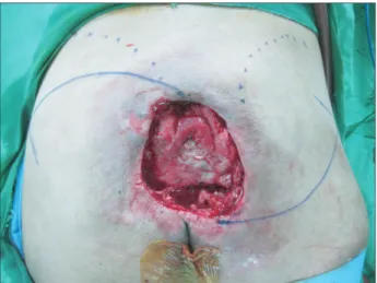
performed for reconstruction of the sacral pressure ulcer under general anesthesia in the prone position. First, the tissues of the ulcer and the scar tissues in the periphery of the ulcer were completely removed. The exposed parts of the sacrum resected and debridement was carried out until the healthy tissues of the skin were exposed. The size of the defective area was measured at 10.0×9.5 cm. The size of the defective area necessitated a resection on both sides of the wound. Initially, the borderline of the iliac crest was marked. Then, a resection line with a convex curve pointing towards the superior direction was drawn beginning from the proximal end of the ulcer, passing through the left lateral direction, and then to the lateral outermost edge of the hip.
On the right side of the ulcer, another resection line with a convex curve pointing to the inferior direction was drawn from the distal end of the ulcer to the lateral outermost edge (Fig. 1).
The incision line arising from the lateral edge to the distal end (left incision line) or the proximal end (right incision line) was drawn to form an acute angle, so that it would improve mobility of the flap later. Accordingly, the gluteus maximus
Hatchet-type Gluteus Maximus Musculocutaneous Flap for Reconstruction of Sacral Pressure Sore
Sang Wook Bae, Tae Kang Lim, Hyong Suk Kim, Baek Yong Song
Department of Orthopedic Surgery, Eulji General Hospital, Eulji University, Seoul, Korea pISSN 1226-2706 eISSN 2288-6184 Arch Reconstr Microsurg 2014;23(1):25-28
CC This is an open-access article distributed under the terms of the Creative Commons Attribution Non-Commercial License (http://creativecommons.org/licenses/by-nc/3.0) which permits unrestricted noncommercial use, distribution, and reproduction in any medium, provided the original work is properly cited.
Copyright © 2014 by the Korean Society for Microsurgery. All Rights Reserved.
Received April 29, 2014 Revised May 7, 2014 Accepted May 9, 2014
Correspondence to: Baek Yong Song Department of Orthopedic Surgery, Eulji General Hospital, Eulji University, 68 Hangeulbiseong-ro, Nowon-gu, Seoul 139-711, Korea
Tel: +82-2-970-8260 Fax: +82-2-970-8254 E-mail: [email protected]
One of the most frequently used flaps for coverage of sacral skin and soft-tissue defects is the gluteus maximus musculocutaneous flap. These authors encountered two cases of sacral pressure sore, for which reconstructive surgery was performed, using the hatchet- shaped gluteus maximus musculocutaneous flap – a modified flap type. We report on our experience in treatment of these two cases, with an excellent outcome.
Key Words: Sacrum, Myocutaneous flap, Pressure ulcer, Gluteus maximus, Hatchet
ARMS
Case ReportArchieves of Reconstructive Microsurgery
Arch Reconstr Microsurg Vol. 23. No. 1. May 2014
26 www.e-arms.org
was exposed as the skin and subcutaneous tissues were dissected. The skin on the left and the tissues of the gluteus maximus constituted a myocutaneous flap with the superior gluteal artery as the pedunculated vessel. This made a direct search for the superior gluteal artery unnecessary. Thus, the formation of a hatchet-shaped musculocutaneous flap was ascertained (Fig. 2). Then, a part of the origin of the gluteus maximus at the medial aspect of the iliac crest and the proximal area of the sacrum was lifted to allow mobility of the flap. However, upon advancing the flap, the covering of the wound was found to be insufficient. Utilizing the similar approach, another musculocutaneous flap was made along the opposite incision line marked on the right side. Then,
both flaps were advanced together to achieve the complete covering of the wound (Fig. 3).
After inserting a drainage tube, the lateral end of the flap on the left side was first sutured using the V-Y advancing method. This made additional skin graft for the donor skin unnecessary. Wound suturing and the ulcerated area covering were possible without having tension of the tissues in both flaps. The hemodynamic status of the operated area and its vicinity for this patient was stably maintained. The drainage tube was removed three days after surgery, and the suture material was removed two weeks after surgery.
The ulcerative wound was successfully sutured and healed by the first operation without postoperative complications, such as hematoma, infection or wound fissure (Fig. 4). No ulcer recurrence was found during the follow-up observation Fig. 1. Th e defect of 49-year-old male patient following the resection
of the sacral sore.
Fig. 2. Th e hatchet-shaped musculocutantous fl ap was prepared and advanced into the defect.
Fig. 3. Th e sutured hatchet fl ap, suffi ciently covering the large defect.
Fig. 4. The defect healed without complications postoperative 6 months. No additional surgical procedure was needed. Note that there are no contour diff erence remaining in the surgical fi eld.
Sang Wook Bae, et al. Hatchet-type Musculocutaneous Flap for Reconstruction of Sacral Pressure Sore
27
www.e-arms.org period of one year after surgery. These authors managed
two cases of sacral pressure ulcer using the same approach.
Another case involved a patient with paralyzed lower extremities, having an ulcer size of 7.0×6.5 cm. Unlike the method utilized for the first case, an attempt was made to use a unilateral flap to cover the defective area. Unfortunately, this patient developed a complication of delayed wound healing in the medial aspect of the flap (the center and distal area of the ulcer), two weeks after surgery. Thus, debridement and closure of the delayed wound were performed one time to attain the successful recovery of the ulcer.
DISCUSSION
Various approaches have been used for surgical management of pressure ulcer of the sacrum or the hip, which include regional flap, skin grafting, perforator flap, free flap and others. Among them, the musculocutaneous flap of the gluteus maximus is one of the most widely used method.1-4 With respect to the gluteus maximus flap, a flap separation is technically easy, while it has an abundance of blood circulation and excellent elasticity due to a large volume of the muscle. Also, with it, it is possible to supplement the defective area and has the merit of facilitation in maintaining muscular functions.2,3 Conventionally, a surgical approach utilizing the gluteus maximus flap designed as an island flap and its mobilization has been widely used.
However, surgery utilizing the hatchet-shaped gluteus maximus musculocutaneous flap, as seen in this case report, has not yet been reported in Korea.
Demirseren et al.5 first reported a hatchet-shaped myocutaneous flap, which had been utilized in the management of a trochanteric femur defect for 4 cases in 2003. At that time, the myocutaneous flap of the tensor muscle of fasci lata had been used. They also reported that previous utilized flaps of the tensor muscle of fasci lata were flap operations involving a posterior transposition.
This method has disadvantages of 1) unsatisfactory blood circulation in the distal portion of the flap, and 2) development of a dog-ear deformation. They concluded that a hatchet-shape design would solve such disadvantages. Later, Jósvay et al.6 reported successful treatment results utilizing a hatchet-shape design for the gluteus maximus myocutaneous
flap for 71 cases involving 54 patients in 2005. They applied this type of flap for various areas including the sacrum, the ischium, and the great trochanter of the femur. Jósvay et al.6 also reported that, based on the successful experience in pressure ulcer cases early on, it had been possible to have an effective treatment of not only pressure ulcers, but also non- pressure ulcers seen in myelomeningocele, Crohn’s disease, and others. Together, they reported the advantages of such method: that designing of the incision line would be easy, the operation time would be short, and there would be less hemorrhage and external deformation after surgery. They also asserted that it would have a merit of utilizing both sided flaps in cases of large-size defects.
Through these previous studies, clinicians may realize that the hatchet-shape musculocutaneous flap is an effective approach for flap operation, which would be applicable to defects of multiple dimensions. Owing to the advantages of easy flap design and not requiring isolations of the pedicle vessels, we also performed the surgery with ease and were able to attain successful covering of the defect without complications, despite the fact that we had limited experience in myocutaneous flap design. We realized that the aspect of incorporating either a unilateral or bilateral flap, depending on the defect size and flap mobility, would be the advantage that could widen our choice of operation. Moreover, owing to the ability of maintaining the merit of the thick and elastic gluteus maximus musculocutaneous flap, the patient with paralyzed lower extremities could safely move around in wheelchair without ulcer recurrence. Favorable results could be attained from patients who had been vulnerable to ulcer development and recurrence. In comparison with island flap, no other scar except for simple skin incision lines remained after surgery. The benefit of the ability to maintain the natural contour of the hip could also be observed. The consideration is that the gluteus maximus musculocutaneous flap employing a hatchet-shaped design is an effective approach for flap surgery, even for inexperienced operators through modification of flap designs while retaining the advantage of the gluteus maximus flap that have been widely used before.
Jósvay et al.6 reported complications of hatchet-type flaps among 71 cases of flap operations, which included seroma (4 cases), hematoma (2 cases), repeated surgery for the management of hemorrhage (1 case), necrosis of the
Arch Reconstr Microsurg Vol. 23. No. 1. May 2014
28 www.e-arms.org
flap border and delayed wound healing (1 case), and ulcer recurrence (2 cases). In view of these complications, the risk of general complications that develop after flap recovering surgery of pressure ulcer is the same as that of other flap operations. One of two cases of this report developed wound fissures. This adverse effect was due to a mistaken decision of estimating the ulcer size being relatively smaller than it was, in designing the gluteus maximus flap, and also due to the result of closure without securing adequate flap mobility. It suggests that the center of the ulcer, at which either a unilateral or bilateral flaps are joined together after flap translocation, can be the most vulnerable area. In an attempt to prevent such adverse effect, clinicians should consider the aspect that flap mobility is sufficiently secured and further attention must be paid to closure of the center of a flap’s medial aspect as well as wound management.
REFERENCES
1. Borman H, Maral T. The gluteal fasciocutaneous rotation- advancement flap with V-Y closure in the management of sacral pressure sores. Plast Reconstr Surg 2002;109:2325-9.
2. Fisher J, Arnold PG, Waldorf J, Woods JE. The gluteus maximus musculocutaneous V-Y advancement flap for large sacral defects. Ann Plast Surg 1983;11:517-22.
3. Ohjimi H, Ogata K, Setsu Y, Haraga I. Modification of the gluteus maximus V-Y advancement flap for sacral ulcers: the gluteal fasciocutaneous flap method. Plast Reconstr Surg 1996;98:
1247-52.
4. Ramirez OM, Orlando JC, Hurwitz DJ. The sliding gluteus maximus myocutaneous flap: its relevance in ambulatory patients. Plast Reconstr Surg 1984;74:68-75.
5. Demirseren ME, Gökrem S, Ozdemir OM, Katircioğlu A, Can Z, Serel S. Hatchet-shaped tensor fascia lata musculocutaneous flap for the coverage of trochanteric pressure sores: a new modification. Ann Plast Surg 2003;51:419-22.
6. Jósvay J, Sashegyi M, Kelemen P, Donáth A. Clinical experience with the hatchet-shaped gluteus maximus musculocutaneous flap. Ann Plast Surg 2005;55:179-82.