– 127 –
대한근전도∙전기진단의학회지 11(2):127~130, 2009.
J Korean EMG Electrodiagn Med
O’Sullivan-McLeod 증후군 1례
성균관대학교 의과대학 강북삼성병원 신경과a, 연세대학교 의과대학 신경과학교실b
백장현a∙서범천a∙김승민b∙선우일남b
– Abstract –
A Case of Patient with O’Sullivan-McLeod Syndrome
Jang-Hyun Baek, M.D.
a, Bum Chun Suh, M.D.
a, Seung Min Kim, M.D.
b, Il Nam Sunwoo, M.D.
bDepartment of Neurology, Kangbuk Samsung Hospital, Sungkyunkwan University School of Medicine, Seoul
a, Department of Neurology, Yonsei University College of Medicine, Seoul
b, Korea
O’ Sullivan-McLeod 증후군은 서서히 진행하는 팔의 위약과 위축을 특징으로 하는 드문 아래운동신경세포 질환이다. 이는 팔에 국한된 척수근위축증의 한 형태로 여겨지는데, 전형적인 증상과 경과를 보인 증례를 경 험하였기에 보고하고자 한다. 60세 남자가 4년 동안 서서히 진행한 우측 손의 쇠약과 내재근 위축을 주소로 내원하였다. 이후 10년 이상의 추적 관찰기간 동안 우측 손가락 및 손목 폄근의 쇠약과 경미한 좌측 손의 쇠약이 추가로 발생하였으나 다리 쇠약이나 팔다리의 감각 증상은 없었다. 팔의 심부건반사는 경미하게 감 소되었으나 다리에서는 정상이었다. 환자에게 특별한 질환력이나 가족력은 없었다. 좌측 새끼벌림근 (abductor digiti minimi)에서 복합근육활동전위의 진폭이 감소한 것 이외에 운동 및 감각신경전도검사는 모두 정상 소견이었으며, 추적 검사에서도 특별한 변화를 보이지 않았다. 그러나 바늘근전도검사에서는 양 측 팔에 국한된 신경원성 변화가 관찰되었고, 이는 추적 기간에 걸쳐 서서히 진행하였다. 면역억제제 (steroid and azathioprine)를 투약하였지만 병의 경과를 멈추거나 호전시키지는 못 하였다.
Key Words: O’Sullivan-McLeod syndrome, Motor neuron disease, Spinal muscular atrophy
Address reprint requests to Il Nam Sunwoo, MD
Department of Neurology, Yonsei University College of Medicine
TEL: 82-2-2228-1602, FAX: 82-2-393-0705, E-mail: [email protected] 투고일: 2009년 7월 15일, 수정일: 2009년 9월 18일, 게재확정일: 2009년 10월 5일
Introduction
O’ Sullivan and McLeod reported six patients with atrophy and weakness confined to the dis- tal muscles of the upper limbs in 1978.
1It is thought to be a variant of spinal muscular atrophy with unique and distinct clinical fea- tures. Here, we report a case of O’ Sullivan- McLeod syndrome in Korean.
Case
In 1997, a 60-year-old man came to the hos-
pital due to weakness of right hand grasp with
muscle wasting of right first dorsal
interosseous muscle. The symptoms were start-
ed from 4 years ago, and he had no specific
occupational or family histories. Initially, right
hand grasp was MRC grade 4 and there were
김승범∙이인식∙임정훈∙고성은∙이종민∙김세원∙박진영
no definite sensory symptoms or signs. All deep tendon reflexes of bilateral upper and lower extremities were normal and symmetrical. No upper motor neuron signs were noticed. The motor and sensory nerve conduction study of right arm was within normal range including the amplitudes of compound motor action potentials (CMAPs) and sensory nerve action potentials (SNAPs) (Table 1). But the needle
electromyographic (EMG) study showed chronic neurogenic change (reduced recruitment of high amplitude and long duration motor unit potentials) with reduced insertion activities in right first dorsal interosseous and abductor dig- iti minimi muscles (Table 2). No definite fibril- lations/positive sharp waves or fasciculations suggesting the active denervating processes were found. Other muscles of right forearm
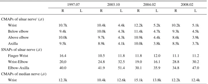
– 128 – Table 1. Serial Changes of Nerve Conduction Studies
1997.07 2003.10 2004.02 2008.02
R L R L R L R L
CMAPs of ulnar nerve1(㎶)
Wrist 10.7k 10.4k 4.4k 12.2k 5.2k 10.2k 5.1k
Below elbow 9.4k 10.0k 4.3k 11.4k 4.7k 9.3k 4.5k
Above elbow 10.0k 9.7k 4.3k 10.9k 4.4k 8.6k 3.9k
Axilla 9.5k 8.9k 4.1k 10.0k 3.8k 8.5k 3.7k
SNAPs of ulnar nerve (㎶)
Finger-Wrist 16.4 10.5 11.8 11.8 12.0 11.1 11.2
Wrist-Elbow 20,0 24.8 32.5 19.0 16.1 28.8 30.2
Elbow-Axilla 40.0 41.9 51.4 30.1 35.9 34.8 47.0
CMAPs of median nerve (㎶)
Wrist 12.3k 10.4k 12.6k 15.1k 13.8k 12.2k 12.4k
1Recorded at abductor digiti minimi muscle. R, right side; L, left side; CMAPs, compound motor action potentials; SNAPs, sensory nerve action potentials
Table 2. Serial Changes of Needle Electromyographic Findings1
1997.07 2003.10 2004.02 2008.02
R L R L R L R L
First dorsal interosseous -2 Y3 + Y + Y + Y + Y + Y
Abductor digiti minimi - Y3 + Y
Flexor carpi ulnaris - N3 - N + Y - Y
Abductor pollicis brevis + N + Y + N + Y
Extensor digitorum communis - N3 + Y + Y + Y
Brachioradialis - N3 + Y
Triceps - N3 - N + N + Y
Biceps brachii - N3 + N + N + Y
Deltoid - N3 + N - N
Vastus lateralis - N - N - N - N
Tibialis anterior - N - N - N - N
Medial gastrocnemius - N - N - N - N
1The electromyographic finding of an individual muscle is indicated by spontaneous activities (fibrillation and/or positive sharp wave) and neurogenic motor unit potentials (MUPs). 2Presence and absence of spontaneous activities are indicated by ‘+’ and ‘-’.
3Presence and absence of neurogenic MUPs are indicated by ‘Y’ and ‘N’. R, right side; L, left side.
and upper arm were normal. At that time, we suspected to have some sort of ulnar neuropa- thy at the distal Guyon’ s canal but not treat- ed. He visited the hospital again in 2003 because of fingers drop (MRC grade 3) with wasting of forearm muscles in right upper extremity. The nerve conduction study was essentially same with the study of 1997 includ- ing normal radial sensory nerve conductions, but low amplitudes of CMAPs in left ulnar nerve were found incidentally. EMG study revealed mild denervation potentials in right first dorsal interosseous, abductor pollicis brevis with giant MUAPs and reduced recruitment.
Moderate denervation potentials were noticed in right extensor digitorum communis and extensor indicis proprius muscles. EMG study of right flexor carpi ulnaris, extensor carpi radialis and triceps muscles was normal. By asking about the left hand weakness, the patient answered that the weakness of left hand grasp appeared a couple of years before, but no definite weakness was seen on manual motor test. Laboratory tests and immunologic studies including anti-ganglioside antibodies (anti-GM1 and anti-GQ1b antibody) were absolutely normal. Follow-up nerve conduction study in 2004 revealed no essential change in both upper extremities and no abnormality in legs. EMG study of right upper limb showed mild denervation potentials with chronic neuro- genic MUAPs in upper arm muscles including triceps, biceps and deltoid in addition to right forearm and hand muscles. In left arm, first dorsal interosseous and abductor pollicis brevis muscles are involved. EMG studies of both legs were normal. Steroid and azathioprine were given for more than three years but did not show any benefit on disease progression. Insidi- ous progression of weakness of right finger flexors and wrist extensors were followed sequentially.
On last examination in 2008, there were severe muscle weakness of right hand including grasping, finger spreading, and fingers exten- sors (MRC grade 2) and moderate weakness of right wrist flexors and extensors (MRC grade 3) with mild weakness of left hand movement
(MRC grade 4). Elbow and shoulder looked not involved clinically. The electrodiagnostic study is similar to the previous study.
Discussion
The clinical characteristics of this man are very slowly progressive, asymmetrical, and dis- tal dominant muscle weakness confined to upper extremities, without specific peripheral nerve distribution. There was no evidence of peripheral neuropathy or myopathy clinically and electrophysiologically. Differential diagno- sis in this patient includes multifocal motor neuropathy (MMN), benign focal amyotrophy (BFA), and a kind of motor neuron disease like amyotrophic lateral sclerosis (ALS) and spinal muscular atrophy (SMA). MMN can show slowly progressing asymmetrical distal hand weakness, which mimics lower motor neuron disease.
2But no evidence for demyeli- nating conduction block on nerve conduction study for more than 10 years and negative anti-GM1 antibody are against the diagnosis of MMN. BFA is also characterized by slowly pro- gressing weakness confined to upper extremi- ties asymmetrically.
2,3However, symptoms of this disease usually begin at late teen or early twenties and progression is arrested within 5 years in 73% of patients.
3Absence of upper motor neuron signs and very longer progressing course are critical points for differential diag- nosis of ALS or its variants.
2Traditionally, the distribution of weakness of SMA is symmetri- cal and worse in proximal leg muscles, Howev- er, many authors reported the heterogeneity in weakness distribution, genetic trait, and onset age of distal SMA.
4-6Several cases for ‘distal SMA’ showed the involvement of both lower and upper extremities, which appeared first and was worse in lower extremities and spread out to upper extremities. All these reports had specific genetic transmission traits: autosomal dominant or recessive, and their onset ages were in childhood or very early adulthood.
Based on these reports, our presenting case seems not to be corresponded with the current concept for ‘distal SMA’ .
O’Sullivan-McLeod Syndrome
– 129 –
김승범∙이인식∙임정훈∙고성은∙이종민∙김세원∙박진영
Harding et al reported 18 cases with chronic asymmetrical SMA, which had uncertain genetic backgroud.
7Among them 5 cases had the weakness confined to distal upper extremi- ties and their mean age of onset was 32 years.
These cases were very similar to the patients reported by O’ Sullivan and McLeod and they considered these patients as a unique disorder distinct from known variants of SMA. O’ Sulli- van-McLeod syndrome is defined as an asym- metric lower motor neuron disease confined to upper extremities with very long and slow pro- gression of unknown etiology and pathogenesis, and its symptom was usually started from mid- dle age, ranging from third to fifth decade.
1, 7-9The lack of a specific laboratory test for sur- vival motor neuron (SMN) gene or imaging studies for cervical spine may be a limitation in differential diagnosis. However, we think the most possible diagnosis of our presenting case is O’ Sullivan-McLeod syndrome, clinically and electrodiagnostically, and agree that O’
Sullivan-McLeod syndrome may be a kind of unique variant of distal asymmetrical SMA.
The pathogenesis of O’ Sullivan-McLeod syn- drome is unknown. O’Sullivan-McLeod syn- drome generally has been assumed to be due to cervical cord compression, however, Kawano et al reported a progressing case of O’ Sullivan- McLeod syndrome even after cervical laminec- tomy.
8They, then, proposed an immunologic contribution in the mechanism of O’ Sullivan- McLeod syndrome based on a patient improved by intravenous immunoglobulin. Even though they reported the improvement of weakness for a few months, treatment for O’Sullivan- McLeod syndrome is also uncertain. Our pre- senting case did not respond to prednisolone and azathioprine at all.
This patient might be a typical case of O’
Sullivan-McLeod syndrome. The clinical char- acteristics include very slowly progressive asymmetrical muscle atrophy and weakness
confined to upper extremities from distal hand, starting at the age of late fifties. Immunother- apy with steroid and azathioprine does not pre- vent the disease progression. This syndrome, we think, may be a kind of distal asymmetri- cal SMA and should be included in differential diagnosis of adult onset motor neuron disease
참고문헌
01. O'Sullivan DJ, McLeod JG: Distal chronic spinal muscular atrophy involving the hands. J Neurol Neurosurg Psychia- try 1978: 41: 653-658.
02. Boonyapisit K, Shapiro BE: Atypical motor neuron dis- eases. In: Katirji B, Kaminski HJ, Preston DC, Ruff RL, Shapiro BE, editors. Neuromuscular disorders in clinical practice, 1st ed, Massachusetts: Butterworth-Heinemann, 2002, pp454-461.
03. Hirayama K: Juvenile muscular atrophy of distal upper extremity (Hirayama disease). Intern Med 2000: 39: 283- 290.
04. Adams C, Suchowersky O, Lowry RB: Congenital autoso- mal dominant distal spinal muscular atrophy. Neuromus- cul Disord 1998: 8: 405-408.
05. Panigrahi I, Kesari A, Phadke SR, Mittal B: Clinical and molecular diagnosis of spinal muscular atrophy. Neurol India 2002: 50: 117-122.
06. Russman BS: Spinal muscular atrophy: clinical classifica- tion and disease heterogeneity. J Child Neurol 2007: 22:
946-951.
07. Harding AE, Bradbury PG, Murray NM: Chronic asym- metrical spinal muscular atrophy. J Neurol Sci 1983: 59:
69-83.
08. Kawano Y, Nagara Y, Murai H, Kikuchi H, Ohyagi Y, Kira J: Slowly progressive distal muscular atrophy of the bilateral upper limbs (O'Sullivan-McLeod syndrome) par- tially alleviated by intravenous immunoglobulin therapy.
Intern Med 2007: 46: 515-518.
09. Petiot P, Gonon V, Froment JC, Vial C, Vighetto A: Slow- ly progressive spinal muscular atrophy of the hands (O'Sullivan-McLeod syndrome): clinical and magnetic resonance imaging presentation. J Neurol 2000: 247: 654- 655.
– 130 –