J Korean Soc Radiol 2018;79(2):63-67 https://doi.org/10.3348/jksr.2018.79.2.63
INTRODUCTION
Intercostal schwannomas, also known as intercostal nerve neurilemmomas or neurinomas, are uncommon encapsulated neoplasms that originate in nerve sheaths of intercostal nerves (1, 2). Fewer than 10% of primary neural tumors of the chest originate peripherally from intercostal nerves; most neural tu- mors of the chest arise in the mediastinum (3). They are almost invariably slow-growing nonaggressive neoplasms and usually manifest clinically as a painless mass without neurologic symp- toms (4). Radiologically, they are well-marginated soft-tissue mass with contrast enhancement, and may contain areas of fat or cystic degeneration (5). Herein, we present a case of inter- costal schwannoma with imaging findings of various modali- ties involving typical magnetic resonance imaging (MRI) and discuss the clinical and radiographic manifestations.
CASE REPORT
A 61-year-old female, having a nonspecific past history, pre- sented to pulmonology department of our hospital with cough and sputum for about 10 days. Physical examination by a pul- monologist showed clear breathing sound. Results of her labo- ratory evaluation and pulmonary function test were unremark- able. However, on chest radiograph, there was nodular lesion abutting on right upper chest wall (Fig. 1A). A contrast en- hanced computed tomography (CT) scan was performed for further evaluation of this lesion. On pre-enhanced CT, about 1.5 cm sized, well-circumscribed homogenous mass was noted in right chest wall, just inferior to the 5th rib, and the mean atten- uation of the lesion was 3 Hounsfield unit (HU). On CT scans acquired after contrast material administration, the mean at- tenuation of mass was increased to 28. 7 HU and it showed pe- ripheral enhancement (Fig. 1B). The lesion-lung interface was smooth, but it was hard to distinguish whether the mass was lo-
MRI Findings of Intercostal Schwannoma: A Case Report
늑간 신경초종의 MRI 소견: 증례 보고Ha Yan Sim, MD
1, Ik Yang, MD
1, Hye-Suk Hong, MD
1, Ji Young Woo, MD
1, Ji-Young Hwang, MD
1, Jin Hee Moon, MD
1, Han Myun Kim, MD
1, Hye Jeong Kim, MD
1, Sook Min Hwang, MD
1,
Mi Kyung Shin, MD
2, Hee Young Kim, MD
1*
Departments of 1Radiology, 2Pathology, Kangnam Sacred Heart Hospital, Hallym University College of Medicine, Seoul, Korea
Intercostal schwannomas are uncommon, encapsulated neoplasms that originate in nerve sheaths of intercostal nerves. They account for less than 10% of primary neu- ral tumors of the chest wall. Herein, we report a pathologically confirmed case of intercostal schwannoma with typical magnetic resonance imaging findings.
Index terms Schwannoma Intercostal Nerve
Magnetic Resonance Imaging
Received February 27, 2018 Revised April 13, 2018 Accepted April 21, 2018
*Corresponding author: Hee Young Kim, MD Department of Radiology, Kangnam Sacred Heart Hospital, Hallym University College of Medicine, 1 Singil-ro, Yeongdeungpo-gu, Seoul 07441, Korea.
Tel. 82-2-829-5241 Fax. 82-2-832-1845 E-mail: younglady@hallym.or.kr
This is an Open Access article distributed under the terms of the Creative Commons Attribution Non-Commercial License (https://creativecommons.org/licenses/by-nc/4.0) which permits unrestricted non-commercial use, distri- bution, and reproduction in any medium, provided the original work is properly cited.
cated at extrapleural space or not. These CT findings of the lesion suggested a benign soft tissue tumor but could not specify the diagnosis, so we recommended MRI scan. On MRI, the mass was clearly on the lateral side of extrapleural fat which showed as thin high signal intensity lining on T1- and T2-weighted imag-
es, so it proved to be a extrapleural tumor that originated from intercostal soft tissue along the course of 5th intercostal nerve in the right side of chest wall. The mass showed low signal in- tensity (similar to that of adjacent muscles) on T1-weighted MR images, and intermediate to high signal intensity with in-
A B
C D
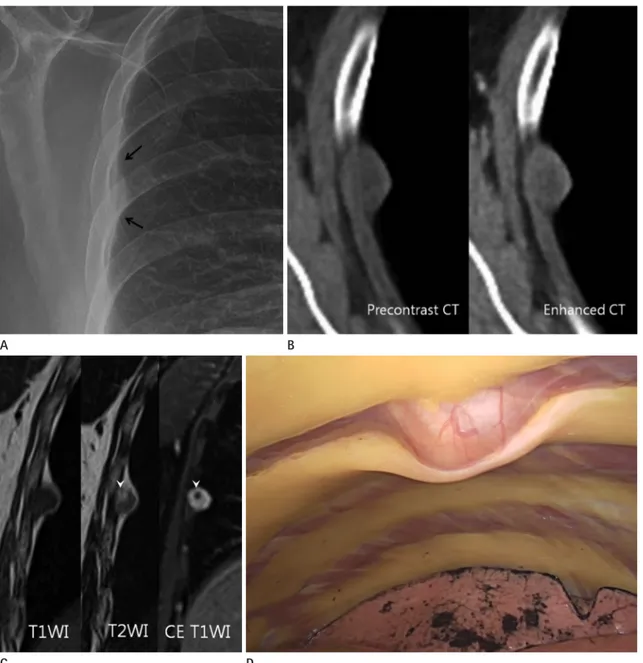
Fig. 1. A 61-year-old woman with intercostal schwannoma involving the right 5th intercostal nerve.
A. Chest radiography shows a nodular lesion abutting on right upper chest wall, just inferior to 5th rib (arrows).
B. Pre-enhanced CT demonstrates a 1.5 cm nodule showing heterogeneous attenuation in the chest wall, just inferior to the 5th rib. After CE, the mass shows thin peripheral enhancement.
C. The mass reveals low signal intensity on T1WI, intermediate signal intensity with internal high signal intensity focus (arrowhead) on T2WI. On gadolinium-enhanced fat-suppressed T1WI, it shows bright enhancement, with tiny non-enhancing area (arrowhead) corresponding to the high signal intensity focus on T2WI.
D. Intraoperative thoracoscopic image demonstrates a well circumscribed mass abutting on inferior margin of the right 5th rib showing mass ef- fect on the chest wall.
CE = contrast enhancement, CT = computed tomography, T1WI = T1-weighted image, T2WI = T2-weighted image
ternal high signal intensity focus on T2-weighted images. On gadolinium-enhanced fat-suppressed T1-weighted images, it showed bright enhancement with small non-enhancing area that was consistent with high signal intensity focus on T2-weighted image, suggesting central cystic degeneration (Fig. 1C). Based on these MRI findings, the radiologic differential diagnosis was assumed as neurogenic tumor that originated in right 5th inter- costal nerve. The patient underwent mass excision via video- assisted thoracoscopic surgery. The mass was 1.5 cm sized and located at inferior aspect of right 5th rib, abutting superficial surface of parietal pleura (Fig. 1D). There was no remarkable bony erosion on right 5th rib. The final pathologic diagnosis was intercostal schwannoma (Fig. 1E, F).
DISCUSSION
Schwannomas are type of benign peripheral nerve sheath neo- plasm. The tumor has a propensity to involve head, neck, flexor surfaces of extremities, posterior mediastinum, and retroperito- neum (6). Chest wall is an uncommon location for schwanno- mas and they arise from spinal nerve roots or intercostal nerves (7), but they may involve any thoracic nerves (including the phrenic or vagus nerve) (5). The incidence of intercostal schwan- nomas is below 10% of all primary neurogenic thoracic tumors (1). Schwannomas typically occur in patients between 20 years and 50 years of age, with no sex predilection (2). They are al- most slow-growing nonaggressive neoplasms and usually man-
ifest clinically as a painless mass without neurologic symptoms, unless the mass has become large enough to compress the adja- cent nerve (4, 8).
Grossly, small tumors tend to be spheroid, firm, and well cir- cumscribed, whereas larger tumors are ovoid or irregularly lob- ulated (2). Chest radiographs do not usually depict small schwan- nomas, but when the lesion is delineated, it shows a smoothly marginated, oval mass (2, 5). Unenhanced CT scans of schwan- noma show a well-circumscribed homogeneous mass with at- tenuation slightly less than or equal to that of muscle (2). Schwan- nomas may contain areas of low attenuation corresponding to fat or cystic degeneration, and calcifications may be primarily in a peripheral pattern, particularly in long-standing lesions with advanced degeneration (8). Adjacent osseous pressure erosion may also be present, but it is relatively rare, and presence of bone erosion without destruction indicates the benign nature and slow growth rate of the lesion (2, 5). On contrast-enhanced CT, the attenuation of the mass is equal to or slightly greater than that of muscle (2). When the mass is small, the mass may show homogeneous enhancement, while larger schwannomas can contain nonenhancing area and show more heterogeneous en- hancement due to cystic and hemorrhagic changes (2, 5).
MRI shows low to intermediate signal intensity (equal to or slightly greater than that of muscle) on T1-weighted images, and shows relatively high signal intensity on T2-weighted images.
On gadolinium-enhanced imaging, schwannomas show intense enhancement of solid components (2, 5). The larger the schwan- Fig. 1. A 61-year-old woman with intercostal schwannoma involving the right 5th intercostal nerve.
E. Photomicrograph shows an Antoni A zone of compact cellular architecture with nuclear palisading (Verocay bodies) and Antoni B zone of spindle or rounded cells within loose myxoid stroma (hematoxylin and eosin stain, × 100).
F. Photomicrograph shows diffuse strong positive finding in S100 immunohistochemical stain (× 100).
E F
noma, the more heterogeneous it will appear on all sequences due to cystic degeneration, hemorrhage, or both. Regions of very high signal intensity on T2-weighted imaging correspond to cystic degeneration and may only show peripheral zone en- hancement or no enhancement associated with the very high signal intensity, and those findings favor schwannoma (5). Sev- eral foci of marked hyperintensity on T2-weighted image due to multifocal cystic degeneration, which is so-called bead-like appearance, are one of the characteristic MRI finding of schwan- noma (9). Also, as in our case, MRI can clearly demonstrate ex- trapleural fat which shows high signal intensity on T1- and T2- weighted images, so it can help distinguish extrapleural tumors from pleural tumors.
These imaging findings of schwannomas are similar to those seen with neurofibromas and, in many cases, cannot be distin- guished (8), so fine-needle cytology or surgical biopsy is often needed to establish the diagnosis (10). However, some features can help differentiate these two lesions. Pathologically, schwan- nomas are fusiform masses that are eccentrically located in re- lation to the involved nerve, so in imaging studies, schwanno- mas tend to be eccentrically positioned and the parent nerve can be identified, whereas centrally located masses suggest neurofi- bromas. In addition, heterogeneous appearance with degenera- tion and cystic cavitation are much more common in schwan- nomas than in neurofibromas (8). Areas designated as Antoni A are more organized and are composed of cellular spindle cells arranged in short bundles or interlacing fascicles. Antoni B ar- eas are hypocellular and contain more myxomatous loosely ar- ranged tissue (7). In our case, upper margin of the mass was just abutting on the lower margin of the rib where the intercos- tal nerve located, and extended mainly to the inferior direction, resulting eccentric position related to intercostal nerve. And cys- tic degeneration was also noted within the mass. From these im- aging features, the diagnosis of our case could be intercostal schwannoma more likely, rather than neurofibroma.
Besides neurofibroma, there are other thoracic neurogenic tumors that should be considered as differential diagnosis, in- cluding neuroblastoma, paraganglioma, ganglioneuroma, and malignant peripheral nerve sheath tumor. There are some im- aging features that can help differentiate these tumors from schwannoma. Neuroblastomas can show coarse or curvilinear calcifications on CT. Paragangliomas show marked enhance-
ment due to increased vascularity and may show salt-and-pep- per appearance from vessel signal flow voids within the mass on T2-weighted MR images. Ganglioneuromas are usually elongated and oriented on a vertical axis following the direction of the sympathetic chain, and may show whorled appearance of low signal intensity bands on T1- and T2-weighted MR images.
And, malignant peripheral nerve sheath tumors (MPNSTs) usually show compression or destruction of adjacent structures and pleural abnormalities indicating malignancy. Some MRI features are useful in diagnosis of MPNSTs, and if a tumor has two or more features, it should be considered highly suspicious of malignancy. Those features are increased largest dimension of the mass (> 5 cm), presence of peripheral enhanced pattern, presence of perilesional edema, and presence of intratumoral cystic change. In our case, though the mass showed tiny focus of cystic change, there was no other features that suggesting malig- nancy, so it is more likely to be benign (5).
Local resection of the mass is sufficient treatment for most of the smaller schwannomas of intercostal nerve, and thoracosco- py is usually the preferred surgical approach. Larger or more aggressive tumors require resection of the chest wall (3). The possibility of recurrence or malignant transformation is consid- ered extremely low (10).
In summary, we present a case of schwannoma of intercostal nerve that shows obvious and typical imaging features, espe- cially on MRI. Although pathologic confirm is recommended for diagnostic confirmation of this entity, characteristic MRI fea- tures may provide a clue to the diagnosis and play an important role in the diagnosis of schwannoma.
REFERENCES
1. Meyer C, Rodepeter F, Bartsch D, Kirschbaum A. Intercostal neurinoma: a rare cause of persistent thoracic pain. Thorac Cardiovasc Surg Rep 2014;3:48-50
2. Tateishi U, Gladish GW, Kusumoto M, Hasegawa T, Yokoya- ma R, Tsuchiya R, et al. Chest wall tumors: radiologic find- ings and pathologic correlation: part 2. malignant tumors.
Radiographics 2003;23:1491-1508
3. McClenathan JH, Bloom RJ. Peripheral tumors of the inter- costal nerves. Ann Thorac Surg 2004;78:713-714
4. Nam SJ, Kim S, Lim BJ, Yoon CS, Kim TH, Suh JS, et al. Imag-
ing of primary chest wall tumors with radiologic-pathologic correlation. Radiographics 2011;31:749-770
5. Pavlus JD, Carter BW, Tolley MD, Keung ES, Khorashadi L, Li- chtenberger JP 3rd. Imaging of thoracic neurogenic tumors.
AJR Am J Roentgenol 2016;207:552-561
6. Hwang ST, Sung DJ, Sim KC, Han NY, Park BJ, Kim MJ, et al.
Radiologic findings of renal schwannoma: a case report and literature review. J Korean Soc Radiol 2018;78:289-294 7. Nakazono T, White CS, Yamasaki F, Yamaguchi K, Egashira
R, Irie H, et al. MRI findings of mediastinal neurogenic tu- mors. AJR Am J Roentgenol 2011;197:W643-W652
늑간 신경초종의 MRI 소견: 증례 보고
심하얀
1· 양 익
1· 홍혜숙
1· 우지영
1· 황지영
1· 문진희
1· 김한면
1· 김혜정
1· 황숙민
1· 신미경
2· 김희영
1*
늑간 신경초종은 늑간 신경에서 기원하는 피막으로 둘러싸인 드문 종양이며, 흉곽에서 발생하는 신경성 종양의 10% 미만 을 차지한다. 우리는 특징적인 자기공명영상 소견을 보이는 병리학적으로 확진된 늑간 신경초종에 대한 증례를 보고한다.
한림대학교 의과대학 강남성심병원 1영상의학교실, 2병리학교실
8. Lin J, Martel W. Cross-sectional imaging of peripheral nerve sheath tumors: characteristic signs on CT, MR imaging, and sonography. AJR Am J Roentgenol 2001;176:75-82 9. Takanashi Y, Urabe N. [Schwannoma of the chest wall show-
ing a bead-like appearance;report of a case]. Kyobu Geka 2013;66:1027-1029
10. Kim KS, Ji SR, Kim HM, Kwon YJ, Hwang JH, Lee SY. Inter- costal nerve schwannoma encountered during a rib-latis- simus dorsi osteomyocutaneous flap operation. Arch Plast Surg 2015;42:800-802