우리나라에서 흔히 볼 수 있는 담도결석을 동반하는 담관염은 대부분 재발성화농성담관염이 그 원인으로 알려져 있으며 ( 1 , 2 ) , 담관결석과 담관협착 및 부종등에 의한 담도폐쇄로 인해 반복적 인 급성염증을 일으키며, 이로 인하여 환자는 패혈증을 유발하여 사망할 수도 있으므로 보다 정확한 급성염증의 진단과 이에 대 한 적극적인 치료가 필요하다 (3,4). 재발성 화농성 담관염의 영 상진단방법으로 초음파검사, 전산화단층촬영술(이하 C T로 약 함), 담관조영술을 이용하고 있다 (3-7). 초음파검사는 이 질환의
진단에 있어 일차검사로 시행하고 있으나, 병리상태를 보다 정확 하고 효과적으로 진단하고자 C T를 많이 이용하며 치료방침의 수립과 추적검사에 도움이된다 (2-6). 최근 나선식 C T를 이용하 면서 병변의 역동조영증강양상을 평가할 수 있게 되어, 간담도계 의 종양의 발견 및 특성화와 염증성질환의 진단에도 이용되고 있다 (8). 재발성화농성담관염의 경우 지금까지 재래식 C T를 이 용한 보고들이 있었으나 (2-4), 나선식 C T를 이용한 병변의 조영 증강양상에 대한 보고는 없다. 저자들은 재발성화농성담관염 환 자에서 흔히 동반되는 급성염증의 소견과 간실질위축의 원인으 로 문맥의 협착정도를 진단하는데 있어, 이중시기(동맥기 및 문 맥기) 나선식 C T의 유용성에 대하여 알아보고자 하였다.
목적 : 재발성화농성담관염 환자에서 급성염증의 동반여부와 간실질위축의 원인이 되는 문
맥의 협착정도를 진단하는데 있어 이중시기 (동맥기 및 문맥기) 나선식 C T의 유용성에 대 하여 알아보고자 하였다.
대상 및 방법 : 이중시기 나선식 C T를 시행하였고, CT, ERCP, 수술에 의해 재발성화농성담
관염으로 진단된 3 0명의 환자를 대상으로 하였다. 모든 환자에서 조영전 C T와, 조영제 주입 시작후 3 0초와 6 0 - 7 0초에 각각 동맥기와 문맥기영상을 얻었다. 급성염증과 C T소견의 연관성 을 알아보기 위하여 동맥기와 문맥기에서 담관주위간실질과 담관벽의 조영증강양상을 분석 하였으며, 급성염증의 진단은 임상소견과 검사실소견을 근거로 하였다. 만성염증에 의한 간 실질위축과 문맥협착정도와의 연관성을 알아보기 위하여 문맥기에서 병변부위의 문맥직경 을 측정하였고, 간위축이 없는 간실질에서 문맥직경과 비교하여 감소된 정도를 3단계 (경 도; 25% 이하, 중등도; 25% - 75%, 고도; 75% 이상)로 분류하였다.
결과 : 총 3 0명중 1 0명의 환자는 임상적으로 급성염증발현기에 C T를 시행하였고, 20명은 C T
시행 시에 급성염증소견이 없었다. 급성염증을 보였던 1 0예중 8예( 80% )는 동맥기에서 담관 주위간실질의 일시적인 조영증강을 보였으나, 급성염증이 없었던 2 0예의 경우 3예( 15 % )에 서만 이런소견을 보여, 급성염증의 발현과 동맥기에서 담관주위간실질의 일시적인 조영증강 사이에 유의한 상관관계가 있었다 (p<0.05). 동맥기에서 담관벽의 조영증강은 급성염증을 보 였던 1 0예중 3예( 30% )에서 보였고, 급성염증이 없었던 2 0예중 7예( 35% )에서도 관찰되어, 급성염증과 담관벽의 조영증강과는 유의한 상관관계가 없었다 (p>0.05). 문맥기에서 담관주 위간실질이나 담관벽의 조영증강은 급성염증이 있었던 예와 없었던 예사이에 유의한 차이 가 없었다 (p>0.05). 병변부위에서 간실질위축은 2 4예에서 보였고, 이중 경도의 문맥협착을 보인 예가 5예( 21%), 중등도협착이 1 4예( 58%), 고도협착이 5예( 21 % )였으며 간실질의 위축 정도와 비례하였다. 간실질위축이 없었던 6예에서는 모두 정상문맥을 보였다.
결론 : 이중시기 나선식 C T는 동맥기영상에서 급성염증을 시사하는 담관주위간실질의 일시
적인 조영증강을 볼 수 있으며, 문맥기영상에서 간실질위축의 원인으로 문맥의 협착정도를 평가하는데 도움이 되어 재발성화농성담관염 환자의 진단에 유용하다.
1충남대학교 의과대학 진단방사선과학교실
이 논문은 1 9 9 9년 7월 6일 접수하여 1 9 9 9년 1 0월 3 0일에 채택되었음.
재발성화농성담관염 : 이중시기 나선식 C T의 유용성1
정기호・조준식・신경숙・이세효・유호준・박진용・김영민
대상 및 방법
1 9 9 5년 1월부터 1 9 9 8년 9월까지 재발성화농성담관염으로 진 단된 환자중 이중시기 나선식 C T를 시행한 3 0명의 환자를 대 상으로 하였다. 이중 남자가 8명 여자가 2 2명이었으며, 연령분 포는 4 0세에서 8 2세로 평균 5 6세였다. 30명 모두에서 혈액생화 학검사, 초음파검사, 이중시기 나선식 CT, 내시경역행췌담관 조영술( E R C P )을 시행하였고, 이중 7명은 수술로, 23명은 임 상 및 영상소견으로 재발성화농성담관염으로 확진하였다.
C T는 HiSpeed advantage scanner(General Electric Medical sys- tem, Milwaukee, U.S.A.)를 사용하였다. 모든 환자에서 조영전 C T를 얻었고, 조영증강 C T는 1 3 0 - 1 50m l의 조영제를 4 m l / s e c 의 속도로 주입후 절편두께 7mm, 재구성 간격 7mm, 1:1 p i t c h로 조영제 주입시작후 3 0초와 6 0 - 7 0초에 각각 동맥기와 문맥기 영상을 얻었다. CT소견은 임상소견을 모르는 상태에 서 두명의 방사선과 의사의 합의하에 후향적으로 분석하였다.
동맥기와 문맥기에서의 담관벽 및 간실질의 조영증강양상과 간실질위축이 있는 예에서는 문맥의 협착정도와의 관계를 평 가하였다. 급성염증의 진단은 임상소견( C h a r c o t’s triad; 황달, 산통, 발열)과 검사실 소견을 근거로 하였다. 만성염증에 의한 간실질의 위축과 문맥의 협착정도와의 연관성을 알아보기 위 하여 병변부위의 문맥직경을 측정하였고, 간위축이 없는 간실 질에서 문맥직경과 비교하여 정상, 경도, 중등도, 고도감소(경 도; 25% 이하, 중등도; 25%-75%, 고도; 75% 이상)로 분류하 였다. 간실질위축은 간전체크기와 간실질두께의 감소 간윤곽 과 담도구조의 변화소견에 의하여 판단하였고 정량적 측정은 하지 않았다 (4,6). 통계적 분석은 x²t e s t를 이용하였고, p <0.05 일 때 통계적으로 유의성이 있는 것으로 평가하였다.
결 과
총 3 0명의 환자중 1 0명은 임상적으로 급성염증발현기에 C T 를 시행하였고, 20명은 C T시행 시에 급성염증소견이 없었다.
급성염증유무에 따른 간실질 및 담관벽 조영증강양상을 분석 한 결과, 급성염증을 보였던 1 0예중 8예( 80% )는 동맥기에서 담관주위간실질의 일시적인 조영증강을 보였으나 (Fig. 1), 급 성염증이 없었던 2 0예의 경우 3예( 15% )에서만 동맥기에서 담 관주위간실질의 일시적인 조영증강을 보여서, 급성염증의 발 현과 동맥기에서 담관주위간실질의 일시적인 조영증강사이에 유의한 상관관계가 있었다(p<0.05). 간실질의 조영증강은 3예 에서 균일한 양상을 보였고 나머지에서는 비균일한 조영증강 을 보였으며, 담관주위간실질에 국한되는 양상을 보였다. 동 맥기에서 담관벽의 조영증강은 급성염증을 보였던 1 0예중 3 예( 30% )에서 보였고, 급성염증이 없었던 2 0예중 7예( 35% )에 서도 관찰되어 (Fig. 2), 급성염증과 동맥기에 담관벽의 조영 증강과는 유의한 상관관계가 없었다(p>0.05) (Table 1). 문맥 기에서 담관주위간실질의 조영증강은 급성염증이 있었던 1 0 예중 2예( 20%), 급성염증이 없었던 2 0예중 5예( 25% )에서 보
Table 1. Correlation between Acute Inflammatory Symptoms with Findings of Two-Phase Spiral CT
Acute Inflammation Presence A b s e n c e Arterial phase
Transient periductal parenchymal enhancement* 8/10(80) 3/20(15) Ductal wall enhancement 3/10(30) 7 / 2 0 ( 3 5 ) Portal phase
Periductal parenchymal enhancement 2/10(20) 5/20(25) Ductal wall enhancement 5 / 1 0 ( 5 0 ) 1 3 / 2 0 ( 6 5 )
* Statistically significant p<0.05 numbers in parenthesis are percentage
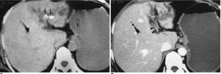
A B
Fig. 1. A 82-year-old woman with recurrent pyogenic cholangitis during acute inflammatory stage.
였으며, 담관벽의 조영증강은 급성염증이 있었던 1 0예중 5예 ( 50%), 급성염증이 없었던 2 0예중 1 3예( 65% )에서 보여, 급성 염증과 문맥기에서의 담관주위간실질 및 담관벽의 조영증강 과는 유의한 상관관계를 볼 수 없었다(p>0.05) (Table 1). 이 중 급성염증이 있고 문맥기에서 간실질조영증강을 보였던 2 예는 동맥기에서 문맥기보다 더 강한 조영증강을 보였고, 동 맥기에서 담관벽의 조영증강을 보였던 예에서는 모두 문맥기 에서도 조영증강을 보였다. 간실질위축은 2 4예에서 보였으며, 이중 1 6예는 좌엽외분절에, 6예는 우엽후분절에, 2예는 좌엽외 분절 및 우엽후분절에서 위축을 보였다. 간실질위축을 보였던 2 4예중 정상 혹은 경도의 문맥협착을 보인 예가 5예( 21 % ) (Fig. 2), 중등도가 1 4예( 58%) (Fig. 3), 고도협착이 5예( 21% ) (Fig. 4) 이었으며 간실질위축정도와 비례하였다. 간실질위축 이 없었던 나머지 6예에서는 모두 정상문맥을 보였다 ( T a b l e 2). 그 외 동반된 C T소견으로는 간내담관확장 3 0예, 간내담관 결석 2 4예, 간외담관결석 5예, 간외담관확장 1 8예, 담관협착 6 예, 담관내 공기 8예등 이었다.
고 찰
재발성화농성담관염(recurrent pyogenic cholangitis)은 동양담 관염(oriental cholangitis), 동양담관간염(oriental cholangiohep-
atitis), 간내담관색소결석증(intrahepatic pigment stone disease), 혹은 Hong Kong disease등 여러 이름으로 불리는 질환으로 (1,3-7,9,10) 우리나라, 홍콩, 일본, 중국등 동아시아에서 많이 발생하며 (2-7,9-11), 미국이나 서양에서도 동양에서 이민온 사 람들의 증가로 발생빈도가 증가하고 있다 (3,4,9). 이 질환은 고열, 오한, 복통, 황달이 계속 재발되는 특징을 갖고 있으며, 담관내에 흔히 결석을 동반한다. 이 때의 결석은 서양에서의 콜레스테롤결석과 달리 진흙 같거나 푸석푸석한 흑색 결석을 보인다 (1,3-5). 재발성화농성담관염의 C T소견으로 간내담관 및 간외담관의 확장, 담관결석, 담관협착, 담관벽의 비후와 조 영증강, 간실질위축, 간분절의 조영증강을 볼 수 있고, 그밖에 담도내 공기, 간농양, 간내담관암등의 동반된 소견을 관찰할 수 있다 (2-7,9,10). 그러나 C T에서 관찰되는 담관확장과 담관
Table 2. Correlation between Hepatic Parenchymal Atrophy with Portal Vein Stenosis
Degree of PV Stenosis Atrophy
Presence (n=24) Absence (n=6)
Normal 6(100%)
Mild 05(21%)
Moderate 14(58%)
Severe 05(21%)
A B
C
Fig. 2. A 72-year-old woman with recurrent pyogenic cholangi- tis without acute inflammatory symptom.
A. Arterial-phase CT scan shows marked dilatation of the intra- hepatic duct in the lateral segment of the left hepatic lobe. Note moderate enhancement of walls of dilated intrahepatic ducts (arrows).
B. Portal-phase CT scan at the same level to A also shows con- trast enhancement of walls of dilated intrahepatic ducts (ar- rows).
C. Portal-phase CT scan at the caudal level to B shows mild atro- phy of the lateral segment of the left hepatic lobe and relatively preserved portal vein (arrowheads) of affected left hepatic lobe.
벽의 조영증강소견은 급성염증을 진단하는데 있어 도움이 될 수 있으나, 만성염증에 의한 경우에도 이러한 소견을 볼 수 있 기 때문에 급성염증상태를 진단하기에는 그 소견이 특징적이 지 않다. 최근 역동조영증강 C T를 이용한 보고에서 급성담낭
염에 의한 염증반응이 담낭주위간실질로 파급되어, 동맥기에 서 일시적인 조영증강을 보였다가 문맥기나 지연기에서는 등 음영으로 변하는 것을 관찰할 수 있었고, 이를 이용하여 급성 염증소견을 진단할 수 있다고 하였다 (12). 이 연구에서도 병
A B
Fig. 3. A 36-year-old woman with recurrent pyogenic cholangitis.
A . Unenhanced CT scan shows two stones (arrows) in dilated left intrahepatic ducts and moderate atrophy of the lateral segment.
B . Portal-phase CT scan shows moderate portal vein stenosis (arrowheads) in affected left hepatic lobe.
A B
Fig. 4. A 56-year-old woman with recurrent pyogenic cholangitis A. Unenhanced CT scan shows severe atrophy of the lateral seg- ment of left hepatic lobe with intrahepatic stone (arrow).
B. Portal-phase CT scan shows obliteration of the left portal vein (arrowheads) in affected left lobe with severe atrophy, due to se- vere portal vein stenosis.
C. Portal-phase CT scan at the caudal level to B shows a main portal vein (arrowheads) with normal caliber.
변부위의 담관 및 그 주위간실질에 급성염증반응이 있을 경우, 동맥기에서 일시적인 조영증강을 볼 수 있었고, 이러한 현상은 급성담낭염 시에 담낭주위간실질이 동맥기에서 일시적인 조 영증강을 보이는 것과 같은 현상으로 볼 수 있다. 동맥기에서 일시적인 담관주위간실질의 조영증강은 급성염증반응이 담관 주위간실질로 퍼진 것을 의미하며, 이러한 변화는 급성염증의 발현시기에 간실질에 염증세포의 침윤에 의한 울혈과 문맥침 범에 의한 이차간동맥혈류증가 및 동정맥단락에 의한 것으로 생각한다 (2,4,13). 간실질의 조영증강은 재래식 C T에서도 간 혹 볼 수 있는 것으로 보고되었으나 (4), 나선식 C T를 시행하 면 동맥기에서 급성염증에 의한 일시적인 조영증강소견을 손 쉽게 볼 수 있으며, 이 연구에서도 급성염증의 발현과 동맥기 에서 담관주위간실질의 조영증강사이에 유의한 상관관계를 보였고 임상증상과 잘 일치하였다. 한편 담관주위간실질의 일 시적인 조영증강소견은 동맥기에서 간혈류의 일시적인 변화 에 따른 가짜병변에서도 볼 수 있어 감별이 필요하다. 저자들 의 경우 간실질조영증강은 병변부위, 즉 담관확장 및 담관결석 을 보이는 담관주위 간실질에 국한되어 조영증강을 보였고, 이 부위는 가성병변이 잘 생기는 위치와 달랐으며, 또한 가성병변 을 일으킬 수 있는 종양, 동정맥 기형이나 단락을 가지고 있지 않으므로 감별이 가능하였다. 담관벽의 조영증강이 급성염증 을 진단하는데 도움이 된다는 보고가 있었으나 (2,4), 저자들 의 연구에서는 담관벽의 조영증강은 급성염증에서 뿐만 아니 라 만성염증에서도 담관벽비후와 섬유화에 의한 조영증강을 볼 수 있었고 유의한 차이가 없었으며, 이러한 담관벽의 조영 증강소견은 급성염증상태를 진단하기에 특징적이지 않았다.
담관확장과 담관결석이 있는 부위에서 간실질위축은 이 연구 에서 좌엽외분절과 우엽후분절에 주로 나타났다. 이러한 간실 질위축은 간경변변화보다는 담관주위의 문맥의 섬유성증식과 혈전의 기질화에 의한 문맥분지의 감소와 폐색에 의한 것으로 알려져 있다 (4). Kusano등 ( 1 1 )은 재발성화농성담관염 환자 에서 간실질위축과 문맥폐쇄와의 연관성을 알아보기 위하여 담관조영술, 초음파, CT, 혈관조영술 소견을 비교하였다. 그 결 과 간실질위축이 없는 경우에는 정상문맥을, 중등도위축이 있 는 경우에는 앙상한 가지모양의 문맥을, 심한 위축이 있는 경 우에는 문맥의 완전폐쇄을 보였다고 하였고, 담관염환자에서 간실질위축과 문맥폐쇄와의 연관성이 있음을 시사하였다. 이 연구에서도 이러한 연관성을 알아보기 위해 간실질위축이 있
었던 예에서 문맥직경의 감소정도를 정상 간실질과 비교하였 고, 간실질위축정도와 문맥폐쇄정도가 비례하여서 연관성이 있음을 알 수 있었다. 결론적으로 나선식C T의 동맥기영상에 서는 급성담관염을 시사하는 담관주위간실질의 조영증강을 볼 수 있었고, 이러한 소견은 임상소견과 잘 일치하였으며, 문 맥기영상에서는 병변부위 간실질위축의 원인으로 문맥협착정 도를 파악하는데 도움이 되었다. 따라서 이중시기 (동맥기 및 문맥기) 나선식 C T는 재발성화농성담관염의 임상상태와 혈류 학적변화를 평가하는데 있어 효과적인 방법으로 생각된다.
참 고 문 헌
1. 임재훈. 동양담관염? 대한방사선의학회지1 9 9 0 ; 2 6 : 8 2 1 - 8 2 4 2. 정승혜, 임재훈, 고영태, 이동호. 재발성화농담관염의전산화단층
촬영소견. 대한방사선의학회지1 9 9 1 ; 2 7 : 5 5 5 - 5 5 8
3. Lim JH. Oriental cholangiohepatitis: pathologic, clinical, and radio- logic features. A J R 1 9 9 1 ; 1 5 7 : 1 - 8
4. Chan FL, Man SW, Leong LLY, Fan ST. Evaluation of recurrent pyogenic cholangitis with CT: analysis of 50 patients. R a d i o l o g y 1 9 8 9 ; 1 7 0 : 1 6 5 - 1 6 9
5 . Herbener TE, et al. Abdominal case of the day. Recurrent pyo- genic cholangitis. A J R 1 9 9 7 ; 1 6 9 : 2 5 4
6 . Shroff M, Khemani R, Shetty P, Harisinghani M. Imaging in infec- tious diseases of the liver and biliary tree. In Gazelle G, Saini S, Mueller P. Hepatobiliary and pancreatic radiology:imaging and inter- v e n t i o n. New York:Thieme, 1998:355-357
7 . Okuno WT, Whitman GJ, Chew FS. Recurrent pyogenic cholangi- tis. A J R 1 9 9 6 ; 1 6 7 : 4 8 4
8 . Oliver JHⅢ, Baron R. Helical biphasic contrast-enhanced CT of the liver: technique, indications, interpretation, and pitfalls.
R a d i o l o g y 1 9 9 6 ; 2 0 1 : 1 - 1 4
9. Memel D, Balfe D, Semelka R. The biliary tract. In Lee J, Sagel S, Stanley R, Heiken J. Computed body tomography with MRI correla- t i o n. 3rd ed. Philadelphia:Lippincott-Raven, 1998:822-824 1 0 . MacCarty R. Noncalculous inflammatory disorders of the biliary tract.
In Gore R, Levine M, Laufer I. Textbook of gastrointestinal radiology.
1st ed. Philadelphia:W.B Saunders, 1994:1739-1740
1 1 . Kusano S, Okada Y, Endo T, Yokoyama H, Ohmiya H, Atari H.
Oriental cholangiohepatitis: correlation between portal vein occlu- sion and hepatic atrophy. A J R 1 9 9 2 ; 1 5 8 : 1 0 1 1 - 1 0 1 4
1 2 . Yamashita K, Jin MJ, Hirose Y, et al. CT finding of transient focal increased attenuation of the liver adjacent to the gallbladder in a- cute cholecystitis. A J R 1 9 9 5 ; 1 6 4 : 3 4 3 - 3 4 6
1 3 . Freeny PC. Actue pyogenic hepatitis: sonographic and angiograph- ic findings. A J R 1 9 8 0 ; 1 3 5 : 3 8 8 - 3 9 1
J Korean Radiol Soc 2000;42:1 15- 1 2 0
Address reprint requests to : Ki-Ho Jeong, Department of Diagnostic Radiology, Chungnam National University Hospital.
#640, Daesa-Dong, Jung-Gu, Taejon 301-040, Korea.
Tel. 82-42-220-7333 Fax. 82-42-253-0061
Re c u r rent Pyo genic Cholangitis : E f f i c a cy of Two-Phase Helical CT1
Ki-Ho Jeong, M.D., June-Sik Cho, M.D., Kyung-Sook Shin, M.D., Se-Hyo Lee, M.D., Ho-Jun Yu, M.D., Jin-Yong Park, M.D., Young-Min Kim, M.D.
1Department of Diagnostic Radiology, Chungnam National University College of Medicine
Purpose : To evaluate the usefulness of two-phase helical CT in patients with recurrent pyogenic cholangitis (RPC) for the detection of acute inflammation and assessment of the degree of portal vein (PV) stenosis as a cause of hepatic parenchymal atrophy.
Materials and Methods : We retrospectively reviewed two-phase CT findings in 30 patients with RPC diag- nosed by CT, ERCP (endoscopic retrograde cholangiopancreatography), and surgery. Two-phase helical CT s- cans were obtained 30 sec (arterial phase, AP) and 70 sec (portal phase, PP) after the start of IV administration of contrast material. Without prior information, we analyzed periductal parenchymal and ductal wall en- hancement during the AP and PP, and the degree of PV stenosis during the PP. Acute inflammation was diag- nosed on the basis of symptoms and laboratory findings. To evaluate the relationship between parenchymal a- trophy and PV stenosis, the degree of PV stenosis in affected parenchyma was classified as one of three types (mild, <25%; moderate, 25-75%; severe, >75 %), as compared with the diameter of normal PV in unaffected parenchyma.
Results :Ten of the 30 patients underwent CT during the acute inflammatory stage and 20 during the remis- sion stage. Of the ten patients with acute inflammation, eight (80%) showed transient periductal parenchymal enhancement during the AP (p<0.05), which correlated closely with acute inflammation. Only three (15%) of the 20 patients with remission, however, showed transient parenchymal enhancement during this phase, at which time ductal wall enhancement was seen in three (30%) of the ten patients with acute inflammation and in seven (35%) of the 20 who showed remission (p>0.05). There was no significant difference in parenchymal and ductal wall enhancement during the PP between patients with acute inflammation and those who showed remission (p>0.05). Hepatic parenchymal atrophy of the lesion was seen in 24 patients. Among these, PV stenosis was mild in five (21%), moderate in 14 (58%), and severe in five (21%). Degree of PV stenosis corre- lated closely with severity of parenchymal atrophy during the PP. In six patients without parenchymal atro- phy, PV caliber was normal.
Conclusion : Our results suggest that in patients with RPC, two-phase helical CT is useful for the detection of transient periductal parenchymal enhancement accompanying acute inflammation during the AP, and for as- sessment of the degree of PV stenosis as a cause of hepatic parenchymal atrophy during the PP.
Index words :C h o l a n g i t i s Liver, CT