合歡皮 藥鍼液의 사람 피부아세포의 콜라게나제 활성 및 프로콜라겐 합성과 티로시나제 활성에 미치는 영향
임강현
세명대학교 한의과대학 본초학교실
목적 : 본 연구는 合歡皮藥鍼液이 사람 피부 섬유아세포의 콜라게나제 활성 및 프로콜라겐 합성에 미치 는 영항과 티로시나제 활성에 미치는 효과를 측정하고자 실시하였다.
방법 : HS68 사람 정상 섬유아세포에 UVB 조사 후 合歡皮 藥鍼液가 type I procollagen 생성과 콜라게 나제 효소활성에 미치는 효능과 티로시나제 효소활성에 미치는 효능을 평가하였다.
결과 : 合歡皮藥鍼液은 UVB 조사된 세포의 콜라게나제 효소활성을 통계적으로 유의하게 억제하였고, 티 로시나제 활성을 통계적으로 유의하게 억제하였다. 그러나 티로시나제 억제활성의 정도는 미백효능으로 활 용하기에 약간 약한 경향이 있었다.
결론 : 合歡皮藥鍼液의 콜라게나제 억제효능은 주름개선 약침치료에 활용이 가능할 것으로 생각된다.
핵심 단어 : 합환피, 자귀나무, 제I형 프로콜라겐, 콜라게나제, 티로시나제
1)
Effects of Albizziae Cortex Pharmacopuncture Extracts on the Collagenase Activity and
Procollagen Synthesis in HS68 Human Fibroblasts and Tyrosinase Activity
Leem Kang-hyun
Dept. of Herbology, College of Oriental Medicine, Semyung University
*
This work was supported by a grant from the Ministry of Knowledge Economy of the Republic of Korea (RIC-07-06-01).
․Acceptance : 2011. 3. 11. ․Adjustment : 2011. 3. 24. ․Adoption : 2011. 3. 24.
․Corresponding author : Leem Kang-hyun, Dept. of Herbology, College of Oriental Medicine, Semyung University, 579 Shinwoul-dong, Jecheon-si, Chungbuk 390-711, Republic of Korea.
Tel. 82-43-649-1341 E-mail : [email protected]
국문초록
Original Article
Ⅰ. Introduction
Albizziae Cortex is the bark of Albizzia julibrissin Durazz., which riches in tannin, saponin, sitosterol, and other elements
1). It is used to treat depression, insomnia, invigorate blood circulation, reduce swelling, promote generation of flesh, and facilitate healing of bone fractures
1,2).
Every man and woman has a desire to look and feel at least ten years younger than their chrono- logical age. Therefore, in these days, aging seems to be treated as not a nature to accept but a disease or a disorder to overcome. Theories of aging fall into two categories, (1) programmatic theory and (2) stochastic theory. The programmed theory pro- poses a clock in our bodies that controls not only our process of development but also triggers our self-destruction. The stochastic theory proposes that the cross-linking of proteins and other cellular mac- romolecules leads to age-dependent diseases and disorders. Processes that are associated with cellular damage and aging are the production of free radicals(a process much enhanced after ultraviolet irradiation) and an increasing number of errors during DNA replication. Cellular manifestations of intrinsic aging include decreased life span of cells, decreased responsiveness of cells to growth signals, which may reflect loss of cellular receptors to growth factors, and increased responsiveness to growth inhibitors. All these findings are more pronounced in cells derived from photo-damaged skin
3). Aging process in skin(extrinsic aging) is generally referred to as photo-aging due to chronic exposure to short wavelength UV light(UVB) and is characterized by severe wrinkling and pigmentary changes, such as solar lentigo and mottled pigmentation on exposed areas such as the face, neck, and forearm
4). It has been shown that UV irradiation leads to the formation of reactive oxygen species(ROS) that activate the mitogen-activated protein(MAP) kinase pathway, which subsequently induces the expression and activation of matrix metalloproteinases(MMPs) in human skin in vivo
5,6). MMPs including collagenase
are considered key factors in the photo-aging process.
Melanogenesis was induced after UV irradation as well. The key regulator in melanogenesis is well known as a type of enzyme, tyrosinase. Tyrosinase is a copper-containing enzyme present in animal tissues that catalyzes the production of melanin
7).
In the present study, the effect of Albizziae Cortex pharmacopuncture extracts(ACPE) on type I procollagen production and collagenase activity in human normal fibroblasts HS68 after UVB(312 nm) irradiation were investigated. The tyrosinase activity after treatment of ACPE was measured as well.
Ⅱ. Materials and methods
A. Sample preparation
Albizziae Cortex was purchased from Omniherb (Korea). Albizziae Cortex Pharmacopuncture Extracts (ACPE) was prepared as follows. 100 g of Albizziae Cortex in 2,000 ml 70% ethanol was heated in a heating extractor for 3 hours. The extract was filtered and concentrated by using the rotary evaporator.
The extracts were lyophilized by using freeze dryer (5.67 g). The lyophilized extract was dissolved in water and filtered three times with microfilter paper (Whatman no. 2, 0.45-0.2 μm). It was placed in a disinfected vial and sealed for further study.
B. Reagents
All reagents were purchased from Sigma-Aldrich except as mentioned below(St Louis, MO, USA).
C. Cell culture
HS68 human fibroblasts(Health Protection Agency
Culture Collections, UK) were cultured in Dulbecco’s
Modified Eagle’s medium(Gibco, USA) containing
10% fetal bovine serum, 1% antibiotics at 37°C in a
humidified atmosphere of 5% CO
2. When cells
reached above confluency, subculture was conducted
at a split ratio of 1:3.
D. UVB irradiation
A UVB lamp(Vilber Lourmat, France) was used as a UVB source. In brief, HS68 cells were rinsed twice with phosphate-buffered saline(PBS), and all irradiations were performed under a thin layer of PBS(200 μl/well)
8). Immediately after irradiation, fresh serum-free medium was added to the cells. After 24 hours incubation period, responses were measured.
Mock-irradiated blanks followed the same schedule of medium changes without UVB irradiation.
E. Cell viability
General viability of cultured cells was determined by reduction of 3-(4,5-dimethylthiazol-2-yl)-2,5- diphenyltetrazolium bromide(MTT) to formazan. The human fibroblast cells(HS68) were seeded in 24- well plates at a density of 2×10
5/ml per a well and cultured at 37°C in 5% CO
2. Cells were pretreated with the sample at a concentration of 100, 10, 1 μg/ml for 24 hours prior to UVB irradiation. After UVB irradiation, cells were retreated with the sample and incubated for additional 24 hours, before being treated with 0.05 mg/ml(final concentration) of MTT. The blank and control group was cultivated without sample treatment. The cells were then incubated at 37°C for additional 4 hours. The medium containing MTT was discarded, and MTT formazan that had been produced was extracted with 200 μl of DMSO. The absorbance was read at 595 nm with a reference wavelength of 690 nm.
The cell viability was calculated as follows:
Cell viability(%)
= [(OD595 of sample) / (OD595 of control)] × 100
F. Assays of collagen type I synthesis and collagenase inhibition
HS68 human fibroblasts were inoculated into 24-well plate(2×10
5cells/well) and cultured at 37°C in 5% CO
2. Cells were pretreated with the sample at a concentration of 10, 30, and 100 μg/ml for 24 hours prior to UVB irradiation. After UVB irradiation,
cells were retreated with the sample and incubated for additional 24 hours. The blank and control group was cultivated without sample treatment. After culturing, the supernatant was collected from each well, and the amount of pro-collagen type I was measured with a procollagen type I C-peptide assay kit(Takara Bio, Japan). The activity of collagenase was measured with a matrix metalloproteinase-1 (MMP-1) human biotrak ELISA system(Amersham life science, USA).
G. Tyrosinase inhibition assay
Tyrosinase activity was determined essentially as previously described
9). The reaction mixtures were prepared by adding 40 U of mushroom tyrosinase to 20 μl of ACPE dissolved in 0.1 M sodium phosphate buffer(pH 6.5), and then adding 40 μl of 1.5 mM L-tyrosine and 220 μl of 0.1 M sodium phosphate buffer. The resulting mixture(300 μl) was incubated for 10 min at 37°C and then absorbance at 490nm was measured. The same mixture, but without ACPE extract, was used as a control.
H. Inhibition of L-DOPA oxidation
The inhibitory effect of ACPE on L-DOPA oxidation was determined according to the method of Joshi with a slight modification
10). 50 μl of ACPE dissolved in 0.1 M sodium phosphate buffer was added to 40 U of mushroom tyrosinase in 900 μl of 0.1 M sodium phosphate buffer(pH 6.5). After 6 min of incubation at 37°C, 3 mM of L-DOPA was added. Then the mixture was incubated at 37°C for 15 min. Activities were quantified by measuring absorbance at 475 nm. The same mixture, but without ACPE extract, was used as a control.
I. Statistical analysis
The results were expressed as means ± standard
error of the mean(SEM). Significances of changes
were determined using the one-way ANOVA with
a Dunnett’s post hoc test. Values of p < 0.05 were
considered statistically significant.
Ⅲ. Results
A. Cytotoxicity on HS68 human fibroblasts
In order to evaluate the cytotoxicity of ACPE, samples were prepared at various concentrations and used to treat human fibroblasts(HS68). The results of this evaluation were shown in Fig. 1 at concentrations of 10, 30, 100 μg/ml. The cell viability was recalculated into 100% of control group. The cell viabilities of ACPE 10 μg/ml treated, ACPE 30 μg/ml treated, ACPE 100 μg/ml treated were 94.4 ± 1.1%, 97.1 ± 1.0%, and 97.1 ± 0.0%, respectively. ACPE showed no cytotoxicity up to the effective concentration for anti-wrinkle activity(less than 100 μg/ml).
Fig. 1. Cell viability of Albizziae cortex pharm- acopuncture extracts(ACPE) on HS68 human fibroblasts
B : blank, distilled water treated group without UVB irradiation.
C : control, distilled water treated group with UVB irradiation.
10, 30, and 100 : ACPE 10, 30, and 100 μg/ml treated group.
Data were expressed as the mean ± SEM of three experiments.
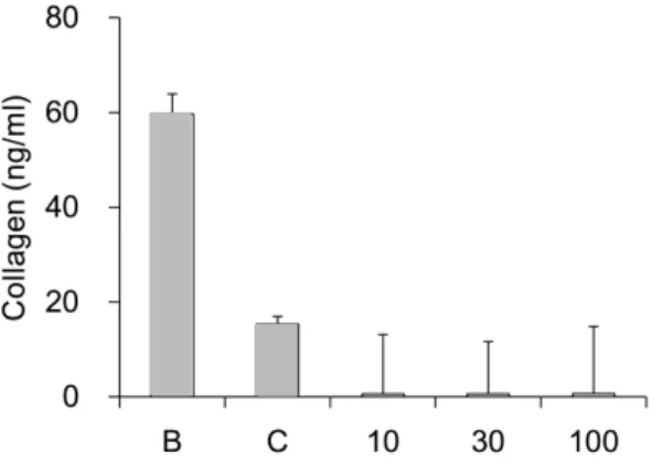
B. Assay of collagen type I synthesis
To evaluate the amount of collagen type I synthesis that occurred upon exposure to the sample, collagen type I was quantitatively detected by using the previously described procollagen type I C-peptide assay kit. Collagens are synthesized as precursor molecules, called procollagens. These molecules contain additional peptide sequences,
usually referred to as ‘propeptides’, at both the amino-terminal end and the carboxy-terminal end.
These propeptides are cleaved from the collagen triple-helix molecule during its secretion, after which the triple-helix collagens are polymerized into extracellular collagen fibrils. Thus, the amount of free propeptide stoichiometrically reflects the amount of collagen molecules synthesized
11). The amounts of type I collagen synthesis of ACPE were shown in Fig. 2. ACPE did not increase the expression of type I collagen at all concentrations of 10, 30, and 100 μg/ml(0.6 ± 12.5 ng/ml, 0.6 ± 11.0 ng/ml, and 0.6 ± 14.2 ng/ml) compared with control group(15.3 ± 1.6 ng/ml, Fig. 2).
Fig. 2. Effect of Albizziae cortex pharmacopunc- ture extracts(ACPE) on collagen type I synthesis in human fibroblast cells
B : blank, distilled water treated group without UVB irradiation.
C : control, distilled water treated group with UVB irradiation.
10, 30, and 100 : ACPE 10, 30, and 100 μg/ml treated group.
Data were expressed as the mean ± SEM of three experiments.
C. Assay of collagenase activity
To evaluate the collagenase activity, matrix
metalloproteinase-1(MMP-1) activity was quantita-
tively measured by using the previously described
matrix metalloproteinase-1 assay kit. The activities
of MMP-1 of ACPE treatment were recalculated
into 100% of control group(Fig. 3). ACPE significantly
reduced the MMP-1 activity at concentrations of 10
μg/ml, 30μg/ml, and 100μg/ml(15.6 ± 1.2%, 6.6 ±
0.3%, and 23.2 ± 1.0%, respectively., Fig. 3).
Fig. 3. Effect of Albizziae cortex pharmacopunc- ture extracts(ACPE) on collagenase activity in human fibroblast cells
B : blank, distilled water treated group without UVB irradiation.
C : control, distilled water treated group with UVB irradiation.
10, 30, and 100 : ACPE 10, 30, and 100 μg/ml treated group.
Data were expressed as the mean ± SEM of three experiments.
* : significantly different from the control(p < 0.05).
D. Tyrosinase activity assay
The activities of ACPE on tyrosinase activity were recalculated into 100% of control group(Fig. 4).
ACPE significantly reduced the tyrosinase activity at concentrations of 10 mg/ml(61.6 ± 3.7%, p < 0.05).
The tyrosinase activity of ACPE 0.1 and 1 mg/ml treated group did not show any significance(90.9 ± 8.6% and 83.4 ± 5.8%, respectively).
Fig. 4. Effect of Albizziae cortex pharmaco- puncture extracts(ACPE) on tyrosinase activity
C : control, distilled water treated group.0.1, 1, and 10 : ACPE 0.1, 1, and 10 mg/ml treated group.
Data were expressed as the mean ± SEM of three experiments.
* : significantly different from the control(p < 0.05).
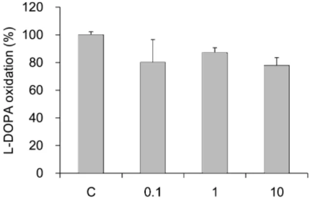
E. L-DOPA oxidation
The activities of ACPE on L-DOPA oxidation were recalculated into 100% of control group(Fig. 5).
ACPE did not show any significance at all concentraions. The effects of 0.1, 1, and 10 mg/ml were 80.1 ± 16.5%, 87.0 ± 3.7%, and 77.8 ± 5.7%, respectively.
Fig. 5. Effect of Albizziae cortex pharmaco- puncture extracts(ACPE) on L-DOPA oxidation
C : control, distilled water treated group.0.1, 1, and 10 : ACPE 0.1, 1, and 10 mg/ml treated group.
Data were expressed as the mean ± SEM of three experiments.