16
Journal of International Society for Simulation Surgery 2014;1(1):16-18
서 론
악안면골 중에서도 특히 안와골은 시력을 담당하는 안와를 감싸고 있는 뼈로, 그 모양이 상하좌우가 모두 다른 독특한 해 부학적 특징을 가지고 있다. 내측벽은 우리 몸의 뼈 중 가장 얇은 뼈로 안와에 대한 외부 충격에 대해 쉽게 부서져서 안와 자체의 파열을 방지하는 역할을 하며, 하벽 또한 비교적 얇은 뼈로 형성 되어 내측벽과 함께 안와 자체의 파열을 방지함과 동시에 안와 를 수직적으로 지탱한다. 외측벽과 상벽은 비교적 두꺼운 관골 과 전두골이 주를 이루며 외부로 부터의 충격으로부터 안와 자 체를 보호하는 역할을 함께 수행한다.1,2 이러한 안와골은 흔히 안와 파열 골절이라는 메커니즘으로 손상받게 되는데, 이는 안 와에 직접적인 타격이 이루어질 경우 주로 내측벽과 하벽이 부
서지면서 안와 자체의 파열을 방지하면 발생한다. 이러한 안와 파열골절의 경우 뼈 자체의 두께가 워낙 얇아서 기존의 골절된 뼈로는 재건이 쉽지 않으며, 따라서 인조뼈 형태의 물질로 기존 의 골절된 뼈를 대체하여 수술하는 방식이 일반적이다.3-6
하지만 이러한 안와파열 골절 복원 수술에 있어 안와골의 복 잡한 형태로 인해 조금만 잘못 복원이 되어도 환자는 쉽게 안구 함몰 증상이 나타날 수 있으며 이는 추가적인 수술로도 복원이 매우 힘든 상황 중에 하나에 속한다. 또한 안구를 움직이는 6개 의 안와주변 근육이 제대로 재배치되지 못하면 복시 등의 증상 으로 큰 불편함을 야기하게 된다.7,8
이러한 정확한 재건수술을 위해 최근 각광받고 있는 것이 3 차원 컴퓨터 시뮬레이션이다. 컴퓨터 시뮬레이션 이미지가 실제 수술에 적용되기 위해서는 surgical guide 형태의 매개체가 필
ISSN 2383-5389
Application of 3D Simulation Surgery to Orbital Wall Fracture : A preliminary Case Study
Jong-Woo Choi, M.D., Ph.D., M.M.M.
Department of Plastic and Reconstructive Surgery, College of Medicine, Ulsan University, Seoul Asan Medical Center, Seoul, Korea
The orbit has a very special anatomical structure. The complex anatomical structure should be restored when we encounter the patient with orbital wall fracture. Unless these specific anatomy were reconstructed well, the patient should suffer from various complications such enophthalmos, diplopia or orbital deformity. In addition, because the patient has a his own specific orbital shape, individualized approach will be necessary. The aim of this trial is to try to restore the original orbit anatomy as possible based on the mirrored three dimensional CT images based on the computer simulation. Preoperative computed tomography (CT) data were processed for the patient and a rapid prototyping (RP) model was produced. At the same time, the uninjured side was mirrored and superimposed onto the traumatized side, to create a mirror-image of the RP model. In order to restore the missing skipped images between the cuts of CT data because of the thinness of the orbital walls, we manipulated the DICOM data for im- aging the original orbital contour using the preoperatively manufactured mirror-image of the RP model. And we fabricated Tita- nium-Medpor to reconstruct three-dimensional orbital structure intraoperatively. This prefabricated Titanium-Medpor was then inserted onto the defected orbital wall and fixed. Three dimensional approach based on the computer simulation turned out to be very successful in this patient. Individualized approach for each patient could be an ideal way to manage the traumatic patients in near future.
Key WordsZZOrbital wall fracture ㆍThree-dimensional surgery ㆍBlow out fracture ㆍPre-surgical simulation ㆍ Three-dimensional simulation.
Received: July 8, 2014 / Revised: July 11, 2014 / Accepted: July 15, 2014 Address for correspondence: Jong-Woo Choi, M.D., Ph.D., M.M.M.
Department of Plastic and Reconstructive Surgery, Seoul Asan Medical Center, PungNapDong, SongPaGu, Seoul, Korea Tel: 02-3010-3604, Fax: 02-476-7471, E-mail: [email protected]
Case Report
www.issisglobal.org 17
Application of 3D Simulation Surgery to Orbital Wall Fracture : A Preliminary Case Study █ Choi JW
요한데, 안와 파열 골절에 있어서는 기존의 골절된 뼈를 대체하 는 다양한 임플란트들이 그 역할을 담당할 수 있다. 또한 이러 한 컴퓨터 시뮬레이션을 위해서는 기준이 되는 이미지가 필요한 데, 안와골절 복원 수술의 경우 반대편 건측 안와가 이러한 기 준 역할을 수행할 수 있다고 판단된다.
따라서 저자들은 이러한 안와파열 골절에 있어 반대편 정상 안와의 CT data를 mirroring을 이용하여 재건하여 이를 통해 Rapid prototype을 작성하고 이에 맞추어 안와파열 골절 복원 수술을 하여 환자가 가진 원래의 안와 모양을 최대한 이상적으 로 복원시키고자 시도하였다.
증례 보고
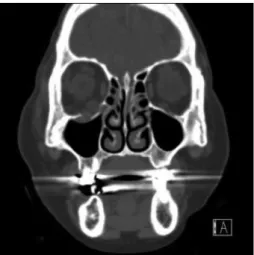
42세의 여자 환자가 넘어짐을 통한 우측 안와하벽골절을 주 소로 본과에 내원하였다. 이에 CT 촬영을 포함한 통상의 술전 검사를 시행하였으며 3차원 물체로 재구성한 환자의 두개안면 골에 대해 악안면기형 진단 및 분석을 시행하였다. 삼차원 CT scan을 촬영하였고, CT data를 Simplant (simplant pro, MaterialsⓇ, Belgium)를 이용하여 분석한 후 컴퓨터 시뮬레이 션 수술을 계획하였다(Fig. 1).
CT DICOM 파일을 기반으로 환자의 안와골을 3차원 물체로
재구성 하였으며, 좌측의 정상안와형태의 CT data를 mirror- ing 기법 (Mimics Z software : Materialize, Leuven, Bel- gium)을 이용하여 우측에도 적용하여 환자가 다치기 전의 양측 안와골의 CT 이미지를 얻었다. 이를 3D 프린팅을 통해 제작함 으로써 실제 수술 중 안와파열 골절을 복원하기 위한 Titani- um-Medpore implant을 위치 시키기 위한 기준으로 사용하였 다 (Fig. 2).
제작된 3차원 Rapid Prototype을 이용하여 통상의 안와파 열 골절 복원수술을 진행하였으며 transconjunctival prese- ptal approach를 이용하여 하안와연을 노출시키고, 하안와 신 경을 하벽으로부터 bipolar coagulator를 이용하여 박리하였 다. 3차원 컴퓨터 시뮬레이션을 이용해 제작한 Rapid proto- type mirroring model의 안와골에 맞추어서 사전에 제작하여 둔 Titanium Mepore implant를 삽입하여 안와골을 복원하 였다. 환자는 수술 후 안구 함몰이나 복시 등의 합병증이 발생 하지 않았으며, 특이한 문제 없이 술 후 3일째 퇴원하였다. 술 후 3일째 6개월째 술후 CT scan을 통해 수상전과 유사하게 복 원되었음을 확인할 수 있었다 (Fig. 3).
고 찰
안면골의 형태는 각 개인, 인종, 성별에 따라 다른 것으로 알 려져 있으며, 안와골 역시 각 개인마다 조금씩 그 크기가 뼈의 위치 및 각도가 다르다. 하지만, 지금까지 일반적으로 수술을 함 에 있어 이러한 환자 개인의 차이가 수술에 반영되기는 어려웠 다. 특히나, 안와골절 복원 수술의 경우에는 절대 손상이 되어 서는 안 되는 시신경과 인접되어 수술이 진행되어야 하고, 수술 시야가 좋지 않아 이러한 주변 안와골의 모양을 충분히 노출 시 킨 후 수술을 하는 것이 현실적으로 쉽지 않다. 따라서 이러한 안와 파열 골절의 복원을 위해 사용되는 임플란트의 경우에는 미리 그 모양을 제작하여 삽입하는 형태로 수술이 진행될 수 있 다면 가장 이상적이다. 하지만, 이러한 사전 제작을 위해서는 환 자의 원래 안와골의 형태를 알 수 있어야 하는데, 동측은 이미 Fig. 1. The CT scan coronal image of the patient with inferior or-
bital wall fracture.
Fig. 3. Postoperative CT coronal image.
Fig. 2. Computer simulation based on the mirroring technique.
18
Journal of International Society for Simulation Surgery █ 2014;1(1):16-18
골절이 되어 정확한 이미지를 얻을 수 없으므로, 반대편 건측 안 와골이 가장 좋은 대안이 될 것이다. 이를 이용한 기법이 Mir- roring technique이다.
본 증례에서 우측 안와 하벽골절의 복원을 위해 이 미러링 기 법을 이용하였는데, 만족할 만한 결과를 얻을 수 있었다. 이는 통상 대부분의 경우 양측 얼굴이 2mm 이내의 대칭성을 가지고 있는 것으로 알려져 있고, 반대측 건측이 가장 좋은 landmark 가 될 수 있기 때문이다. 문제는 CT촬영을 통해 이러한 안와골 의 거울영상을 획득하더라도 안와골이 통상 매우 얇은 뼈로 이 루어져 있어서, 안와골이 CT 영상에 skip 되어 결손 형태로 Rapid Prototype이 제작되는 것이다. 이러한 문제를 해결하기 위해 저자들은 3차원 컴퓨터 시뮬레이션 조작을 통해 skip된 CT image를 연결하는 기법을 이용하여 결손부위 없는 연속성 있는 안와골 RP model을 제작할 수 있었다. 이는 수술 시 사용 하게 되는 Titanium-Medpore implant의 이상적인 곡면을 만 드는데 있어 필수적이었으며, 매우 유용하였다.
또한, Titanium-Medpore implant를 이용하여 안와벽을 재건함으로써 술후 CT scan에서 반대편 건측과 유사하게 우측 의 안와 하벽이 잘 재건되었음을 확인할 수 있었고, 본 거울영 상을 이용한 3차원 컴퓨터 시뮬레이션 기법을 이용해 환자 본인 의 안와골을 최대한 원래에 가깝게 복원할 수 있었으며, 이러한 접근은 미래의 individualized medicine의 현실적인 접근방법 의 하나로 간주 될 수 있을 것으로 판단된다.
결 론
안와파열 골절은 전통적으로 술자의 경험에 의존하여 주변 안와골을 노출 시킨 후, 결손 부위를 다양한 임플란트로 대체
하는 형태의 수술이 주로 이루어져왔으나, 실제 안와골의 복잡 한 안와골 형태가 재건되지 못하면, 안구함몰, 복시 등의 심각한 합병증이 발생함을 감안하였을 때, 이러한 3차원 컴퓨터 시뮬레 이션을 이용한 안와재건은 individualized medicine의 첫걸음 이자, 매우 유용한 이상적인 수술 방식임을 확인 할 수 있었다.
참 고 문 헌
1. Kakizaki H, Takahashi Y, Asamoto K, Nakano T, Selva D, Leibo- vitch I. Anatomy of the superior border of the lateral orbital wall:
surgical implications in deep lateral orbital wall decompression surgery. Ophthalmic plastic and reconstructive surgery. 2011 Jan- Feb;27(1):60-3. PubMed PMID: 20736872
2. Beden U, Edizer M, Elmali M, Icten N, Gungor I, Sullu Y, et al.
Surgical anatomy of the deep lateral orbital wall. European journal of ophthalmology. 2007 May-Jun;17(3):281-6. PubMed PMID:
17534804
3. Oh SA, Aum JH, Kang DH, Gu JH. Change of the orbital volume ratio in pure blow-out fractures depending on fracture location.
The Journal of craniofacial surgery. 2013 Jul;24(4):1083-7. PubMed PMID: 23851745
4. Lee H, Ahn J, Lee TE, Lee JM, Shin H, Chi M, et al. Clinical char- acteristics and treatment of blow-out fracture accompanied by can- alicular laceration. The Journal of craniofacial surgery. 2012 Sep;23(5):1399-403. PubMed PMID: 22948636
5. Lee S, Yen MT. White-eyed blow-out fracture. Journal of pediatric ophthalmology and strabismus. 2011 Sep-Oct;48(5):320. PubMed PMID: 21913629
6. Park JS, Lew H, Lee SY. Role of inferior orbital wall morphologic properties in isolated orbital blow-out fracture. Ophthalmic re- search. 2012;47(1):1-6. PubMed PMID: 21691135
7. Hayakawa I. [Surgical treatment of the orbital blow-out fracture (author's transl)]. No shinkei geka Neurological surgery. 1976 Oct;4(10):933-40. PubMed PMID: 1033468
8. Kugelberg U. Diplopia in down-gaze after a blow-out fracture.
Acta ophthalmologica Scandinavica. 1998 Oct;76(5):629-31.
PubMed PMID: 9826057