• 검색 결과가 없습니다.

The Serum Concentrations of YKL-40, IL-6, and TNF-α in Retired Workers Exposed to Inorganic Dusts

N/A
N/A
Protected

Academic year: 2021

Share "The Serum Concentrations of YKL-40, IL-6, and TNF-α in Retired Workers Exposed to Inorganic Dusts"

Copied!
6
0
0

로드 중.... (전체 텍스트 보기)

전체 글

(1)

Biomedical Science Letters 2014, 20(1): 8~13 eISSN : 2288-7415

The Serum Concentrations of YKL-40, IL-6, and TNF-α in Retired Workers Exposed to Inorganic Dusts

Kyung Myung Lee 1,3 , Jae Hoon Shin 2 , JooHwan Hwang 2 , Jong Seong Lee 2 , Byung-Soon Choi 2 and In Sik Kim 3,†

1

Forensic DNA Division, National Forensic Service, Wonju 220-170, Korea

2

Occupational Lung Diseases Institute, KCOMWEL, Ansan 426-858, Korea

3

Graduate School of Health Science, Eulji University, Daejeon 201-746, Korea

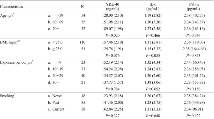
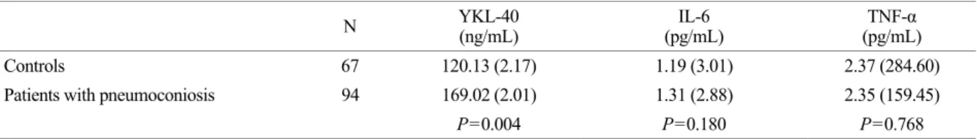
Occupational long-term exposure to inorganic dusts may cause a variety of lung diseases such as pneumoconiosis and chronic obstructive pulmonary disease (COPD). Diagnosis of pneumoconiosis and COPD, however, is currently dependent on radiological findings and pulmonary test, which are both late diagnostic tools. Therefore, there is a need to identify novel biomarkers in pneumoconiosis and COPD. Hence, in this current study we investigated the serum concentrations of YKL-40, interleukin-6 (IL-6) and tumor necrosis factor-alpha (TNF-α) as biomarkers for pneumoconiosis and COPD in 161 retired male workers exposed to inorganic dusts. The serum concentration of YKL-40 was significantly increased with age, pneumoconiosis, and airflow limitation. The serum concentration of IL-6 was significantly higher in airflow limitation. These results suggest that serum concentration of YKL-40 is associated with age, pneumoconiosis, and airflow limitation. Also, serum concentration of IL-6 is associated with airflow limitation.

Key Words: YKL-40, IL-6, TNF-α, COPD, Pneumoconiosis

INTRODUCTION

Chronic occupational exposure to inorganic dusts such as coal and crystalline silica may cause a variety of interstitial lung diseases such as progressive massive fibrosis (PMF), coal workers pneumoconiosis (CWP), and chronic obstructive pulmonary disease (COPD), including paren- chymal destruction (emphysema) and small airway disease (obstructive bronchiolitis) (Schins and Borm, 1999). Inhaled coal mine and silica dust may result in abnormal inflam- matory response of the lung and leads to progressive airflow limitation that is characteristics of COPD (Vestbo

et al., 2013). Diagnosis of pneumoconiosis is performed by radiological findings with occupational exposure history and pulmonary function test. Unfortunately, as current diagnostic tools of pneumoconiosis are only limited fibrosis in the lung which is usually irreversibly progressive, there have been some limitations in the detection of the early stage of pneumoconiosis. Therefore, it is necessary to study reliable and prospective biomarkers for pneumoconiosis before irreversible damage of the lung (Gulumian et al., 2006).

COPD is a complex disease involving more than airflow limitation and the "spill-over" of the inflammatory mediators into the circulation may result in systemic manifestations and comorbidities (Barnes and Celli, 2009). As in many patients with COPD have systemic inflammation, the levels of cytokines such as interleukin-6 (IL-6) and tumor necrosis factor-alpha (TNF-α) and acute phase proteins such as C-reactive protein (CRP) and YKL-40 are increased in the systemic circulation of COPD patients and abnormalities in circulating inflammatory cells such as lymphocytes have

Original Article

*

Received: February 2, 2014 / Revised: March 27, 2014 Accepted: March 29, 2014

Corresponding author: In Sik Kim. Department of Biomedical Laboratory Science School of Medicine, Eulji University 143-5, Yeuongdu-dong, Jung-gu Daejeon 301-746, Korea.

Tel: +82-42-259-1753, Fax: +82-42-259-1759 e-mail: [email protected]

C

The Korean Society for Biomedical Laboratory Sciences. All rights reserved.

(2)

been reported (Agusti et al., 2003; Gan et al., 2004; Wouters et al., 2007).

YKL-40 belongs to the family of chitinase-like proteins and regulates mitogenesis, differentiation, and extracellular homeostasis in mammalian cells and has been associated with inflammation, tissue remodeling, fibrosis, and several malignancies (Johansen, 2006). In previous reports, serum concentrations of YKL-40 was up-regulated in patients with COPD (Létuvé et al., 2008) and associated with decline of lung function in the general population (Guerra et al., 2013).

Cytokines regulates various biological effects such as inflammation, metabolism, cell growth and proliferation, fibrosis, and homeostasis (Elias and Zitnik, 1992), Among these cytokines, IL-6 and TNF-α have been reported to be a prospective biomarkers to estimate the progression or exacerbation of pneumoconiosis and COPD (Di Francia et al., 1994; Razzaque and Taguchi, 2003; Vanhee et al., 1995;

Yende et al., 2006). Although there were a few reports of the relationships between inflammatory mediators and occupational lung diseases such as pneumoconiosis and COPD (Lee et al., 2009; Lee et al., 2010), there was no report between YKL-40 in retired workers exposed to inorganic dusts in Korea. Therefore, there was a need to identify novel biomarkers in patients with pneumoconiosis and COPD who were exposed to chronic exposed to inorganic dusts. The present study was aimed to investigate the serum concentrations of YKL-40, IL-6, and TNF-α as biomarkers for pneumoconiosis and COPD in retired coal miners.

MATERIALS AND METHODS Study Subjects

The study subjects contained 161 retired male workers exposed to inorganic dusts. We collected serum and stored at -80℃ until assay. Personal information including age, height, and weight as well as job history and smoking status were obtained by a structured questionnaire. All subjects provided informed consent and the study was approved by the Research Ethics Committee of Occupational Lung Diseases Institute.

Analysis of YKL-40, IL-6, and TNF-α

The concentrations of serum YKL-40 (MicroVue

TM

YKL-40 EIA, QUIDEL, USA), IL-6 (Human Interleukin-6 ELISA, BioVendor, Czech), and TNF-α (Human TNF-alpha ELISA, BioVendor, Czech) were analyzed by sandwich enzyme immunoassay.

Pulmonary function test

We carried out pulmonary function test in accordance with recommended guideline of ATS/ERS Task Force (Brusasco et al., 2005) by spirometry (Vmax22, Sensor- Medics, USA). We measured forced vital capacity (FVC), forced expiratory volume in one second (FEV

1

), and FEV

1

/ FVC ratio.

Chest radiographs

chest radiographs were obtained and reviewed by the Table 1. General characteristics of study subjects

Characteristics N (%) Mean

*

SD

*

Range

Age, yrs 161 62.8 8.0 38~82

~59 54

(33.5)

60~69 75

(46.6)

70~ 32

(19.9)

BMI, kg/m

2

161 23.8 2.8 15.4~30.5

Exposure period, yrs 161 18.0 8.7 2~46

~ 9 23

(14.3)

10~19 77

(47.8)

20~29 40

(24.8)

30~ 21

(13.0)

Smoking, N 161

Never 18

(11.2)

Past 85

(52.8)

Current 58

(36.0)

*

Arithmetic mean and arithmetic standard deviation

BMI, body mass index

(3)

pneumoconiosis review committee of Korea Workers' Compensation & Welfare Service. The radio opacity category of above 1/0 was defined as pneumoconiosis according to Internal Labor Office (ILO, 2002) classification.

RESULTS General characteristics of study subjects

General characteristics of study subjects are shown in Table 1. In this study, 67 study subjects were classified as controls and 94 (41.6%) were classified as patients with pneumoconiosis (58.4%) (Table 2).

Serum concentrations of YKL-40, IL-6, and TNF-α according to general characteristics

The mean concentration of serum YKL-40 was signifi- cantly higher in subjects above 70 year (180.07 ng/mL) than that of less than 59 year (120.00 ng/mL) (P=0.020) and tended to increase with increment of years. There were no significant differences between mean concentrations of

IL-6 and TNF-α in serum and general characteristics such as BMI, exposure period, and smoking status (Table 3).

Serum concentrations of YKL-40, IL-6, and TNF-α according to pneumoconiosis

The mean concentration of serum YKL-40 was signifi- cantly higher in subjects with pneumoconiosis (169.02 ng/mL) than that of controls (120.13 ng/mL) (P=0.004).

However, there were no significant differences between mean concentrations of IL-6 and TNF-α and pneumo- coniosis (Table 4).

Table 2. ILO categories of study subjects ILO category N % Profusion (N) 0 (Normal) 67 41.6 0/0 (54); 0/1 (13) Pneumoconiosis 94 58.4

Small opacity 78 48.4

I 52 32.3 1/0 (19); 1/1 (19); 1/2 (14) II, III 26 16.1 2/1 (13); 2/2 (8); 2/3 (4); 3/3 (1) Large opacity 16 9.9 4A (13); 4B (3)

Table 3. Mean concentration of serum YKL-40 , IL-6, and TNF-α according to general characteristics

Characteristics N YKL-40

(ng/mL) IL-6

(pg/mL) TNF-α

(pg/mL)

Age, yrs

*

a. ~59 54 120.00 (2.10) 1.19 (2.82) 2.38 (402.75)

b. 60~69 75 151.98 (2.11) 1.30 (3.20) 2.34 (143.89)

c. 70~ 32 189.07 (1.98) 1.27 (2.58) 2.36 (163.16)

P=0.020 P=0.466 P=0.746

BMI, kg/m

2†

a. < 25.0 110 157.46 (2.19) 1.31 (2.81) 2.36 (119.00)

b. ≥ 25.0 51 125.76 (1.91) 1.15 (3.32) 2.35 (1644.66)

P=0.076 P=0.055 P=0.853

Exposure period, yrs

*

a. ~ 9 23 152.19 (2.14) 1.32 (4.34) 2.44 (560.40)

b. 10~19 77 154.24 (2.28) 1.24 (2.83) 2.36 (158.05)

c. 20~29 40 134.57 (2.07) 1.20 (2.60) 2.35 (301.22)

d. 30~ 21 137.73 (1.57) 1.38 (3.06) 2.23 (153.92)

P=0.784 P=0.652 P=0.156

Smoking

*

a. Never 18 123.93 (2.18) 1.20 (2.67) 2.38 (384.24)

b. Past 85 141.46 (2.00) 1.23 (2.75) 2.36 (338.98)

c. Current 58 162.84 (2.25) 1.31 (3.33) 2.34 (96.91)

P=0.327 P=0.648 P=0.922

*Calculated by ANOVA test (Tukey HSD test), geometric mean (geometric standard deviation)

†Calculated by student's t-test, geometric mean (geometric standard deviation)

(4)

Serum concentrations of YKL-40, IL-6, and TNF-α according to pulmonary function test

The mean concentration of serum YKL-40 was signifi- cantly higher in subjects with airflow limitation (less than %FEV

1

/FVC ratio < 70) (175.39 ng/mL) than that of above 70% (129.48 ng/mL) (P=0.011). Also, the mean concentration of serum IL-6 was significantly higher in subjects with airflow limitation (1.40 pg/mL) than that of above 70% (1.17 pg/mL) (P=0.012). There was no significant difference between mean concentration of TNF-α and FEV

1

/FVC ratio (Table 5).

DISCUSSION

YKL-40 is expressed and secreted by activated mono- cytes (Hashimoto et al., 1999), late stage of macrophages (Renkema et al., 1998), vascular smooth muscle cells (Nishikawa and Millis, 2003), and cancer cells (Johanson et al., 2006). YKL-40 is important role in cell proliferation and differentiation (Brochner et al., 2011), inflammation (Kawada et al., 2012), extracellular tissue remodeling (Lee et al., 2011), and protection against apoptosis (Lee et al., 2009).

Plasma YKL-40 levels correlated with age in both sexes (Johansen et al., 2009). High levels of YKL-40 in circulation

and bronchoalveolar lavage fluid (BALF) is associated with asthma (Chupp et al., 2007), COPD (Letuve et al., 2008), and idiopathic pulmonary fibrosis (IPF) (Korthagen et al., 2011). Furthermore, YKL-40 is negatively correlated to %FEV

1

predicted in patients with COPD and asthma (Sakazaki et al., 2011). In recent study, high plasma YKL- 40 is associated with mortality in patients with moderate and very severe COPD, suggesting a role for as a potential biomarker of mortality in this patient group (Holmgaard et al., 2013). In present study, high serum concentrations of YKL-40 showed significant association with age, pneumo- coniosis, and airflow limitation, a main characteristics of COPD and tended to increase with age.

Pneumoconiosis results from chronic inhalation to inorganic dusts and is characterized by a pulmonary fibrosis in the lung and irreversible lung damage (Schins and Borm, 1999). Inhaled inorganic dusts may induce release of proinflammatory cytokines such as TNF-α (Lasky et al., 2005) and IL-6 stimulates production of acute-phase protein such as YKL-40, but not TNF-α (Ferrari et al., 2013). Thus, IL-6 could be an upstream activator of YKL-40 independent of IL-6 (Nielsen et al., 2011). The concentration of IL-6 was increased in BALF and alveolar macrophages in patients with pneumoconiosis and associated with disease pro- gression (Gosset et al., 1991; Reuben et al., 2004; Vallyathan Table 5. Concentration of serum YKL-40, IL-6, and TNF-α according to pulmonary function test

N YKL-40

(ng/mL) IL-6

(pg/mL) TNF-α

(pg/mL)

%FEV

1

/FVC ratio ≥ 70.0 95 129.48 (2.06) 1.17 (3.21) 2.35 (293.27)

%FEV

1

/FVC ratio < 70.0 66 175.39 (2.13) 1.40 (2.71) 2.37 (127.50)

P=0.011 P=0.012 P=0.649

%FEV

1

/FVC ratio < 70.0: Airflow limitation

* Calculated by student's t-test, geometric mean (geometric standard deviation)

Table 4. Concentration of serum YKL-40, IL-6, and TNF-α according to pneumoconiosis

N YKL-40

(ng/mL) IL-6

(pg/mL) TNF-α

(pg/mL)

Controls 67 120.13 (2.17) 1.19 (3.01) 2.37 (284.60)

Patients with pneumoconiosis 94 169.02 (2.01) 1.31 (2.88) 2.35 (159.45)

P=0.004 P=0.180 P=0.768

Control: ILO Classification 0/0 or 0/1, Pneumoconiosis: ILO classification 1/0 or more

*Calculated by student's t-test, geometric mean (geometric standard deviation)

(5)

et al., 2000; Zhai et al., 2002). Although pneumoconiosis is strongly related with serum IL-6 and TNF-α, we found that measured serum cytokines were not related with the results of pneumoconiosis in this study, high serum concentrations of IL-6 showed significant association with patients with airflow limitation.

This study has several limitations. One is that mean age of study subjects was over 60 years. Thus, systemic inflam- mation may be still in existence results from other systemic comorbidities such as diabetes, hypertension, and rheuma- toid arthritis. It was necessary to compare with normal controls without occupational history of inorganic dusts and comorbidities.

In conclusion, high serum concentration of YKL-40 is associated with age, pneumoconiosis, and airflow limitation.

Also, high serum concentration of IL-6 is associated with airflow limitation. Further studies will be required to in- vestigate the potential serum acute-phase proteins and cytokines in patients with pneumoconiosis and COPD using lung specific specimens such as BALF, exhaled breath condensate, and lung tissue.

REFERENCES

Agustí AG, Noguera A, Sauleda J, Sala E, Pons J, Busquets X.

Systemic effects of chronic obstructive pulmonary disease.

Eur Respir J. 2003. 21: 347-360.

Barnes PJ, Celli BR. Systemic manifestations and comorbidities of COPD. Eur Respir J. 2009. 33: 1165-1185.

Brøchner CB, Johansen JS, Larsen LA, Bak M, Mikkelsen HB, Byskov AG, Andersen CY, Møllgård K. YKL-40 is differ- entially expressed in human embryonic stem cells and in cell progeny of the three germ layers. J Histochem Cytochem.

2012. 60: 188-204.

Brusasco V, Crapo R, Viegi G. Series "ATS/ERS Task Force:

Standardisation of lung function testing. Eur Respir J. 2005.

26: 319-338.

Chupp GL, Lee CG, Jarjour N, Shim YM, Holm CT, He S, Dziura JD, Reed J, Coyle AJ, Kiener P, Cullen M, Grandsaigne M, Dombret MC, Aubier M, Pretolani M, Elias JA. A chitinase- like protein in the lung and circulation of patients with severe asthma. N Engl J Med. 2007. 357: 2016-2027.

Di Francia M, Barbier D, Mege JL, Orehek J. Tumor necrosis

factor-alpha levels and weight loss in chronic obstructive pulmonary disease. Am J Respir Crit Care Med. 1994. 150:

1453-1455.

Elias JA, Zitnik RJ. Cytokine-cytokine interactions in the context of cytokine networking. Am J Respir Cell Mol Biol. 1992. 7:

365-367.

Ferrari R, Tanni SE, Caram LM, Corrêa C, Corrêa CR, Godoy I.

Three-year follow-up of Interleukin 6 and C-reactive protein in chronic obstructive pulmonary disease. Respir Res. 2013.

20: 14-24.

Gan WQ, Man SF, Senthilselvan A, Sin DD. Association between chronic obstructive pulmonary disease and systemic inflam- mation: a systematic review and a meta-analysis. Thorax.

2004. 59: 574-580.

Gosset P, Lassale P, Vanhee D, Wallaert C, Aerts C, Voisin C, Tonne AB. Production of tumor necrosis factor alpha and interleukin-6 by human macrophages exposed in vitro to coal mine dust. Am J Resp Cell Mol Biol. 1991. 5: 431-436.

Guerra S, Halonen M, Sherrill DL, Venker C, Spangenberg A, Carsin AE, Tarès L, Lavi I, Barreiro E, Martínez-Moratalla J, Urrutia I, Sunyer J, Antó JM, Martinez FD. The relation of circulating YKL-40 to levels and decline of lung function in adult life. Respir Med. 2013. 107: 1923-1930.

Gulumian M, Borm PJ, Vallyathan V, Castranova V, Donaldson K, Nelson G, Murray J. Mechanistically identified suitable bio- markers of exposure, effect, and susceptibility for silicosis and coal-worker's pneumoconiosis: a comprehensive review.

J Toxicol Environ Health B Crit Rev. 2006. 9: 357-395.

Hashimoto S, Suzuki T, Dong H-Y, Yamazaki N, matsushima K.

Serial analysis of gene expression in human monocytes and macrophages. Blood. 1999. 94: 837-844.

Holmgaard DB, Mygind LH, Titlestad IL, Madsen H, Pedersen SS, Johansen JS, Pedersen C. Plasma YKL-40 and all-cause mortality in patients with chronic obstructive pulmonary disease. BMC Pulm Med. 2013. 13: 77.

ILO. Guidelines for the use of the ILO international classification of radiographs of pneumoconiosis. 2002. Geneva, Inter- national labor Organization.

Johansen JS. Studies on serum YKL-40 as a biomarker in diseases with inflammation, tissue remodelling, fibroses and cancer.

Dan Med Bull. 2006. 53: 172-209.

Johansen JS, Jensen BV, Roslind A, Nielsen D, Price PA. Serum YKL-40, a new prognostic biomarker in cancer patients?

Rev Cancer Epidemiol Biomarkers Prev. 2006. 15:194-202.

Johansen JS, Bojesen SE, Mylin AK, Frikke-Schmidt R, Price PA,

(6)

Nordestgaard BG. Elevated plasma YKL-40 predicts increased risk of gastrointestinal cancer and decreased survival after any cancer diagnosis in the general population. J Clin Oncol.

2009. 27: 572-578.

Kawada M, Seno H, Kanda K, Nakanishi Y, Akitake R, Komekado H, Kawada K, Sakai Y, Mizoguchi E, Chiba T. Chitinase 3-like 1 promotes macrophage recruitment and angiogenesis in colorectal cancer. Oncogene. 2012. 31: 3111-3123.

Korthagen NM, van Moorsel CH, Barlo NP, Ruven HJ, Kruit A, Heron M, van den Bosch JM, Grutters JC. Serum and BALF YKL-40 levels are predictors of survival in idiopathic pulmonary fibrosis. Respir Med. 2011. 105: 106-113.

Lasky JA, Ortiz LA, Brody AR. Lung injury-mechanisms, pathophysiology, and therapy: Mediators and mechanisms in chronic lung injury and fibrosis (Notter, RH., et al. Eds).

2005. Taylor & Francis Group, FL, pp. 175-226.

Lee CG, Da Silva CA, Dela Cruz CS, Ahangari F, Ma B, Kang MJ, He CH, Takyar S, Elias JA. Role of chitin and chitinase/

chitinase-like proteins in inflammation, tissue remodeling, and injury. Annu Rev Physiol. 2011. 73: 479-501.

Lee CG, Hartl D, Lee GR, Koller B, Matsuura H, Da Silva CA, Sohn MH, Cohn L, Homer RJ, Kozhich AA, Humbles A, Kearley J, Coyle A, Chupp G, Reed J, Flavell RA, Elias JA.

Role of breast regression protein 39 (BRP-39)/chitinase 3- like-1 in Th2 and IL-13-induced tissue responses and apoptosis.

J Exp Med. 2009. 206: 1149-1166.

Lee JS, Shin JH, Lee JO, Lee WJ, Hwang JH, Kim JH, Choi BS.

Blood levels of IL-1β, IL-8, TNF-α, and MCP-1 in pneumo- coniosis patients exposed to inorganic dusts. Toxicol Res.

2009. 25: 217-224.

Lee JS, Shin JH, Lee JO, Lee KM, Kim JH, Choi BS. Serum levels of interleukin-8 and tumor necrosis factor-alpha in coal workers' pneumoconiosis: One-year follow-up study. Safety and health at Work. 2010. 1: 69-79.

Létuvé S, Kozhich A, Arouche N, Grandsaigne M, Reed J, Dombret MC, Kiener PA, Aubier M, Coyle AJ, Pretolani M.

YKL-40 is elevated in patients with chronic obstructive pulmonary disease and activates alveolar macrophages. J Immunol. 2008. 181: 5167-5173.

Nielsen AR, Plomgaard P, Krabbe KS, Johansen JS, Pedersen BK.

IL-6, but not TNF-alpha, increases plasma YKL-40 in human subjects. Cytokine. 2011. 55: 152-155.

Nishikawa KC, Millis AJT. gp38k (CHI3L1) is a novel adhesion and migration factor for vascular cells. Exp Cell Res. 2003.

287: 79-87.

Razzaque MS, Taguchi T. Pulmonary fibrosis: cellular and

molecular events. Pathol Int. 2003. 53: 133-145.

Renkema GH, Boot RG, Au FL, Donker-Koopman WE, Strijland A, Muijsers AO, Hrebicek M, Aerts JM. Chitotriosidase, a chitinase, and the 39-kDa human cartilage glycoprotein, a chitin-binding lectin, are homologues of family 18 glycosyl hydrolases secreted by human macrophages. Eur J Biochem.

1998. 251: 504-509.

Reuben JS, Guo RF, Ward PA. Oxygen/Nitrogen radicals-Lung injury and disease: Mediators of lung inflammation-Role of reactive oxygen and nitrogen species (Vallyathan et al., Ed.).

2004. Marcel Deckker, NewYork, pp. 91-110.

Sakazaki Y, Hoshino T, Takei S, Sawada M, Oda H, Takenaka S, Imaoka H, Matsunaga K, Ota T, Abe Y, Miki I, Fujimoto K, Kawayama T, Kato S, Aizawa H. Overexpression of chitinase 3-like 1/YKL-40 in lung-specific IL-18-transgenic mice, smokers and COPD. PLoS One. 2011. 6: e24177.

Schins RP, Borm PJ. Mechanisms and mediators in coal dust induced toxicity: a review. Ann Occup Hyg. 1999. 43: 7-33.

Vallyathan V, Goins M, Lapp LN, Pack D, Leonard S, Shi X, Castranova V. Changes in bronchoalveolar lavage indices associated with radiographic classification in coal miners.

Am J Respir Crit Care Med. 2000. 162: 958-965.

Vanhée D, Gosset P, Boitelle A, Wallaert B, Tonnel AB. Cytokines and cytokine network in silicosis and coal workers' pneumo- coniosis. Eur Respir J. 1995. 8: 834-842.

Vestbo J, Hurd SS, Agustí AG, Jones PW, Vogelmeier C, Anzueto A, Barnes PJ, Fabbri LM, Martinez FJ, Nishimura M, Stockley RA, Sin DD, Rodriguez-Roisin R. Global strategy for the diagnosis, management, and prevention of chronic obstructive pulmonary disease: GOLD executive summary. Am J Respir Crit Care Med. 2013. 187: 347-365.

Wouters EF, Groenewegen KH, Dentener MA, Vernooy JH.

Systemic inflammation in chronic obstructive pulmonary disease: the role of exacerbations. Proc Am Thorac Soc. 2007.

4: 626-634.

Yende S, Waterer GW, Tolley EA, Newman AB, Bauer DC, Taaffe DR, Jensen R, Crapo R, Rubin S, Nevitt M, Simonsick EM, Satterfield S, Harris T, Kritchevsky SB. Inflammatory markers are associated with ventilatory limitation and muscle dysfunction in obstructive lung disease in well functioning elderly subjects. Thorax. 2006. 61: 10-16.

Zhai R, Liu G, Ge X, Bao W, Wu C, Yang C, Liang D. Serum

levels of tumor necrosis factor-alpha (TNF-α), interleukin-6

(IL-6), and their soluble receptors in coal workers pneumo-

coniosis. Respir Med. 2002. 96: 829-834.

수치

Table 3. Mean concentration of  serum YKL-40 , IL-6, and TNF-α according to general characteristics
Table 4. Concentration of  serum YKL-40, IL-6, and TNF-α according to pneumoconiosis

참조

관련 문서

IL, interleukin; MCP, monocyte chemoatactant protein; MIG, monokine induced by gamma interferon; TIMP, tissue inhibitor of metalloproteinases; TNF, tumor

This dissertation is composed of 6 chapters. ChapterⅠ deals with the purpose and the methods of this study. ChapterⅡ refers the methods and the problems of

Second, exploratory factor analysis and Cronbach's α were conducted to analyze the validity and reliability of the questionnaire regarding participation

This study, made up of 6 chapters in all, aims to handle the details and problems of the spouse inheritance system under existing law, as well as to review the draft of

JST일자리지원본부 취업과 창업 업무를 통합해 원스톱 일자리 서 비스를 지원한다. JST일자리종합센터 인천의 구직자 및 구인 업체에 취업 지원 서 비스를

The electrode potential and the surface concentrations of О and R are described by an equation of the Nernst form, regardless of the current flow. In effect, the potential

Effects of various concentrations of Scutellaria baicalensis (SB) extract in the diets on growth, body composition, serum chemistry and challenge test of far eastern

Thus, in the present study, we aimed to compare protein pro- files between achalasia patients and healthy controls using the serum proteomic analysis for the detection of