https://doi.org/10.9721/KJFST.2020.52.3.244
244
©The Korean Society of Food Science and Technology
Resource conservation using whole body autophagy:
Self-digestion of shedded gut lining cells in the small intestine
Phil Jun Lee1,2,†, Namki Cho3,†, Hee Min Yoo4,†, Sun-Young Chang1,2, Hyun-Jeong Ko5, and Hong Pyo Kim1,2,*1College of Pharmacy and Ajou University
2Research Institute of Pharmaceutical Science and Technology, Ajou University
3College of Pharmacy and Research Institute of Drug Development, Chonnam National University
4Microbiological Analysis Team, Group for Biometrology, Korea Research Institute of Standards and Science (KRISS), 5Laboratory of Microbiology and Immunology, College of Pharmacy, Kangwon National University
Abstract To retain valuable resources, organisms adopt several strategies including coprophagy. Cells covering the outer skin and internal digestive lumen are actively recycled to maintain their integrity. In present study, we suggested that the small intestine can consume dead cells in a manner similar to how it consumes protein from the diet. We examined the eluates from five segments of the mouse small intestine and cecum and 2 segments of the large intestine and small intestine tissue, and detected immunoreactivity with eukaryotic caveolin-1 and β-actin antibodies only in the cecum and 2 segments from the large intestine. Bacterial agitation of the mouse intestine with Shigella disrupted the architecture and absorptive function of the small intestine. Small intestine eluates were immunoreactive with murine caveolin-1 and contained heme as determined by dot blot analysis. We concluded that the body conserves resources in the small intestine by disposing of and recycling shedded cells.
Keywords: caveolin-1, small intestine, autophagy, heme, resource conservation
Introduction
Strategies for preserving valuable organismal resources are well-recognized phenomena in nature; examples include coprophagy of undigested food and placenta feeding in mammals (Martin, 2011). Cells covering the outer skin and internal digestive lumen are actively recycled to maintain their integrity and protect the body cavity from harsh environments (Williams et al., 2015) Intestinal enterocytes form a barrier between the gut lumen and animal body. Various biological mechanisms maintain the barrier function of the small intestine. It has been reported that an estimated 1,400 mature mouse enterocytes (EC) are shed from a villus tip in each 24 h period, equating to 2×10⁸ cells/day in the small intestine (Lodish, 2000; Bullen et al., 2006). In humans, the extent of daily shedding of ECs has been estimated at 1010-1011 cells (Lodish, 2000; Bullen et al., 2006). Assuming the weight of a murine hepatocyte is 3.5×10⁹ g and that daily shedding of ECs in humans may account for a total weight of 35-350 g, then the daily loss from the small intestine is approximately 14-140 g, which is very high considering the daily consumption of food by the average human and the volume of human ECs (1,400 µm3) is less than that
of human hepatocytes (3,400 µm3). It has been reported that apoptosis and anoikis initiate enterocyte shedding. Interestingly, mice lacking apoptotic machinery (bcl-2 or bax) have an apparently normal villus structure, indicating that alternative methods are used to dispose of exfoliated enterocytes (Bullen et al., 2006; Pritchard et al., 1999). Interestingly, a decade ago, the Fujita group suggested that shed (effete) cells from the ileum are damaged by intraepithelial lymphocytes and subsequently phagocytosed by subepithelial macrophages (Iwanaga et al., 1993). More recently, Hausmann proposed that the life cycle of intestinal enterocytes is terminated by apoptosis and/or shedding (Hausmann, 2010). We postulated that the small intestine can consume dead cells in a manner similar to how it consumes protein from the diet. To test this hypothesis, we examined the eluates from five segments of the mouse small intestine and cecum and 2 segments of the large intestine, rather than the tissue itself, to detect tissue-derived proteins by immuno-reacting with antibodies specific to mouse proteins.
Materials and Methods
Mice C57BL/6 mice (5 weeks old) purchased from Charles River Laboratories (Orient Bio, Inc., Seongnam, Korea) were housed under a standard specific pathogen-free environment with a 12 h dark/light cycle and free access to water and food. All animal experiments were conducted in accordance with the Guidelines for the Care and Use of Laboratory Animals and were approved by the Animal Ethics Review Committees of Ajou University (Permission number: 2013-0006). All mice used in the study were over 8 weeks of age. Virulent Shigella flexneri 2a (YSH6000) was a generous gift from Dr. Chihiro Sasakawa (University of Tokyo,
†These authors contributed equally to this work.
*Corresponding author: Hong Pyo Kim, School of Pharmacy, Uni-versity of Ajou, Suwon 16499, Republic of Korea
Tel: 82-31-219-3452 Fax: 82-31-219-3435 E-mail: [email protected]
Received March 10, 2020; revised April 9, 2020; accepted April 14, 2020
Japan). Mice were infected intragastrically by gavage with 5×10⁹ colony-forming units per mouse for 3 h. Following that, the small intestine of the mouse was separated using surgery. The small intestine was cut into five parts, and the large intestine was cut into cecum and colon 1/2. The cut parts were clearly washed with a PBS. The washed solution was collected.
Western blotting and antibodies
Cells were lysed with radioimmunoprecipitation assay buffer containing 1× phosphate-buffered saline, 1% (v/v) Nonidet P-40, 0.5% (w/v) sodium deoxycholate, 0.1% (w/v) sodium dodecyl sulfate, 0.1 mg/mL phenylmethylsulfonyl fluoride, 30 µL/mL aprotinin, and 1 mM sodium orthovanadate and protease inhibitor cocktail. The cell lysates were centrifuged, and the resulting supernatants were collected. Proteins were separated by 8-15% sodium dodecyl sulfate-polyacrylamide gel electrophoresis and transferred onto a polyvinylidene difluoride membrane. Each membrane was blocked in Tris-buffered saline containing 0.1% Tween-20 (TBST) and 5% non-fat dry milk for 1 h at room temperature, followed by overnight incubation with a primary antibody in TBST containing 1% non-fat dry milk at 4oC. Anti-caveolin-1 and anti-β-actin were purchased from Santa Cruz
Biotechnology (Dallas, TX, USA). Membranes were washed with TBST and incubated with goat anti-rabbit or anti-mouse horseradish peroxidase-conjugated IgG secondary antibody for 2 h. Signals were measured using a chemiluminescence system (GE Healthcare, Little Chalfont, UK).
Detection of Heme level by Chemiluminescence
Each eluate sample was added to radioimmunoprecipitation assay buffer containing 1× phosphate-buffered saline, 1% (v/v) Nonidet P-40, 0.5% (w/v) sodium deoxycholate, 0.1% (w/v) sodium dodecyl sulfate, 0.1 mg/mL phenylmethylsulfonyl fluoride, 30 µL/mL aprotinin, and 1 mM sodium orthovanadate and protease inhibitor cocktail. The cell lysates were centrifuged, and the resulting supernatants were collected. Dot blot was performed on PVDF membranes and incubated with chemiluminescence (CL) which detects the presence of heme-associated horseradish peroxidase activity. Signals were measured using a chemiluminescence system (GE Healthcare).
TUNEL assay
TUNEL analysis was conducted as previously described with an In Situ Cell Death Detection Kit® (Roche, Basel, Switzerland)
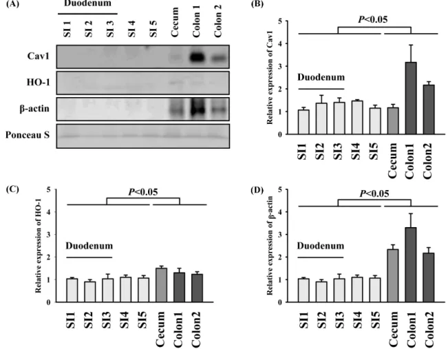
Fig. 1. Immunoreactivity of eluates from small and large intestine to murine antigens. (A) Segmental eluates were probed with antibodies against cav-1, HO-1, and actin (B) Quantification of Cav-1 band intensity (C) Quantification of HO-1 band intensity (D) Quantification of β-actin band intensity Segmental eluates were probed with antibodies against cav-1 and β-actin basally. Loading of the samples was quantitatively validated with Ponceau S staining. The statistical significance of the results was analyzed using one-way ANOVA with Bonferroni correction.
(Jeon et al., 2014) The paraffin slides with embedded lung tissue were deparaffinized with xylene, rehydrated in graded ethanol, and rehydrated with running water for 5 min. The tissues were denatured for 10 min in boiling 10 mM citric acid (pH 6.0), and allowed to stand at room temperature for 10 min. The sections were post-fixed in ethanol-acetic acid (2:1), followed by rinsing. The sections were incubated with proteinase K (100μg/mL), rinsed, incubated in 3% H2O2, permeabilized with 0.5% Triton X-100, rinsed again, and incubated in the TUNEL-reaction mixture. The sections were rinsed and visualized using Converter-POD with 0.05% 3,3-diaminobenzidine. The slides were air-dried overnight at room temperature, and coverslips were mounted using Permount® (Fisher Scientific, Waltham, MA, USA).
Statistical analysis
Analysis and data graphing were done with Prism 4.0 (GraphPad Software, San Diego, CA, USA). Data are expressed as means± SEM of at least three independent experiments. Statistical analysis
was performed by one-way ANOVA for multiple group comparisons followed by Bonferroni correction. A value of p<0.05 was considered statistically significant.
Results and Discussion
Based on previous reports estimating the daily weight loss in the small intestine (Lodish, 2000; Bullen et al., 2006; Iwanaga et al., 1993), the purpose of this study was to carry out lost cells in the small intestine, cecum and colon1/2. This study showed that the small intestine can consume dead cells in a manner similar to how it consumes protein from the diet. Caveloin-1 is found in caveolae, which are 0-100 nm cell surface plasma membrane invagination (Jin et al., 2009) Because caveolin-1 and caveolae are present in nearly all cell types, the presence of caveolin-1 can indicate the presence of a cell (Lee et al., 2015). Additionally, the intracellular functions of caveolin-1 and caveolae are diverse (Lee et al., 2018). ß-actin is a cytoskeletal protein related to cell structure and
Fig. 2. Measurement of intracellular cav1 and heme level in time-dependent. (A) Segmental eluates were probed with antibodies against eukaryotic cav-1 (B) Relative band intensity of Fig 1A (C) Segmental eluates were probed with antibodies against eukaryotic HO-1 (D) Relative band intensity of Fig. 1C. Segmental eluates were probed with antibodies against eukaryotic cav1-Heme levels in each segment were analyzed using a chemiluminescence (CL) assay after bacterial infection, which detected heme-associated horseradish peroxidase activity. SI, small intestine; cav-1, caveolin-1. The statistical significance of the results was analyzed using one-way ANOVA with Bonferroni correction.
motility and is ubiquitously expressed. Specifically, many studies have considered β-actin as a housekeeping gene (Lin et al., 2012). Therefore, in our study, cav-1 and β-actin antibodies were applied to determine whether cells remained in the elutes. As shown in Fig. 1A-D, immunoreactivity with eukaryotic caveolin-1 antibody was detected only in the cecum and 2 segments from the large intestine. Thus, this protein was not detected in eluate from small intestine, compared to eluate from cecum and large intestine, which indicated that few cell was in eluate from small intestine. Likewise, mouse β-actin antibodies also reacted with eluates only from the cecum and large intestine. Reportedly, dead cells shed from the lining of the large intestine are in part disposed of by bacterial feeders, which produce vitamins and help digest fiber (Cummings et al., 1997). Some dead cells were detected with antibodies against eukaryotic epitopes. To understand this result, we agitated the lining of the small intestine with virulent Shigella flexneri 2 by intragastrical administration (Chang et al., 2013). As shown in Fig. 2, after 3 h of bacterial infection, we detected undisposed cells in the eluates from the small intestine by western blotting with caveolin-1 antibodies. However, immunoreactivity against caveolin-1 disappeared in the small intestine after 24 h. This indicates that proteins isolated from dead cell were reabsorbed into the small intestine during diet. Diets in experiments are considered as a variable factor such as the size and type of food, which can lead to inconsistent results. Therefore, we examined elutes from the small intestine after bacterial infection (Fig. 3). Specifically, the eluate of segment 5 from the small intestine after 3 h of infection contained caveolin-1-positive clumps with
DAPI-stained nuclei (Fig. 3A). An emerging field in biology considers the number of cells as a novel factor for scaling of biological structures and phenomena (Phillips et al., 2007). Depending on the number of enterocytes that are shed daily and their weight, eliminating them entirely would constitute a considerable loss of valuable resources from the body. Shedded enterocytes may be disposed in the same way as the food that an animal eats every day, in addition to by apoptosis. Bacterial agitation not only facilitates apoptosis or shedding of enterocytes, but also impedes barrier function, including the villi function (Chang et al., 2013). At an early time point (1 h after bacterial infection), we observed no caveolin-1 immunoreactivity in the small intestine, even though the gut lining underwent partial injury, indicating that the intestine’s absorptive function was still operational. We also measured heme levels in the eluates from each segment (Fig. 2A). Heme is a highly conserved molecule ubiquitously distributed in organisms from all three domains, Prokaryote, Archaea, and Eukaryote (Warren and Smith, 2009). Heme may have originated from the dead bodies of intestinal bacteria or damaged enterocytes. Basal level of heme was faintly observed in the distal parts of both the small and large intestines. One hour after bacterial infection, heme levels were somewhat increased in the distal part of the small intestine and cecum. However, heme levels were dramatically increased after 3 h of bacterial infection in the small and large intestine. Careful examination of images for DNA staining revealed that the level of prokaryotic DNA was negligible in the eluates, suggesting that enterocytes damaged by bacterial infection are responsible for the increased heme levels. Heme oxygenase (HO)-Fig. 3. Shedding of enterocytes from the small intestine after bacterial infection. (A) Eluates from the small intestine (SI segment 5) were stained immunohistochemically with DAPI (nuclei, blue) and a cav-1 antibody (upper). (B-C) H&E staining and TUNEL assay were carried out in the ileum (SI segment 5).
1 catalyzes degradation of heme to formation of bilirubin and carbon monoxide (CO) (Lee et al., 2020). Heme offers severe cellular oxidative damage by promoting ROS formation, lipid peroxidation, DNA and protein damage (Lee et al., 2015). To overcome its toxicity, HO-1 upregulation can be accompanied in heme. In line with this, we measured HO-1 and confirmed that HO-1 level was dependent to heme (Fig. 1A). Additionally, destruction of villus architecture was obvious at 1-3 h and recovered 24 h after bacterial infection (Fig. 3B). Hence, we reasoned that the absorptive function of the villi was not greatly affected by bacterial infection, which was accompanied by a minor increase in terminal deoxynucleotidyl transferase dUTP nick-end labeling (TUNEL) staining 1 h after bacterial agitation (Fig. 3C). The villous architecture divided from the small intestine by bacterial infection was visually confirmed. Nevertheless, we did not distinctly observe protein compared to that in the cecum and colon 1/2, indicating that the small intestine can consume proteins from dead cells. In agreement with this, this study suggests that the body conserves resources by disposing of and recycling shedded cells in the small intestine. Therefore, our study may provide new insights into evolutionary biology. Collectively, given the results of our studies in a murine model, we suggest that the body conserves resources in the small intestine by disposing of and recycling shedded cells (Fig. 4).
Conflicts of interest
The authors declare no conflict of interest.
References
Bullen TF, Forrest S, Campbell F, Dodson AR, Hershman MJ, Prit-chard DM, Turner JR Montrose MH, Watson AJ. Characterization of epithelial cell shedding from human small intestine. Lab. Invest. 86: 1052-1063 (2006)
Chang SY, Lee SN, Yang JY, Kim DW, Yoon JH, Ko HJ, Ogawa M, Sasakawa C, Kweon MN Autophagy controls an intrinsic host defense to bacteria by promoting epithelial cell survival: A murine model. PLoS One 8: e81095 (2013)
Cummings JH, Macfarlane GT. Role of intestinal bacteria in nutrient metabolism. JPEN J. Parenter. Enteral Nutr. 21: 357-365 (1997) Hausmann M. How bacteria-induced apoptosis of intestinal epithelial
cells contributes to mucosal inflammation. Int. J. Inflam. 2010: 574568 (2010)
Iwanaga T, Han H, Adachi K, Fujita T. A novel mechanism for dis-posing of effete epithelial cells in the small intestine of guinea pigs. Gastroenterology 5: 1089-1097 (1993)
Jeon JW, Lee JI, Shin HP, Cha JM, Joo KR, Kim SH. Adenosine A2A-receptor agonist polydeoxyribonucleotide promotes gastric ulcer healing in Mongolian gerbils. Animal Cells Syst. 18: 399-406 (2014)
Jin Y, Kim HP, Cao J, Zhang M, Ifedigbo E, Choi AM. Caveolin-1 regulates the secretion and cytoprotection of Cyr61 in hyperoxic cell death. FASEB J. 23: 341-350 (2009)
Lee PJ, Cho N, Yoo HM, Kim HP. Active turnover of heme in hibernation period in mammals. Front Physiol. 10: 1586 (2020) Lee PJ, Park HJ, Cho N, Kim HP.
3,5-Diethoxy-3'-Hydroxyresvera-trol (DEHR) ameliorates liver fibrosis via caveolin-1 activation in hepatic stellate cells and in a mouse model of bile duct ligation injury. Molecules 23: 2833 (2018)
Lee PJ, Woo SJ, Jee JG, Sung SH, Kim HP. Bisdemethoxycurcumin induces apoptosis in activated hepatic stellate cells via cannab-inoid receptor 2. Molecules 20: 1277-1292 (2015)
Lin J. Redies C. Histological evidence: housekeeping genes beta-actin and GAPDH are of limited value for normalization of gene expression. Dev. Genes Evol. 222: 369-376 (2012)
Lodish H. Molecular Cell Biology. W. H. Freeman, New York, NY, USA (2000)
Martin R. How we do it. The evolution and future of human repro-duction. Basic Books, New York, NY, USA. pp. 118-119 (2011) Phillips R, Milo R. Cell Biology by the Numbers. Garland Science,
Boca Raton, FL, USA (2015):
Pritchard DM, Potten CS, Korsmeyer SJ, Roberts S, Hickman JA. Damage-induced apoptosis in intestinal epithelia from bcl-2-null and bax-null mice: investigations of the mechanistic determinants of epithelial apoptosis in vivo. Oncogene 18: 7287-7293 (1999) Warren MJ, Smith A. Tetrapyrroles: Birth, Life, and Death. Springer,
Berlin, Germany (2009)
Williams JM, Duckworth CA, Burkitt MD, Watson AJ, Campbell BJ, Pritchard DM. Epithelial cell shedding and barrier function: a matter of life and death at the small intestinal villus tip. Vet. Pathol. 52: 445-455 (2015)
Fig. 4. The concept of resource conservation for efficient use of energy.