13
대장균에서 Chlamydia psittaci MOMP 유전자의 과발현과 순수분리
하정순·이도부·한상훈·임윤규1·윤병수*
경기대학교 생물학과
1제주대학교 수의학과 (게재승인: 2006년 1월 22일)
Over-expression of Chlamydia psittaci MOMP in Escherichia coli and its purification
Jung-Soon Ha, Do-Bu Lee, Sang-Hoon Han, Yoon-Kyu Lim
1, Byoung-Su Yoon*
Department of Biology, Kyonggi university, Suwon 443-760, Korea
1
Department of Veterinary medicine, Cheju National University, Cheju 690-754, Korea
(Accepted: January 22, 2006)
Abstract
:Generally known psittacosis or ornithosis is a disease of birds caused by the bacterium
Chlamydia psittaci. Humans are accidential hosts and are most commonly infected from avian sources. It raises hepatitis or neurosis. As major outer membrane protein (MOMP) of
Chlamydia psittacihas been known to play a role in the avoidance of host immune defenses, research on developing a
Chlamydiavaccine has focused on the MOMP. In this study, the gene encoding the major outer membrane protein (MOMP) of the
Chlamydia psittacistrain 6BC was cloned and expressed in
Escherichia colistrain M-15. The recombinant DNA was cloned by fusion prokaryotic expression vector pQE30-GFPII. Expression of the recombinant protein was performed in
E. coliand was induced by IPTG. The size of expressed recombinant protein is 74.220 kDa (MOMP, 43.260 kDa; GFP expression region, 30 kDa; 6
×His tag, 960Da). This protein was purified by using his-tagging-inclusion body. Recombinant protein was reconfirmed through ELISA test and western blot with antibody against pQE30-GFPII. It will be useful antibody development.
Key words :
Chlamydia psittaci,Major outer membrane protein, pQE30-GFPII
서 론
조류의
Chlamydia psittaci
감염은앵무새에서처음 으로발견되었기때문에앵무병(psittacosis)
이라불리고있으며
,
인체에감염된경우에도같은병명을사용하 고있다.
학자에따라서는,
사람과조류에있어서의감 염을avian chlamydiosis(AC)
라부르고사육되는동물이거나야생조류의경우에
ornithosis
로불린다.
이두 질병은비슷하나포유류에서의Chlamydia
증은약간다 른병인체에의해일어난다. AC
의주는사람에서심각한병증을유발하고죽음에이르게할수있으며사 람에서의
ornithosis
가psittacosis
보다경증으로진행된 다.
칠면조로부터전염된질병은앵무새로부터유래된 것보다종종중증으로진행된다[18, 19].
다행히대부분의
Chlamydia
의감염증은아직항생제에의한치료가가능하나 병원균에의한자연면역은 매우일시적이기에치료후연속적인감염이될수있 다
.
계속되는감염보다악화된증상을보이는경우가 많으나재감염을피하기위한유용한vaccine
은유전적 인그리고병리학적어려움등의이유로아직개발되*Corresponding author: Byoung-Su Yoon
Department of Biology, Kyonggi university, Suwon 443-760, Korea
[Tel: +82-31-249-9645, Fax: +82-31-243-1707, E-mail: bsyoon@kyonggi.ac.kr]
지못하고있다
[7, 8].
Chlamydia
는감염력이 있는elementary body(EB)
와세포내생존상이며감염력이없는
reticulate body(RB)
로확실히구분된다
[8, 9, 13]. EB
의세포막은일반 세균공통의세포벽구성요소인peptidoglycan layer
가없으며일부막단백질이
disulfide cross-linkage
를이루 고있다[7].
이막단백질은훨씬복잡한분자구조를 형성하여EB
의삼투압적안정성을부여한다[9].
막단백질의 대부분은
major outer membrane protein (MOMP)
이 한 개 내지 여러 개의cysteine-rich pro- teins(CRPs),
그리고작은CRP
로구성되어있다[5, 10, 11, 12].
MOMP
는constant region
과variable region
으로구분 되며,
혈청형에의한serotype
은진단을위해이용되고있다
[6, 20].
이러한유전적요소들은Chlamydia
증에반응하는숙주의보호면역에중요한역할을하며
[20,
21]
이것은vaccine
개발에이용되고있다[13].
본연구는
Chlamydia psittaci
에존재하는MOMP
유 전자를PCR
방법으로증폭하고[15], fusion vector
인pQE30-GFPII
를사용하여단백질을생성하였다.
대장 균내에서의이단백질의대량생산가능성여부를조 사하여 순수분리를실시하였고,
최종적으로protein vaccine
개발에사용될예정이다.
재료 및 방법
Chlamydia psittaci의 분리
본실험에사용된
Chlamydia psittaci strain 6BC
는잘보존된
lab-strain
으로써비교적다양한host cell type
에 쉽게적응되어성장하며덜치명적이다.
본연구에서 사용된C. psittaci strain 6BC
는C. psittaci strain Francis
와약
98%
의상동성을가지며[2]
미국Jeorge Washing- ton
대학의J. Stokes
교수에게서분양받았다.
Chlamydia psittaci MOMP 유전자 확보 및 발현 vector로의 molecular cloning
Chlamydia psittaci
의chromosomal DNA
를template
로사용하였으며
primer
로는forward
로Bam HI
자리가삽 입된MOMP-F(GAGGGCTCCATGAAAAAA CTCTTG) 100pmol
과reverse
는Sal I
자리가 삽입된MOMP-R (ACTGTCGACTTAGAATCTGAATTG) 100 pmol
을사 용하였다.
총양은100
µl
로1.5 mM MgCl
2와0.25 mM dNTP, 2.5 unit Taq polymerase(Gene clone Co., Korea)
를사용하였으며
Gene Amp 9600(ABI Co., U.S.A)
을 사용하여94
oC 3
분의predenaturation
과94
oC 30
초, 53
oC 30
초, 72
oC 1
분을30
회반복한후final elongation 72
oC
5
분을더주어증폭하였다.
확보된
PCR
산물들은pBluescriptKS(+)
유래의pBlueXcm vector
를사용하여molecular cloning
을수행하였으며염기서열의분석을통하여
Chlamydia psittaci MOMP
유전자임을 확인하였다.
차후에primer
내의restriction enzyme
자리를이용하여발현vector
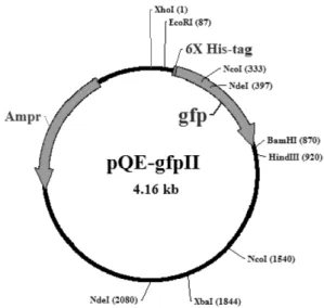
인pQE 30-GFPII(Fig. 1)
에molecular cloning
을실시하였다[1].
IPTG 농도에 따른 Chlamydia psittaci MOMP 유 전자 발현 변화
Chlamydia psittaci MOMP
유전자와 퓨전vector
인pQE30-GFPII
로재조합된plasmid DNA
를삽입하여E.
coli
숙주인M-15
에형질변환시켜이clone
을LB
배 지20 ml
에100
µg/ ml ampicillin
을첨가한배지에배 양한균액을1 ml
넣고37
oC, 180 rpm
에서4
시간계대 배양을수행하였다. Chlamydia psittaci MOMP
의재조합된
clone
단백질의과발현유도를위하여계대배양액에
100 mM Isopropylthio-
β-D-galactoside(IPTG)
를0 mM, 0.003 mM, 0.007 mM, 0.015 mM, 0.03 mM, 0.07 mM, 0.15 mM, 0.3 mM
를첨가하여각각37
oC, 180 rpm
에서
6
시간배양하여induction
을실시하였다.
최적조 건에서자란20 ml
배양액을50 ml conical tube
에넣 고3,300
×g
에서15
분간원심분리를실시하였고PBS
로2
회washing
을실시한후전체700
µl
에부유시키고Fig. 1. A Schematic diagram of the fusion prokaryotic
expression vector, pQE30-GFPII, for the expression of
Chlamydia psittaciMOMP protein. pQE30-GFPII vector
has multiple cloning site (MCS), which is able to enhance
the molecular cloning. Ampicillin represents ampicillin
resistance gene. N-terminal has a 6*His-tag which is able
to purify the fusion protein.
파쇄한 후 추출한
total protein sample
은Bio-rad
의MINI-PROTEAN III protein electrophoresis system
을사용하여
12% SDS-polyacrylamide gel
을사용하여분자 량별로분리하여확인하였다.
pQE30-GFPII-Chlamydia psittaci MOMP 단백질 정제 가장 강하게 발현된
0.3 mM
의 단백질700
µl
에100 mM PMSF
를최종농도1 mM
로하여첨가하고ice
에서
10
분간정치한후amplitude 26%, pulse 0.5sec
로3
분간파쇄하였고, 5
분간ice
에정치시킨후다시같은 조건으로파쇄하였다.
이때이단백질의inclusion body
형성여부를관찰하기위해서
1
회, 2
회, 3
회로나누어 각각다르게파쇄하였다.
각각의상층액을추출하여1.2
µm
로여과한후His trap kit(Amersham Biosciences, U.S.A)
를이용하여정제를하였다.
나머지pellet
은세 척액(20 mM Tris/Cl pH8.0, 0.5% tween20,
최종pH8.5
로조정
)
으로1
회세척후14,000
×g, 4
oC, 10
분동안원 심분리하여 생성된pellet
을증류수로1
회 세척하고14,000
×g, 4
oC, 10
분동안원심분리한후에700
µl
의평형용액
(50 mM NaH
2PO
4,300 mM NaCl, 5 mM immi- dazole)
으로냉장에서16
시간보관하였다.
용해한단백 질을ice
에10
분정치한후ice
속에서sonication
을수행하였고
14,000
×g, 4
oC, 10
분동안원심분리한후에상 층액을 회수하여1.2
µm
로 여과한 후His trap kit (Amersham Biosciences, U.S.A)
을이용하여정제하였다.
정제한단백질은100% TCA
를이용하여농축하였고 추출한total protein sample
은Bio-rad
의MINI- PROTEANIII protein electrophoresis system
을사용하여12% SDS-polyacrylamide gel
을사용하여분자량별로 분리하여확인하였다.
ELISA를 통한 pQE30-GFPII-Chlamydia psittaci MOMP 단백질 발현 확인
96-well(flat form) plate
에coating buffer
로희석시킨항원단백질용액을
50
µl/well
씩가하고37
oC
에서1
시 간정치시켰다.
이후plate
를털어서용액을제거하고well
의나머지 표면을blocking
하기 위하여blocking solution(0.1% BSA in PBS) 100
µl
를각well
에가하고1
시간동안4
oC
에서정치시켰다.
이후PBS
용액으로3
회세척하였다.
Anti-pQE30-GFPII
항체생성세포의배양상층액을PBST
로1/10
로희석하고50
µl/well
로가하고실온에서30
분동안정치한 후제거하고PBS
로3
회세척하였다
. HRPO conjugated anti-mouse immunoglobulin(anti- mouse IgG)
는PBST
에1/10000
로희석하여50
µl/well
씩가하였다
.
이를실온에서30
분간정치시킨후그용액을제거하고
PBS
로3
회세척하였다.
기질효소반응 을시키기위해서0.04% OPD
기질용액을well
에50
µl
를넣고상온에서
15
분-30
분간정치하고반응을정지 시키기위해서2.5N H
2SO
450
µl
를가하였으며,
이때반응하여발색된정도를흡광도
490 nm
에서측정하였다
[4].
이때
M-15
에형질전환된pQE30, pQE30-GFPII, r- pQE30-GFPII-MOMP
를IPTG
로발현을유도하였으며negative control
은pQE30, r-pQE30-GFPII-MOMP
에IPTG
를첨가하지않은것과BSA
를사용하였다.
각항 원의농도는bradford reagent
를이용한micro assay
를수행하여정량하였고 항원의농도는 각각
100
µg/ml, 10
µg/ml, 500 ng/ml
로조정하였다.
Western blotting을 통한 pQE30-GFPII-Chlamydia psittaci MOMP 단백질 발현 확인
SDS PAGE gel
전기영동을 마친gel
을transfer buffer(Tris base 25 mM, Glycine 150 mM, Methanol 10%, pH 8.3
보정한후증류수로1 l
조정)
에10-15
분정도담가두었으며
PVDF membrane
을methanol
에서3
초간정치시키고증류수에서1-2
분동안세척한다음, transfer buffer
에서5
분간 정치시켰다. Whatman paper (No. 3)
는상부와하부각5
장씩transfer buffer
에적셨 다. Transfer blotter(Biometra, Germany)
에하부Whatman paper(No. 3)
를5
장깔고그위에PVDF memebrane
을올리고
SDS-PAGE gel
을올린다음상부에whatman paper(No. 3)
를5
장올렸다. Transfer
는gel
의면적에따 라5 mA/cm
2의전류로120
분동안transfer
를수행하였으며
power supply
는BIO-RAD
사의Power PAC 300
제 품을 사용하였다. Transfer
한PVDF membrane
을blocking solution (3% Skim milk-TBS)
에넣고상온에서
1
시간 정치시켰다.
그 후TBST(TBS-Tween20 0.05%)
로흔들면서5
분간3
회씩세척하였다. Primary antibody(anti-pQE30-GFPII serum
을3% Skim milk- TBS-Tween20 0.05%
에1/10
으로 희석하여사용)
를넣 고30
분동안shaking incubation
을한후, 3
회TBST
로 세척하였다. Secondary antibody reaction
은anti-mouse- IgG
를3% Skim milk-TBST
에1/10000
으로 희석하여30
분동안shaking incubation
을한후, 3
회TBST
로세 척하였다. DAB solution
을사용하여발색을확인하였으며증류수로세척하여 반응을종결시켰고건조후 사진을찍어보관하였다
.
결 과
Chlamydia psittaci MOMP
의유전자는이용한PCR
을통하여
1209 bp PCR product
을얻을수있었고,
이PCR product
와pBlueXcm
을사용하여 재조합plasmid DNA
를 제작하였다.
재조합된DNA
의sequencing reaction
을통하여Genebank accession NO. X56980
의 염기서열시작코돈364 bp
부터종결코돈1572 bp
까지
1209 bp
의염기서열이100%
일치함을확인할수 있었다.
확인된염기서열은NEB
사의제한효소프로그 램을통하여분석한후분석된결과를토대로하여재 조합DNA
를절단한후확인하였고,
차후에Chlamydia psittaci MOMP
단백질의발현을위해서pBlue Xcm
에 재조합된DNA
를제한효소Bam HI, Sal I
을이용하여발현
vector
인pQE30-GFPII
로의sub cloning
을수행하였다
(Fig. 2).
재조합된DNA
를r-pQE30-GFPII-MOMP
로명명하였으며이를발현숙주인
M-15
로형질전환하여
IPTG
농도에따른단백질의발현을유도하였다.
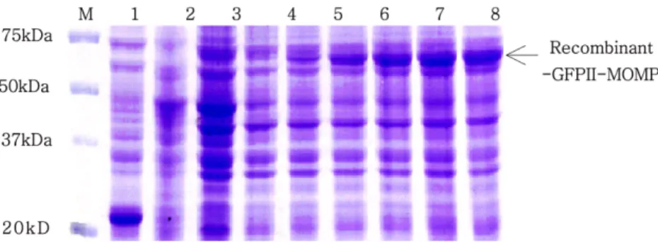
그결과
IPTG
농도의증가에따라단백질발현의양이증가함을알수있었으며
(Fig. 3),
최적의발현조건은
IPTG 0.3mM
에서6
시간발현을유도하였을때였다.
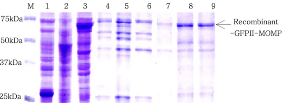
이단백질의
inclusion body
형성여부를관찰하기위 한실험에서sonication
의횟수가증가할수록단백질의양이증가됨이관찰되었고상등액과
pellet
의정제된단 백질관찰에서상등액에는발현단백질이없음이확인 되었고grea
를이용한pellet
의정제에서발현된단백질이관찰되었다
(Fig. 4).
이를통하여recombinant-GFPII- MOMP(r-GFPII-MOMP
로 명명)
단백질이inclusion body
를형성한다고판단하였다.
r-GFPII-MOMP
단백질의발현을확인하기 위해서항체인
anti-pQE30-GFPII
의배양상층액을 이용하여ELISA
를 수행하였을 때negative control
인BSA, pQE30
에IPTG 0.5 mM
첨가한 것과r-GFPII-MOMP
에
IPTG
를첨가하지않은것에서는0.2
이하의역가를나타내었고 이것은
anti-pQE30-GFPII
가특이적으 로 작용하는 것을시사한다. Positive control
인 항원pQE30-GFPII-IPTG 0.5 mM
에서는항원농도10
µg/ml
에서최고
2.27
까지의높은값을나타내었으며r-GFPII- MOMP-IPTG 0.5mM
에서는항원농도100
µg/ml
에서0.875
의 값을나타내었다(Fig. 5).
이를 통해pQE30- GFPII
보다는소량이지만r-GFPII-MOMP
에서단백질 이발현되었다고판단되었고또한IPTG
농도에따른r-GFPII-MOMP
의ELISA
수행시IPTG
농도가증가함에 따라 역가가 증가됨이 확인되었다
(Fig. 6). r- GFPII-MOMP
의발현은Western blot
을통하여재확인 되었다(Fig. 7).
Fig. 2. Restriction analysis of recombinant DNA. Recom- binant DNA of
Chlamydia psittaciMOMP gene was analyzed by using NEB cutter program (not showed). Then, recombinant DNA fragments were confirmed through restriction enzyme
BamHI/
SalI. Lane M, DNA size marker;
lane1, pQE30-GFPII 4.1 kbp; lane 2, r-pBlueXcm-MOMP 2.9 kbp/1.2 kbp; lane 3, r-pQE30-GFPII-MOMP 4.1 kbp/
1.2 kbp.
Fig. 3. Expression patterns of r-GFPII-MOMP in
E. colistrain M-15 under different IPTG concentration. SDS-PAGE was performed with 12% polyacrylamide gel containing 0.1% SDS and 1×TGS (Tris/Glycine/SDS) running buffer. All sample was loaded after sonication. Lane 1, pQE30-GFPII IPTG 0.3 mM; lane 2 to 8, r-GFPII-MOMP IPTG 0 mM, 0.003 mM, 0.007 mM, 0.015 mM, 0.03 mM, 0.07 mM, 0.15 mM, 0.3 mM. The molecular weight is 74.220 kDa (MOMP, 43.260 kDa;
GFP expression region, 30 kDa; 6×His tag, 960 Da). The fusion protein is increasing with IPTG concentration.
고 찰
Chlamydia
의감염증은항생제에의한치료가가능하지만항생제의오남용이초래되어보다악화된증상을 보이는경우가많다
[6, 11].
현재Chlamydia
의배양은 동물세포주에감염및공배양으로만이루어지고있으 며조제된 배지에의한배양은성공하지 못하였기에 아직유용한vaccine
의개발이되지않았다[7, 8].
본연구에서는
r-GFPII-MOMP
단백질을제작하였고GFP
의과발현을이용하여MOMP
단백질을발현시켰으며
pQE30-GFPII
항체를이용하여확인하였다.
여기 서 사용된GFP
는E. coli
와 선충류인Caenorhabditis elegans
에서각각발현되는특징을가지며[3],
그것의발현이원핵세포와진핵세포모두에서세포의성장과 기능에아무런간섭을끼치지않는다
.
이러한특성은 여러유전자의발현을monitoring
할수있는다재다능한도구로써의가능성을시사한다
.
Vanrompay
등[16]
은칠면조에서Chlamydia psittaci MOMP
유전자를이용한plasmid DNA
백신을개발하였고이는
T-helper cell
과B memory cell
의향상을유 도하였다고하였지만진핵세포로의transfection
시높 은면역반응을유도하지는못하였음을보고하였다.
따라서본연구에서는
Chlamydia psittaci MOMP
유 전자를이용한protein
백신을제작을위하여Chlamydia psittaci MOMP
단백질의발현을 도모하였다. MOMP
단백질의 과발현을위해
GFP
단백질을 사용하였으며이로인하여
MOMP
단백질의발현여부를확인할수가있었다
.
제작된r-GFPII-MOMP
의단백질을이용하여차후에단클론항체를생성할예정이며제작된백
Fig. 4. Expression and purification of r-GFPII-MOMP in M-15. Lane 1, r-pQE30-GFPII IPTG 0.3 mM; lane 2, r-GFPII- MOMP IPTG 0 mM; lane3, r-GFPII-MOMP IPTG 0.3 mM; lane 4 to 9 are total lysates of r-GFPII-MOMP that were induced under 0.3 mM final concentration. The concentration of r-GFPII-MOMP increased with sonication times, respectively. Lane 4 to 6, the supernatant of expressed protein were purified by His trap kit. Lane 7 to 9, the pellets of expressed protein are handled by using 9 M urea followed by being purified by His trap kit.
Fig. 5. Specific binding of mouse anti-pQE30-GFPII to expressed materials was confirmed through ELISA analysis of each antigen. ELISA test accomplished under OD 490 nm. Negative control; BSA; pQE30, IPTG 0.5 mM; r- GFPII-MOMP, IPTG 0 mM. Positive control; pQE30-GFPII- MOMP, IPTG 0.5 mM; r-GFPII-MOMP, IPTG 0.5 mM.
Detectable value of negative controls was 0.2 or less.
However, optimal density value of r-GFPII-MOMP IPTG 0.5mM (antigen concentration, 100
µg/ml) was 0.875. It showed that primary antibody, culture supernatant of anti- pQE30-GFPII producing hybridoma cells detected antigen of r-GFPII-MOMP.
Fig. 6. Expression patterns of r-GFPII-MOMP in M-15 with IPTG concentration were confirmed by ELISA analysis. Concentration of IPTG: 0 mM; 0.003 mM; 0.007 mM; 0.015 mM; 0.03 mM; 0.07 mM; 0.15 mM; 0.3 mM;
0.5 mM. The greatest expression quantities of r-GFPII-
MOMP was obtained under at IPTG 0.5mM of IPTG
concetration.
신은항생제오남용으로인한 여러가지폐해들이발 생하고있는
psittacosis
증의전파와예방을위하여사 용될것으로기대한다.
결 론
Chlamydia
의백신개발을위하여항원제작에필요한
Chlamydia psittaci MOMP
단백질의 과발현은pQE30-GFPII
에서 이루어졌으며,
생성된r-GFPII- MOMP
단백질은inclusion body
를형성하였다.
따라서 정제시9 M urea
처리한후His tagging
을이용하여 정제하였다.
발현된r-GFPII-MOMP
는anti-pQE30- GFPII
를이용한ELISA
수행시매우특이적으로반응 하였고유도물질인IPTG
의농도증가에따라r-GFPII- MOMP
의역가가증가됨이확인되었다.
이는Western blot
분석을 통하여 재확인되었다.
발현된r-GFPII-
MOMP
의단백질을이용하여차후에단클론항체를생성하여백신을제작하여
psittacosis
의전파와예방을위하여사용될수있을것으로사료된다
.
참고문헌
1.
윤병수.
Chlamydia 형질전환을 위한GFP-plasmid vector
의개발. In:
윤병수(ed.).
분자생물학연구방법론
2. pp. 70-78,
경기대학교연구지원팀, 1999.
2.
한상훈,
정규회, Stokes GV,
윤병수.
Chlamydiapsittaci
strain fransis
의plasmid pCpA1
과 C. psittacistrain 6BC
의plasmid
의염기서열상동성분석.
경기대학교논문집


