DOI 10.17480/psk.2017.61.1.12
소포체 스트레스를 매개한 curcumin의 폐암세포주 사멸효과
오지윤 · 김소현 · 곽재환* · 정영석# 부산대학교 약학대학, *경성대학교 약학대학
(Received December 25, 2016; Revised January 15, 2017; Accepted January 18, 2017)
Curcumin-induced Apoptotic Cell Death Mediated by ER Stress in Lung Carcinoma Cell Lines
Ji Youn Oh, Sou Hyun Kim, Jae-Hwan Kwak*, and Young-Suk Jung# College of Pharmacy, Pusan National University, Busan 46241, Korea
*College of Pharmacy, Kyungsung University, Busan 48434, Korea
Abstract — In the present study, we investigated the effect of curcumin, a major well-known compound isolated from rhi- zomes of Curcuma longa, on the apoptotic cell death and the potential action mechanism of this effect in human lung car- cinoma cell lines. The results showed that curcumin inhibited the cell viability in a concentration-dependent manner in both H292 and H358 human lung carcinoma cells. Treatment of curcumin activated caspase-3 evidenced by increased cleaved protein of caspase-3, and subsequently, induced cleavage of PARP. In line with this, Annexin V/PI staining clearly showed that curcumin-induced apoptosis in both cell lines. Interestingly, CCAAT-enhancer-binding protein homologous protein (CHOP), a key molecule involved in endoplasmic recticulum (ER) stress-mediated apoptosis, was significantly increased by curcumin treatment. Collectively, the results suggest that ER stress via CHOP induction might play a significant role in cur- cumin-induced apoptotic death of human lung carcinoma cells.
Keywords Lung cancer, Curcumin, Apoptosis, ER stress
암은 종양억제 유전자의 돌연변이와 세포성장에 관여하는 세 포내 신호경로의 과도한 활성화에 의해 세포가 비정상적으로 증 식하며 발생한다.1,2)특히, 폐암의 경우 전 세계적으로 가장 높은 사망률을 보이고 있으며, 우리나라 10대 암의 5년 상대생존율을 비교하였을 때, 폐암에 의한 사망이 가장 높은 것으로 알려져 있 다. 폐암은 병리학적으로 비소세포폐암(non-small cell lung cancer), 소세포폐암(small cell lung cancer)으로 분류되며, 비소 세포폐암은 편평상피세포암종(squamous cell carcinoma), 선암 (adenocarcinoma)과 대세포암(large cell carcinoma)으로 구분된다.
이들 중 비소세포폐암은 전체 폐암의 75%, 소세포폐암은 11.2%
를 차지하고 있다.3)
천연물에서 분리된 flavonoid와 polyphenol등과 같은 피토케미 컬(phytochemical)이 다양한 종류의 암 예방 및 치료에 효과가 있음이 밝혀짐에 따라 피토케미컬의 항암기전 연구가 활발히 진 행되고 있다.4) Curcumin을 함유하고 있는 강황(Curcuma longa) 은 인도 등 동남아시아에서 향신료와 같은 식품의 재료 뿐만 아 니라, 염증질환을 호전시키는 민간요법에 사용되었다. 이러한 이 유로 연구자들의 관심을 받게된 curcumin은 실제로 Nrf2 활성 화에 의한 항산화 효과,5) NF-κB 활성 억제를 통한 항염증효과6) 와 더불어 강력한 항암효과7)또한 보고되고 있다. 현재까지 curcumin의 항암효과는 암세포의 세포주기 진행을 방해하고 apoptosis를 일으킴으로써 암세포의 증식을 억제하는 것에 기인 한다고 알려져 있으며, 이를 매개하는 기전적인 연구는 부족한 실정이다.8,9,10)
소포체는 단백질의 합성과 폴딩(folding)에 관여하는 세포내 중 요 기관이며, 신생단백질의 당화(glycosylation), 이황화결합
#
Corresponding Author Young-Suk Jung
College of Pharmacy, Pusan National University, Busan 46241, Korea
Tel.: 051-510-2816 Fax.: 051-513-6754
E-mail: [email protected]
Short Report
종설(disulfide bonds) 등의 기능도 담당한다.11-13)소포체 기능이 손 상될 경우 폴딩되지 않거나 잘못 폴딩된 단백질(unfolded or misfolded proteins)이 소포체 내에 축적되고, 기능회복을 위한 소포체 스트레스(endoplamic reticulum stress) 반응과정이 활성 화 되어진다.11,12,14)즉, 소포체 스트레스는 다양한 자극에 대해 세포를 보호하기 위하여 활성화 되고, 지속적인 스트레스가 가 해질 경우 기능이 손상된 세포를 제거하기 위한 세포사멸 과정 이 유도된다.11,14,15)특히, 소포체 스트레스 과정에서는 C/EBP계 열의 전사인자에 속한 C/EBP homologous protein(CHOP)과 growth arrest- and DNA damage-inducuble gene 153(GADD153) 이 세포사멸을 매개한다.11,12,16)
본 연구에서는 인간의 비소세포폐암에서 유래한 세포주를 이 용하여 curcumin이 암세포의 사멸에 미치는 영향을 평가하고, 이를 매개하는 기전을 제시하고자 하였다.
재료 및 방법
시약
Curcumin, 3-[4,5-dimethylthiazol-2-yl]-2,5-diphenyltetrazolium bromide(MTT), dimethylsulfoxide(DMSO)는 Sigma-Aldrich(St.
Louis, MO, USA)에서 구입하였다. Penicillin/streptomycin은 GenDEPOT(Baker, TX, USA)에서 구입하였다. RIPA buffer는 Thermo scientific(Pierce, Rockford, IL, USA)에서 구입하였으 며, BCA assay kit는 (Waltham, MA, USA)에서 구입하였다.
RPMI 배지와 4-(2-hydroxyethyl)-1-piperazineethanesulfonic acid(HEPES), fetal bovine serum(FBS)은 Capricorn scientific (Ebsdorgegrund, Hessen, Germany)에서 구입하였다. Polyvinylidene difluoride(PVDF) membrane은 Millipore(Billerica, MA, USA)에서 구 입하였다. Anti-caspase-3, anti-PARP, anti-CHOP 항체들은 cell signaling(Cell Signaling, Beverly, MA, USA)에서 구입하였다.
Anti-GAPDH, HRP-conjugated anti-rabbit IgG(H+L)은 Santa Cruz(Dallas, TX, USA)에서 구입하였고 ECL solution은 Advansta (Menlo Park, CA, USA)에서 구입하였다. Propidium iodide(PI) 와 Annexin V는 Becton-Dickinson Biosciences(San Jose, CA, USA)에서 구입하였다.
세포배양
인간의 비소세포폐암에서 유래한 세포주인 NCI-H292(본 논문 에서는 H292로 표기함), NCI-H358(본 논문에서는 H358로 표기 함)를 한국세포주은행(Korea Cell Line Bank, Seoul, Korea)에 서 구입하여 사용하였다. 5% FBS, 항생제(100 U/ml penicillin, 100μg/ml streptomycin)와 HEPES가 포함된 high-glucose RPMI 배지를 이용하여 5% CO2, 37oC의 환경에서 배양하였다.
세포형태학적 관찰
H292, H358 세포를 6well plate에 배양하여 0, 5, 10, 30 μM의 curcumin을 24시간 처리후 위상차 현미경(ZEISS, oberkochen, Germany)을 이용하여 세포의 모양을 관찰하였다.
세포생존율 측정
H292, H358 세포를 96well plate에 배양하여 0, 1, 5, 10, 20, 50μM의 curcumin을 24시간동안 처리한후 세포 생존율을 측정 하였다. 1 mg/ml 농도의 MTT 용액을 50 μl씩 각 well에 분주하 여 37oC에서 1시간 반응시키고, DMSO로 세포를 용해시킨 후 MULTISKAN GO reader(Thermo Scientific, Waltham, MA, USA)를 이용하여 540 nm에서 흡광도를 측정하였다. 세포생존율 은 100%로 표시한 대조군에 대하여 백분율로 나타내었다.
Flow Cytometry Analysis
세포에서 일어나는 apoptosis를 확인하기 위해 Annexin V와 PI로 이중염색하고 flow cytometry를 이용하여 실험하였다. H292, H358 세포를 60 mm plate에서 배양한 뒤에 30 μM의 curcumin 을 처리하고 24시간 경과 시 세포의 생존여부를 평가하였다. 세 포를 회수하여 원심분리 한 후, 세포 pellet을 FITC가 결합된 Annexin V와 PI 염색용액에서 15분간 반응시켰다. 염색된 세포 는 Accuri C6(BD Biosciences, Ann Arbor, MI, USA)를 이용하 여 측정하였다.
Western blot Analysis
H292, H358 세포를 6well plate에서 배양하여 curcumin을 처 리하고 정해진 시간에 RIPA buffer를 이용하여 lysis하였다. 단 백질은 BCA assay법으로 정량한 후 15 μg을 4X SDS loading buffer와 섞어 5분 동안 95oC에서 끓인 후 10% 또는 15% SDS- PAGE gel을 사용하여 전기영동 하였다. 분리된 단백질은 80 V, 2시간 transfer하여 PVDF membrane에 옮긴 후 상온에서 5%
Skim milk에 blocking하고 1차 항체인 caspase-3, PARP, CHOP, GAPDH와 함께 4oC에서 overnight으로 각각 반응시켰다. 그 다 음 상온에서 TBS-T buffer로 10분씩 3번 세척하고, 5% skim milk에 HRP가 결합된 적합한 이차항체를 반응시킨 후 ECL solution을 반응시켜 C300 image analyzer(Azure, CA, USA)에 서 발현양을 확인하였다.
통계 분석
실험결과에서 얻은 모든 값은 평균±표준편차로 나타내었다.
통계적 유의적 차이의 정도는 Student's t-test 또는 Dunnett’s test for multiple comparison를 사용하여 P<0.05, P<0.01, P<0.001인 값에 대해 유의적인 것으로 처리하였다.
실험결과 및 고찰
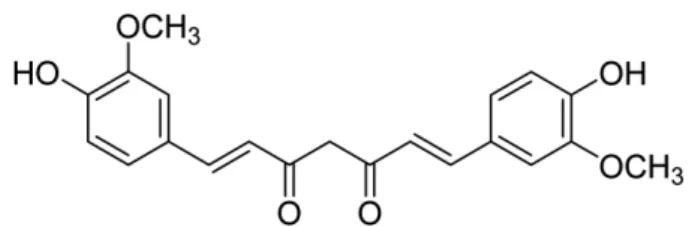
본 연구에서는 전체 폐암의 75%를 차지하는 비소세포폐암 세 포주인 H292세포와 H358세포를 이용하여 curcumin(Fig. 1)이 세포증식과 사멸에 미치는 영향을 확인하였다. Curcumin의 농 도를 5, 10, 30 μM로 24시간 처리하였을 때 농도 의존적으로 세 포의 증식억제 및 사멸이 유도되는 것을 형태학적 관찰을 통해
확인하였다(Fig. 2A). 또한 MTT assay를 이용한 세포생존율 평 가에서 curcumin의 농도를 1, 5, 10, 20, 50 μM로 24시간 처리 하여 확인한 결과 H292, H358세포 모두 20 μM 이상의 농도에 서 현저한 생존율 감소가 관찰되었다(Fig. 2B). 이러한 실험결과 는 curcumin이 H292세포와 H358세포의 증식억제 및 사멸에 효 과적으로 작용하는 것을 보여준다.
Apoptosis는 주변 조직에 상해를 주지 않으며 손상된 조직 및 세포들을 제거하는 정상적인 과정으로 체계적인 분자신호 전달 에 의해 진행되는 programmed cell death로 알려져 있다.17) Caspase는 대표적인 proapoptotic cysteine protease로서 PARP 와 함께 apoptosis의 핵심 지표이다.18,19)즉, caspase 활성화와 이를 통한 PARP 단백질의 분절은 세포의 DNA 복제 및 유전자 발현과 관련된 신호전달에 영향을 주어 apoptosis를 통한 세포사 멸에 기여한다. 따라서, Fig. 2에서 관찰된 curcumin의 효과가 apoptosis에 의해 유도된 것인지 알아보기 위해 신호전달 과정에
Fig. 1 − Chemical structure of curcumin.
Fig. 2 − Changes in cell morphology and viability of curcumin-treated H292 and H358 cells. (A) Cells were treated with indicated
concentration of curcumin for 24hr, and then photographed by microscopy at X400 magnification. (B) Cell viability was assessed by
the MTT assay and expressed as a percentage compared to the untreated cells. The results are expressed as mean±SD (n=8) of
three separate experiments. **,***Significantly different from untreated cells at P<0.01, or P<0.001, respectively (one-way ANOVA
followed by Dunnett’s test).
서 중요한 역할을 담당하고 있는 caspase-3와 PARP의 발현을 확인하였다. H292(Fig. 3A), H358(Fig. 3B) 세포에 0, 3, 10, 30μM의 curcumin을 24시간 처리하였을 때, 두 세포 모두 30 μM 의 curcumin 처리군에서 caspase-3와 PARP의 분절이 현저하게 증가한 것을 확인하였다. 이러한 결과는 curcumin에 의해 caspase-3 활성화가 유도되어 PARP의 활성화에 기여하였음을 의미한다. Curcumin에 의한 apoptosis 유도를 검증하기 위해 Annexin V/PI staining을 진행하였다. Apoptosis가 유도되면 세 포막의 안쪽에 위치하고 있던 phosphatidyl serine(PS)이 세포막 밖으로 이동하여 노출되고, 이때 칼슘 의존적 인지질 결합단백 질(calcium dependent phospolipid binding protien)인 Annexin V를 이용하여 apoptosis가 유발된 세포를 확인할 수 있다. 초기 apoptosis과정의 세포는 세포막 불균형이 일어나 Annexin V positive 부분의 세포수가 증가되고, PI에 의한 세포 염색은 일어 나지 않아 PI negative 부분에 세포수가 증가한다. 그리고, 후기 apoptosis과정의 세포는 세포막이 파괴되어 PI가 핵과 결합하면 서 PI와 Annexin V의 positive 세포수가 증가하게 된다. 30 μM 의 curcumin을 처리하였을 때 H292, H358세포 모두에서 apoptosis가 현저하게 유도됨을 Annexin V positive 부분과 PI positive, PI negative 부분의 세포수 증가를 통하여 확인하였다 (Fig. 4A). 세부적으로 30 μM의 curcumin 처리에 의해 H292세
포에서는 78%(Fig. 4B), H358세포에서는 90%이상의 세포에서 apoptosis가 일어난 것을 확인하였다(Fig. 4C). 이러한 결과는 caspase-3, PARP의 단백질 발현을 통해 관찰한 apoptosis 신호 경로의 활성화와 더불어 curcumin이 apoptosis에 관여한다는 것 을 뒷받침하는 결과이다.
소포체에 존재하는 IRE1, PERK, ATF6의 신호전달 과정20-23) 은 세포의 생존에 관련되어 있을 뿐만 아니라 apoptosis과정에도 관여하는데 세포에 대한 스트레스 상황이 매우 강하거나 지속적 으로 일어나면 소포체의 기능이 비정상적으로 작용하여 apoptosis 가 유도된다.12,24,25)특히 소포체 스트레스에 의한 apoptosis 유 도는 CHOP에 의한 신호전달을 통해 매개되어짐이 잘 알려져
있다.26,27) 30μM의 curcumin을 처리후 3, 6, 9, 12, 18, 24시간
경과시 CHOP의 발현을 관찰한 결과, H292세포에서는 6시간 경 과시 2.5배 증가된 후 지속되어 24시간 경과시 12.6배 증가하였 고(Fig. 5A), H358세포에서는 6시간 경과시 2배 증가된 후 지속 되어 24시간 경과시 4.4배 증가됨을 확인하였다(Fig. 5B). 종합 적으로 본 연구에서 확인된 curcumin에 의한 비소세포폐암세포
Fig. 3 − Effect of curcumin on the protein expression of caspase-3
and PARP in H292(A) and H358(B) cells. Cells were treated 0, 3, 10, 30 µM of curcumin for 24hr and cell lysates were collected, followed by western blot analysis for
caspase-3 and PARP. Fig. 4 − Effect of curcumin on the apoptosis. (A) Cells were treated with 30 µM curcumin for 24hr and stained with Annexin V/
PI. Cells were analyzed by two-color flow cytometry and the apoptotic cells were quantified. (B and C) Bar graph represents the mean percentages of apoptotic cells±SD.
***Significantly different between untreated control and
curcumin-treated cells. ( P<0.001, Student's t-test).
사멸효과는 CHOP를 매개로 하는 지속적이고 강력한 소포체 스 트레스에서 기인할 수 있음을 보여준다.
결 론
본 연구에서는 인간에서 유래한 비소세포폐암 세포주를 이용 하여 curcumin의 암세포 사멸효과를 확인하고, 이를 유발하는 기전으로 소포체 스트레스에 주목하였다. Curcumin의 처리는 caspase-3와 이의 하위단계인 PARP활성화를 야기하였으며, 최 종적으로 apoptosis가 유발됨을 Annexin V/PI 염색을 통해 확인 하였다. 그리고 curcumin은 CHOP의 발현을 지속적으로 유도하 여 curcumin에 의한 H292, H358 폐암세포주의 사멸이 소포체 스트레스에 의해 유도될 가능성을 제시하였다.
감사의 말씀
이 논문은 부산대학교 기본연구지원사업(2년)에 의하여 연구 되었음.
References
1) Jena, N. R. : DNA damage by reactive species: Mechanisms, mutation and repair. J. Biosci. 37, 503 (2012).
2) Mates, J. M., Segura, J. A., Alonso, F. J. and Marquez, J. : Oxidative stress in apoptosis and cancer: an update. Arch.
Toxicol. 86, 1649 (2012).
3) Jackman, D. M. and Johnson, B. E. : Small-cell lung cancer.
Lancet. 366, 1385 (2005).
4) Sarkar, F. H. and Li, Y. : Cell signaling pathways altered by natural chemopreventive agents. Mutat. Res. 555, 53 (2004).
5) Sharma, O. P. : Antioxidant activity of curcumin and related compounds. Biochem. Pharmacol. 25, 1811 (1976).
6) Satoskar, R. R., Shah, S. J. and Shenoy, S. G. : Evaluation of anti-inflammatory property of curcumin (diferuloyl methane) in patients with postoperative inflammation. Int. J. Clin.
Pharmacol. Ther. Toxicol. 24, 651 (1986).
7) Huang, M. T., Newmark, H. L. and Frenkel, K. : Inhibitory effects of curcumin on tumorigenesis in mice. J. Cell. Biochem.
Suppl. 27, 26 (1997).
8) Ng, A. P., Chng, W. J. and Khan, M. : Curcumin sensitizes acute promyelocytic leukemia cells to unfolded protein responseinduced apoptosis by blocking the loss of misfolded N-CoR protein. Mol.
Cancer. Res. 9, 878 (2011).
9) Pae, H. O., Jeong, S. O., Jeong, G. S., Kim, K. M., Kim, H. S., Kim, S. A., Kim, Y. C., Kang, S. D., Kim, B. N. and Chung, H.
T. : Curcumin induces pro-apoptotic endoplasmic reticulum stress in human leukemia HL-60 cells. Biochem. Biophys. Res.
Commun. 353, 1040 (2007).
10) Syng-Ai, C., Kumari, A. L. and Khar, A. : Effect of curcumin on normal and tumor cells: Role of glutathione and bcl-2. Mol.
Cancer. Ther. 3, 1101 (2004).
11) Schröder, M. and Kaufman, R. J. : The mammalian unfolded protein response. Annu. Rev. Biochem. 74, 739 (2005).
12) Oyadomari, S., Araki, E. and Mori, M. : Endoplasmic reticulum stress-mediated apoptosis in pancreatic beta-cells. Apoptosis.
7, 335 (2002).
13) Lin, J. H., Walter, P. and Yen, T. S. : Endoplasmic reticulum stress in disease pathogenesis. Annu. Rev. Pathol. 3, 399 (2008).
14) Credle, J. J., Finer-Moore, J. S., Papa, F. R., Stroud, R. M. and Walter, P. : On the mechanism of sensing unfolded protein in the endoplasmic reticulm. Proc. Nat1. Acad. Sci. U.S.A. 102, 18773 (2005).
15) Harding, H. P., Calfon, M., Urano, F., Novoa, I. and Ron, D. : Transcriptional and translational control in the Mammalian unfolded protein response. Annu. Rev. Cell. Dev. Biol. 18, 575 (2002).
16) Oyadomari, S. and Mori, M. : Roles of CHOP/GADD153 in endoplasmic reticulum stress. Cell. Death. Differ. 11, 381 (2004).
17) Kerr, J. F., Winterford, C. M. and Harmon, B. V. : Apoptosis. Its significance in cancer and cancer therapy. Cancer. 73, 2013
Fig. 5 − Effect of curcumin on the protein expression of CHOP in
H292(A) and H358(B) cells. The cells were treated with
30 µM curcumin for 0, 3, 6, 9, 12, 18, 24hr. Total lysates of
cells were prepared at the indicated time and analyzed by
western blot. GAPDH was used a loading control.
(1994).
18) Jurgensmeier, J. M., Xie, Z., Deveraux, Q., Ellerby, L., Bredesen, D. and Reed, J. C. : Bax directly induces release of cytochrome c from isolated mitochondria. Proc. Natl. Acad. Sci.
U.S.A. 95, 4997 (1998).
19) Antonsson, B. and Martinou, J. C. : The Bcl-2 protein family.
Exp. Cell. Res. 256, 50 (2000).
20) Araki, E., Oyadomari, S. and Mori, M. : Impact of endoplasmic reticulum stress pathway on pancreatic beta-cells and diabetes mellitus. Exp. Biol. Med (Maywood). 228, 1213 (2003).
21) Wang, X. Z., Harding, H. P., Zhang, Y., Jolicoeur, E. M., Kuroda, M. and Ron, D. : Cloning of mammalia Ire1 reveals diversity in the ER stress responses. EMBO. J. 17, 5708 (1998).
22) Yoshida, H., Haze, K., Yanagi, H., Yura, T. and Mori, K. : Identification of the cis acting endolasmic reticulum stress response element responsible for transcriptional induction of mammalian glucose-regulated proteins. J. Biol. Chem. 22, 273
(1998).
23) Harding, H. P., Zhang, Y., Bertolotti, A., Zeng, H. and Ron, D. : PERK is essential for translational regulation and cell survival during the unfolded protein response. Mol. Cell. 5, 897 (2000).
24) Mori, K. : Tripartitie management of unfolded proteins in the endoplasmic reticulum. Cell. 101, 451 (2000).
25) Kaufman, R. J., Scheuner, D., Schroder, M., Shen, X., Lee, K., Liu, C. Y. and Arnold, S. M. : The unfolded protein response in nutrient sensing and differentiation. Nat. Rev. Mol. Cell. Biol. 3, 411 (2002).
26) Tabas, I. and Ron, D. : Integrating the mechanisms of apoptosis induced by endoplasmic reticulum stress. Nat. Cell. Biol. 13, 184 (2011).
27) Wang, W. A., Groenendyk, J. and Michalak, M. : Endoplasmic reticulum stress associated responses in cancer. Biochim.
Biophys. Acta. 1843, 2143 (2014).