korean j intern med 2012;27:30-38
http://dx.doi.org/10.3904/kjim.2012.27.1.30 pISSN 1226-3303 eISSN 2005-6648
http://www.kjim.or.kr
Optimization of Stent Deployment by intravascular Ultrasound
Hyuck-Jun Yoon and Seung-Ho Hur
Department of Internal Medicine, Keimyung University Dongsan Medical Center, Daegu, Korea
Intravascular ultrasound (IVUS) is a useful diagnostic method that provides valuable information in addition to angiog- raphy regarding the coronary vessel lumen, dimensions, plaque burden, and characteristics. The major use of IVUS in coronary intervention is to guide interventional strategies and assess optimal stent deployment. Since the introduction of the drug-eluting stent (DES), concerns about restenosis have decreased. However, high-risk lesion subsets are being routinely treated with DESs, and the incidence of suboptimal results after stent deployment, such as stent underexpan- sion, incomplete stent apposition, edge dissection, geographic miss, and the risk of stent thrombosis, have correspond- ingly increased. Thus, optimization of stent deployment under IVUS guidance may be clinically important. In this review, we focus on the potential role of IVUS in stent optimization during percutaneous coronary intervention and its clinical benefits.
Keywords: Angioplasty, balloon; Coronary stenosis; Mortality; Stents; Therapy; Ultrasonography
Received : January 31, 2012 Accepted : February 2, 2012
Correspondence to Seung-Ho Hur, M.D.
Division of Cardiology, Department of Internal Medicine, Keimyung University Dongsan Medical Center, 56 Dalseong-ro, Jung-gu, Daegu 700-712, Korea Tel: 82-53-250-7998, Fax: 82-53-250-7034, E-mail: [email protected]
IntRoDuCtIon
The coronary angiogram (CAG) remains the gold-stan- dard method for assessing coronary artery disease (CAD).
However, the CAG has inherent pitfalls, such as only show- ing the vessel lumen as an X-ray shadow image, created by the injection of contrast medium, and of often visualizing a “side-view.” Thus, the apparent degree of coronary ste- nosis can be affected by the projection angle due to lesion eccentricity. Additionally, diffuse coronary disease, lesion foreshortening, angulations, calcification, and vessel over- lap can be challenges in the angiographic assessment of le- sion severity. In some cases, an angiographically normal- looking coronary artery actually shows various degrees of atherosclerotic plaque by intravascular ultrasound (IVUS)
[1,2].
IVUS is an invasive imaging technique used to visualize coronary cross-sectional anatomy and is superior to CAG in assessing vessel size, calcium content, and lesion sever- ity [3]. It also provides complementary procedural infor- mation in lesions requiring percutaneous coronary inter- vention (PCI) when determining adequate stent sizing, and confirming optimal stent deployment and apposition without edge tearing in real time. Thus, a growing number of interventional cardiologists attain optimal procedural results with reduced complications when using IVUS in PCI.
Although the routine use of IVUS in daily PCI remains
controversial, stent optimization by IVUS during stenting
procedures, especially in the era of drug-eluting stents
(DESs), may have an important role in improving long- term clinical outcomes such as stent restenosis and stent thrombosis [4]. In this review, we focus on the potential roles of IVUS in stent optimization during PCI and its clinical benefits.
ReAlIty of StentIng pRoCeDuReS In DAIly pRACtICe
PCI has been the fastest growing method for the treat- ment of ischemic CAD over the past three decades.
Coronary stents have emerged as the predominant form of PCI and are currently used in more than 90% of PCI procedures. Procedural success of PCI is usually deter- mined by visual estimation by the operator, and usually, angiographic success after PCI is defined as the attain- ment of residual diameter stenosis of less than 30%, which is generally associated with at least a 20% improvement in diameter stenosis and relief of ischemia [5]. However, such subjective estimation of the severity of coronary artery ste- nosis is thought to be of limited reliability. Previous IVUS studies have demonstrated that visual estimation or quan- titative angiographic analyses of vessel dimension for stent deployment appear inaccurate [6-9]. The post-dilatation clinical comparative (POSTIT) trial was designed to assess the achievement of optimal stent deployment by IVUS, according to normal-to-high pressure balloon dilation after bare metal stent implantation. Among 256 patients, only 14% of cases achieved optimal stent deployment with
under 12-atmosphere pressure dilation and only 36% even with higher deployment pressures (> 14 atmospheres) [9].
Another IVUS study with an early-generation DES in 200 patients assessed stent expansion depending on the manu- facturer’s compliance chart as a guideline. In that study, the DES obtained only 75% of predicted minimal stent di- ameter and 66% of the predicted minimal stent area (MSA) [10] (Fig. 1). Based on these observations, angiographic success cannot always be linked with optimal stent ex- pansion, despite higher pressure balloon inflation during the stenting procedure. In turn, stent optimization using a high-pressure balloon without IVUS guidance also has been associated with an increased risk of arterial perfora- tion, probably secondary to vessel-balloon mismatch [11].
Thus, the operator should consider using IVUS guidance for high-pressure balloon inflation during stent deploy- ment.
Apposition of stent struts to the vessel wall is also an important facet of stent optimization. Adequate stent expansion and adequate stent strut apposition have been reported to be important factors in reducing repeated re- vascularization due to stent restenosis or stent thrombosis [12,13]. In the DES era, incomplete stent apposition has been regarded as an important local factor in DES failure, probably due to reduced drug delivery to the vessel wall [14-17]. In a recent report, incomplete stent apposition was significantly associated with vessel/stent mismatch rather than stent underexpansion immediately after stent im- plantation [18]. Thus, adequate stent sizing by IVUS may be clinically important in preventing incomplete stent ap-
4.5
4
3.5
3
2.5
2
1.5
1
1 1.5
Manufacturer’s predicted stent diameter, mm
IVUS measured MSD, mm
2 2.5 3 3.5
SES PES
4 4.5
SES PES 14
12
10
8
6
4
22 4
Manufacturer’s predicted stent area, mm2
IVUS measured MSD, mm2
6 8 10 12 14
Figure 1. Intravascular ultrasound (IVUS)-measured minimum stent diameter (MSD, A) and minimum stent area (MSA, B) vs. pre- dicted measurements from each manufacturer’s compliance charts. SES, sirolimus-eluting stent; PES, paclitaxel-eluting stent. Reprint with permission from Elsevier Health Science Journals [10].
A b
position and in optimizing initial stent deployment.
IMpoRtAnCe of Stent optIMIzAtIon
IVUS has been used to detect suboptimal results after apparently angiographically successful stent deployment in both the DES and bare metal stent (BMS) eras (Fig. 2).
IVUS predictors that are associated with increased ad- verse outcomes include smaller MSA, stent underexpan- sion, stent edge dissection, incomplete stent apposition, and incomplete lesion coverage [19-26]. In the BMS era, a major problem after stent implantation was stent resteno- sis, and the main mechanism of this phenomenon was a smaller MSA or stent underexpansion [21,26-30]. Several studies in the BMS era showed a beneficial effect of IVUS guidance on post-procedural angiographic results and stent restenosis during long-term follow-up, resulting from a larger MSA with a higher post-dilation balloon pressure [7,19,27,31]. Stent underexpansion, identified by IVUS, can be treated with appropriate post-balloon dilation. IVUS allows more aggressive intervention using a larger diam- eter balloon with confidence in terms of safety; thus, BMS implantation under IVUS guidance can provide a bigger MSA and more favorable clinical outcomes compared with angiography-guided PCI. DESs have led to a marked reduction in the rate of stent restenosis and the need for repeated revascularization compared with BMSs [32,33].
Because of their efficacy, high-risk lesions and clinical conditions, including bifurcation lesions, long lesions, cal- cified lesions, left main disease, diabetes, and multivessel
disease, are now being treated routinely with DESs [34,35].
Thus, the risk of stent underexpansion, incomplete stent apposition, and incomplete lesion coverage increases and these suboptimal stent deployment conditions have been reported to be potent IVUS predictors of stent resteno- sis and stent thrombosis [13,17,24], suggesting that stent implantation under IVUS guidance still has a pivotal role even in the DES era. An important aspect of IVUS is de- termining appropriate reference segments that provide the landing zone for stent deployment. IVUS examination typically reveals a considerable amount of plaque, even in segments of the vessel that appear “normal” on the angio- gram, known as “reference vessel” disease. Quantitative IVUS studies have demonstrated that the segment chosen as the “normal” reference site for the calculation of angio- graphic percent stenosis has an average of 30-50% of its cross-sectional area occupied by plaque [36]. By IVUS, the definition of reference segment is a cross-sectional image adjacent to the lesion that has < 40% plaque burden [37].
A previous study reported the association between clinical outcomes and longitudinal positioning of the stent in 162 consecutive patients with 180 lesions treated with siroli- mus-eluting stent (SES) implantation [38]. In that study, stepwise IVUS criteria primarily targeting plaque burden
< 50% were shown to be feasible and improved the rates of stent restenosis and target lesion revascularization (TLR) at 8 months follow-up.
How to optIMIze Stent DeployMent
IVUS is used frequently in PCI, but the use of IVUS cannot be directly related to stent optimization. Defini- tive guidelines for IVUS-guided stent optimization are not available and it is still performed at the operator’s dis- cretion. So, how do we perform stent optimization using IVUS, and what IVUS criteria are acceptable for current practice?
Although many IVUS criteria for stent optimization have been suggested, the basic concepts underlying them can be summarized briefly as minimizing the occurrence of IVUS-related predictors of adverse events after PCI, including stent underexpansion, incomplete stent apposi- tion, edge dissection, and lesion undercoverage.
Stent underexpansion is defined as an area of inad- equate stent expansion compared with the adjacent refer-
Incompletestent apposition
Stent
underexpansion Edge
dissection Figure 2. Stent-related complications after stent deployment.
ence segments. However, a consensus definition of “ad- equate” expansion is still lacking.
In the BMS era, several randomized trials used various IVUS criteria for stent deployment optimization, yield- ing mixed results. The first large multicenter study, called MUSIC (multicenter ultrasound stenting in coronaries study), sought to define specific criteria for optimal stent deployment and demonstrate the feasibility and safety of IVUS-guided stent optimization [39]. In the MUSIC cri- teria, “adequate” expansion was defined as > 90% of the average reference cross-sectional area (CSA), or > 100%
of a smaller reference CSA with complete apposition and symmetric expansion. Another large multicenter trial was AVID (angiographic versus IVUS direct stent placement);
its defined IVUS optimization required complete stent apposition with stent CSA > 90% of the distal reference lumen area [40]. The results of the AVID trial showed a significant benefit of IVUS guidance in vessels 2.5-3.5 mm in size and in saphenous vein graft PCI. The TULIP (thrombotic activity evaluation and effects of ultrasound guidance in long intracoronary stent placement) Study also showed significant angiographic and clinical benefits of IVUS guidance using criteria such as stent CSA > distal reference lumen [41]. In contrast to the results mentioned above, the OPTICUS trial (optimization with IVUS to re- duce stent restenosis) using the MUSIC criteria for IVUS guidance did not show a significant difference in 6- or 12-month clinical outcomes [42]. However, in this study,
only 56% of stents met all three MUSIC criteria. Many other trials in the BMS era used similar IVUS criteria, and two meta-analyses showed better outcomes of IVUS- guided PCI than angiography-guided PCI, especially in terms of in-stent restenosis and target vessel revascular- ization (TVR), but not in mortality or myocardial infarc- tion [43,44].
In contrast to the BMS era, for IVUS-guided PCI with DES, few randomized studies showing clinical efficacy, preventing TVR or restenosis, or stent optimization under IVUS guidance have been carried out. In the HOME DES trial, optimal stent deployment was defined as complete apposition of the stent struts, no edge dissection, and ad- equate stent expansion, defined as either MSA > 5.0 mm
2or > 90% of the distal reference lumen area [45]. In that study, no significant benefit in terms of TVR or clinical events was reported. A similar finding was also noted in the AVIO (angiography versus IVUS optimization) study in which optimal stent expansion was > 70% of the CSA of the chosen balloon [46]. However, attention should be paid to avoid stent underexpansion. Increasing evidence indi- cates that IVUS-guided PCI may reduce the risk of stent thrombosis (ST) [47]. Stent underexpansion was demon- strated to be one of the major causes of this disastrous complication, although the cause of ST is multifactorial [13,24,48,49]. In a substudy of the SIRIUS trial, adequate patency was defined as a follow-up IVUS MSA > 4.0 mm
2. When the adequate post-interventional MSA of SESs was
Angiographic restenosis, %
100 90 80 70 60 50 40 30 20 10 0
Stent CAS, mm2
3.5 4.0 4.5 5.0 5.5 6.0 6.5 7.0 7.5
100 90 80 70 60 50 40 30 20 10 0
Stent length, mm
10 15 20 25 30 35 40 45 50 55 60 65 70
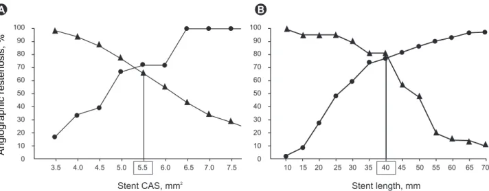
Figure 3. Sensitivity and specificity curves identified optimal cut-off values of final minimum stent cross-sectional area (CSA, A) and stent length (B) that predicted angiographic restenosis after sirolimus-eluting stent implantation: 5.5 mm2 for final minimum stent CSA and 40 mm2 for intravascular ultrasound-measured stent length. Triangle, sensitivity; circles, specificity. Reprinted with permission from Oxford University Press [22].
A b
defined as > 5.0 mm
2, the positive predictive value of pa- tency was 90%, but the optimal cutoff value of BMS was defined as 6.5 mm [50]. Another study concerning SES failure also supported that MSA < 5.5 mm
2and < 5.0 mm
2were the most important predictors of SES failure (Fig.
3) [22,51]. Even if SES had a considerably lower optimal MSA threshold than BMS, these studies showed that un- derexpansion remained the main cause of stent failure in DESs; at least a MSA < 5.0 mm should be avoided in non- -left main (LM) lesions. In LM lesions, optimal MSA was reported in the MAIN-COMPARE (revascularization for unprotected left main coronary artery stenosis: compari- son of percutaneous coronary angioplasty versus surgical revascularization) study to be > 8.7 mm
2to prevent TLR [34].
Edge dissection, which is complicated by lumen nar- rowing < 4 mm
2or dissection angle ≥ 60°, has been as- sociated with an increased incidence of early ST [49]; thus, additional stents may be needed to prevent ST. However, a minor dissection, detected by IVUS, may not be associated with an increased incidence of ST [52,53]. Although no consensus exists on an optimal strategy, in minor dissec- tion, careful observation without stenting can be helpful.
Overall, the results discussed above encourage ensuring good apposition of stent struts to the vessel wall, such that the stent struts are not surrounded by lumen, adequate stent expansion to obtain MSA at least > 6.5 mm
2for BMSs and > 5.0 mm
2for DESs or MSA > 90% of the distal reference lumen CSA, and lack of major dissections, intra- mural hematomas, and geographic misses.
outCoMeS of IVuS-guIDeD VeRSuS AngIogRApHy-guIDeD pCI
Numerous studies have evaluated the clinical benefits of IVUS-guided PCI compared with angiography-guided PCI in the BMS era [41-44]. The OPTICUS trial showed no sig- nificant difference between IVUS- and angiography-guid- ed PCI groups in terms of 6- and 12-month rates of death, myocardial infarction (MI), and TLR in 550 patients meeting the MUSIC criteria [42]. In contrast, the TULIP study demonstrated favorable angiographic and clinical outcomes in patients with long coronary lesions (> 20 mm) treated with a BMS (> 3 mm) under IVUS guidance [41]. In a meta-analysis of 2,193 patients from seven randomized
trials, the rates of 6-month angiographic restenosis and target vessel revascularization were significantly lower in the IVUS-guided PCI group than the angiography-guided group (22% vs. 29%, p = 0.02 and 13% vs. 18%, p < 0.001, respectively), with no difference in the rates of death (2.4%
vs. 1.6%, p = 0.18) or MI (3.6% vs. 4.4%, p = 0.51) [44], consistent with a previous meta-analysis [43].
To date, few studies have investigated the clinical ben- efits of DES optimization under IVUS guidance compared with that of BMSs. The HOME DES IVUS (long-term health outcome and mortality evaluation after invasive coronary treatment using drug-eluting stents with or without IVUS guidance) study was a randomized trial to investigate clinical outcomes of IVUS-guided PCI with DESs. Although the IVUS-guided strategy resulted in the frequent use of adjunctive balloons and a larger size bal- loon with higher pressure, no significant difference was observed in major adverse cardiac events or stent throm- bosis in the study [45]. A similar retrospective study of IVUS-guided stent optimization also showed no signifi- cant difference in the incidence of in-stent restenosis or neointimal volume between IVUS- versus angiography- guided PCI [54]. Conversely, a study with a propensity- matched analysis in 884 patients treated with DESs showed a significant reduction in the stent thrombosis rate at both 30 days (0.5% vs. 1.4%, p = 0.046) and 12 months (0.7% vs. 2.0%, p = 0.014) in the IVUS-guided PCI group [47].
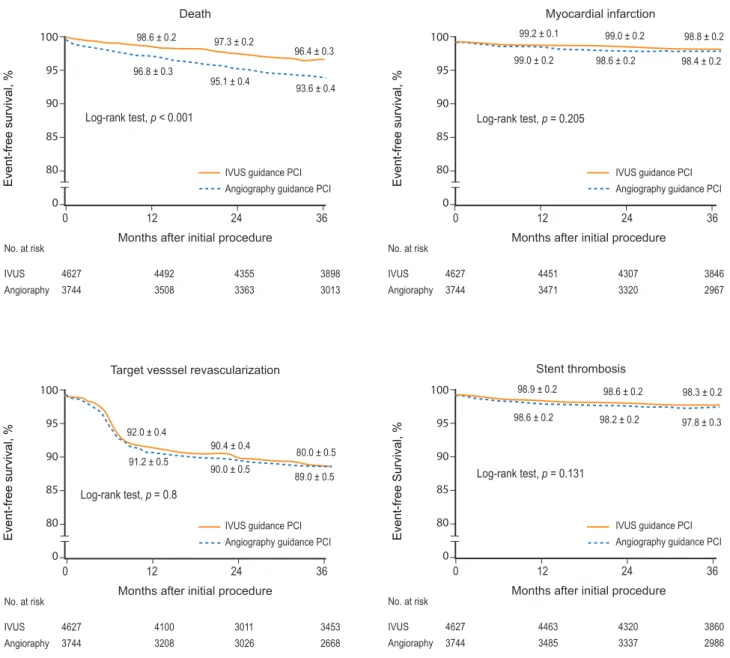
Recently, a large “real world” registry from two Korean centers reported long-term outcomes of both IVUS- and angiography-guided PCI using BMS or DES implantation [55]. In total, 8,371 patients who underwent coronary stenting under IVUS guidance (4,627 patients) or angi- ography guidance (3,744 patients) were consecutively enrolled, and 3-year adverse clinical outcomes were com- pared between the groups using a Cox regression model and propensity score matching. In the overall population, the 3-year adjusted incidence of mortality was signifi- cantly lower in the IVUS-guided PCI group compared with the angiography-guided PCI group (hazard ratio [HR], 0.70; 95% confidence interval [CI], 0.56 to 0.87;
p = 0.001) (Fig. 4). In 2,715 matched pairs of the overall
population, the IVUS-guided PCI group also had a lower mortality risk (HR, 0.71; 95% CI, 0.56 to 0.90; p = 0.005).
However, IVUS-guided PCI did not influence the rates of
myocardial infarction, target-vessel revascularization, or
stent thrombosis in the overall or in the 2,715 matched- pair populations. In the DES subpopulation, IVUS guid- ance significantly reduced the 3-year adjusted mortality rate (HR, 0.55; 95% CI, 0.36 to 0.78; p = 0.001), which was not the case in the BMS subpopulation (HR, 0.79; 95% CI, 0.59 to 1.05; p = 0.10). A propensity score matching analy- sis of 201 matched patients from the MAIN-COMPARE study also demonstrated the importance of IVUS-guided PCI in unprotected left main disease [34]. In this analy- sis, significantly lower incidence of 3-year mortality was
noted in the IVUS-guided PCI group compared with the angiography-guided PCI group (4.7% vs. 13.6%, p = 0.048), but no significant difference was detected in the rates of TVR or MI. Notably, this benefit was found only for DES, and the benefit in mortality appeared to be primarily as- sociated with reduced sudden cardiac death related to late stent thrombosis. Similar findings were also observed in patients undergoing PCI of non-left main bifurcations with DESs [35]. Taken together, the benefit of IVUS guid- ance contributed primarily to decreased rates of stent
10095 90 85 80 0
Event-free survival, %
Months after initial procedure
Death Myocardial infarction
Log-rank test, p < 0.001 Log-rank test, p < 0.001
No. at risk No. at risk IVUS Angioraphy IVUS Angioraphy
4627 3744 4627 3744
4492 3508 4492 3508
4355 3363 4355 3363
3898 3013 3898 3013 0
0 1212 2424 3636
98.6 ± 0.2
98.6 ± 0.2 97.3 ± 0.297.3 ± 0.2
96.4 ± 0.3 96.4 ± 0.3 93.6 ± 0.4 93.6 ± 0.4 95.1 ± 0.4
95.1 ± 0.4 96.8 ± 0.3
96.8 ± 0.3
IVUS guidance PCI Angiography guidance PCI IVUS guidance PCI Angiography guidance PCI
100 95 90 85 80 0
Event-free survival, %
Months after initial procedure Target vesssel revascularization
Log-rank test, p = 0.8 Log-rank test, p = 0.8
No. at risk No. at risk IVUS Angioraphy IVUS Angioraphy
4627 3744 4627 3744
4100 3208 4100 3208
3011 3026 3011 3026
3453 2668 3453 2668 0
0 1212 2424 3636
92.0 ± 0.4 92.0 ± 0.4
90.4 ± 0.4
90.4 ± 0.4 80.0 ± 0.580.0 ± 0.5 91.2 ± 0.5
91.2 ± 0.5 90.0 ± 0.590.0 ± 0.5
89.0 ± 0.5 89.0 ± 0.5
IVUS guidance PCI Angiography guidance PCI IVUS guidance PCI Angiography guidance PCI
100 95 90 85 80 0
Event-free survival, %
Months after initial procedure Log-rank test, p = 0.205
Log-rank test, p = 0.205
No. at risk No. at risk IVUS Angioraphy IVUS Angioraphy
4627 3744 4627 3744
4451 3471 4451 3471
4307 3320 4307 3320
3846 2967 3846 2967 0
0 1212 2424 3636
99.2 ± 0.1
99.2 ± 0.1 99.0 ± 0.299.0 ± 0.2 98.8 ± 0.298.8 ± 0.2 98.4 ± 0.2 98.4 ± 0.2 98.6 ± 0.2
98.6 ± 0.2 99.0 ± 0.2
99.0 ± 0.2
IVUS guidance PCI Angiography guidance PCI IVUS guidance PCI Angiography guidance PCI
Stent thrombosis 100
95 90 85 80 0
Event-free Survival, %
Months after initial procedure Log-rank test, p = 0.131
Log-rank test, p = 0.131
No. at risk No. at risk IVUS Angioraphy IVUS Angioraphy
4627 3744 4627 3744
4463 3485 4463 3485
4320 3337 4320 3337
3860 2986 3860 2986 0
0 1212 2424 3636
98.9 ± 0.2
98.9 ± 0.2 98.6 ± 0.298.6 ± 0.2 98.3 ± 0.298.3 ± 0.2 97.8 ± 0.3 97.8 ± 0.3 98.2 ± 0.2
98.2 ± 0.2 98.6 ± 0.2
98.6 ± 0.2
IVUS guidance PCI Angiography guidance PCI IVUS guidance PCI Angiography guidance PCI
Figure 4. Kaplan-Meier event-free 3-year survival curves for death, myocardial infarction, target-vessel revascularization, and stent thrombosis in 8,371 patients following intravascular ultrasound (IVUS)- (n = 4,627) or angiography- (n = 3,744) guided percutaneous coronary intervention (PCI). Reprinted with permission from John Wiley and Sons [55].
restenosis and repeated revascularization in the BMS era, whereas reduction of the stent thrombosis rate with pos- sible improvement in mortality have predominated in the DES era.
ConCluSIonS
IVUS can provide direct cross-sectional images as well as longitudinal images of the coronary vessel wall. It has also contributed to our understanding of mechanisms in coronary atherosclerotic plaques and provided real- time information at stented segments after coronary interventions. Possible criteria for optimal stent deploy- ment by IVUS are complete stent apposition to the vessel wall, adequate stent expansion, and full lesion coverage without edge dissection (Fig. 5). Recent data suggest that IVUS-guided PCI may reduce long-term mortality when compared with angiography-guided PCI, particularly after DES implantation; thus, the clinical importance of IVUS- guided PCI raised in the BMS era persists in the DES era.
Optimization of stent deployment by IVUS during PCI may be considered as a routine practice in daily PCI, espe- cially for complex lesion intervention.
Conflict of interest
No potential conflict of interest relevant to this article was reported.
RefeRenCeS
1. Nissen SE, Gurley JC, Grines CL, et al. Intravascular ultra- sound assessment of lumen size and wall morphology in normal subjects and patients with coronary artery disease. Circulation 1991;84:1087-1099.
2. Nissen SE, Gurley JC. Application of intravascular ultrasound for detection and quantitation of coronary atherosclerosis. Int J Card Imaging 1991;6:165-177.
3. Waller BF, Pinkerton CA, Slack JD. Intravascular ultrasound: a histological study of vessels during life: the new ‘gold standard’
for vascular imaging. Circulation 1992;85:2305-2310.
4. Schiele F, Meneveau N, Seronde MF, et al. Medical costs of intravascular ultrasound optimization of stent deployment:
results of the multicenter randomized ‘REStenosis after Intra- vascular ultrasound STenting’ (RESIST) study. Int J Cardiovasc Intervent 2000;3:207-213.
5. Smith SC Jr, Feldman TE, Hirshfeld JW Jr, et al. ACC/AHA/
SCAI 2005 Guideline Update for Percutaneous Coronary Inter- vention-summary article: a report of the American College of Cardiology/American Heart Association Task Force on Practice Guidelines (ACC/AHA/SCAI Writing Committee to Update the 2001 Guidelines for Percutaneous Coronary Intervention). Cir- culation 2006;113:156-175.
6. Nakamura S, Colombo A, Gaglione A, et al. Intracoronary ul- trasound observations during stent implantation. Circulation 1994;89:2026-2034.
7. Gorge G, Haude M, Ge J, et al. Intravascular ultrasound after low and high inflation pressure coronary artery stent implanta- tion. J Am Coll Cardiol 1995;26:725-730.
Complete apposition
expansion Well
No edge dissection
Full lesion coverage
• Apposition of stent struts to the vessel wall, not surrounded by lumen
• Detection of reference site with plaque burden of < 50%
• MSA at least
- 5.0-5.5 mm2 (non-LM) & 8.7 mm2 (LM): DES - 6.5-7.5 mm2: BMS (not in small vessels) - >90% of distal ref. lumen area or or >80% of ave.
ref. lumen area
• Post-procedure IVUS for evaluation of edge dissection
IVUS
Figure 5. The possible intravascular ultrasound criteria for optimal stent deployment. LM, left main; DES, drug-eluting stent; BMS, bare metal stent .
8. Mudra H, Klauss V, Blasini R, et al. Ultrasound guidance of Palmaz-Schatz intracoronary stenting with a combined intra- vascular ultrasound balloon catheter. Circulation 1994;90:1252- 1261.
9. Brodie BR, Cooper C, Jones M, Fitzgerald P, Cummins F; Post- dilatation Clinical Compartative Study (POSTIT) Investigators.
Is adjunctive balloon postdilatation necessary after coronary stent deployment? Final results from the POSTIT trial. Catheter Cardiovasc Interv 2003;59:184-192.
10. de Ribamar Costa J Jr, Mintz GS, Carlier SG, et al. Intravascu- lar ultrasound assessment of drug-eluting stent expansion. Am Heart J 2007;153:297-303.
11. Alfonso F, Goicolea J, Hernandez R, et al. Arterial perforation during optimization of coronary stents using high-pressure bal- loon inflations. Am J Cardiol 1996;78:1169-1172.
12. Mintz GS, Weissman NJ. Intravascular ultrasound in the drug- eluting stent era. J Am Coll Cardiol 2006;48:421-429.
13. Cook S, Wenaweser P, Togni M, et al. Incomplete stent apposi- tion and very late stent thrombosis after drug-eluting stent im- plantation. Circulation 2007;115:2426-2434.
14. Hwang CW, Wu D, Edelman ER. Physiological transport forces govern drug distribution for stent-based delivery. Circulation 2001;104:600-605.
15. Papafaklis MI, Chatzizisis YS, Naka KK, Giannoglou GD, Mi- chalis LK. Drug-eluting stent restenosis: Effect of drug type, release kinetics, hemodynamics and coating strategy. Phar- macol Ther 2011 Dec 22 [Epub]. http://dx.doi.org/10.1016/
j.pharmthera.2011.12.006.
16. Hwang CW, Edelman ER. Arterial ultrastructure influences transport of locally delivered drugs. Circ Res 2002;90:826-832.
17. Windecker S, Meier B. Late coronary stent thrombosis. Circula- tion 2007;116:1952-1965.
18. Kume T, Waseda K, Ako J, et al. Intravascular ultrasound as- sessment of postprocedural incomplete stent apposition. J In- vasive Cardiol 2012;24:13-16.
19. Fitzgerald PJ, Oshima A, Hayase M, et al. Final results of the Can Routine Ultrasound Influence Stent Expansion (CRUISE) study. Circulation 2000;102:523-530.
20. Uren NG, Schwarzacher SP, Metz JA, et al. Predictors and out- comes of stent thrombosis: an intravascular ultrasound regis- try. Eur Heart J 2002;23:124-132.
21. Doi H, Maehara A, Mintz GS, et al. Impact of post-intervention minimal stent area on 9-month follow-up patency of paclitaxel- eluting stents: an integrated intravascular ultrasound analysis from the TAXUS IV, V, and VI and TAXUS ATLAS Workhorse, Long Lesion, and Direct Stent Trials. JACC Cardiovasc Interv 2009;2:1269-1275.
22. Hong MK, Mintz GS, Lee CW, et al. Intravascular ultrasound predictors of angiographic restenosis after sirolimus-eluting stent implantation. Eur Heart J 2006;27:1305-1310.
23. Cheneau E, Leborgne L, Mintz GS, et al. Predictors of subacute stent thrombosis: results of a systematic intravascular ultra- sound study. Circulation 2003;108:43-47.
24. Fujii K, Carlier SG, Mintz GS, et al. Stent underexpansion and residual reference segment stenosis are related to stent throm- bosis after sirolimus-eluting stent implantation: an intravascu- lar ultrasound study. J Am Coll Cardiol 2005;45:995-998.
25. Kimura M, Mintz GS, Carlier S, et al. Outcome after acute in- complete sirolimus-eluting stent apposition as assessed by se- rial intravascular ultrasound. Am J Cardiol 2006;98:436-442.
26. Hong MK, Lee CW, Kim JH, et al. Impact of various intra- vascular ultrasound criteria for stent optimization on the six- month angiographic restenosis. Catheter Cardiovasc Interv 2002;56:178-183.
27. Albiero R, Rau T, Schlüter M, et al. Comparison of immediate and intermediate-term results of intravascular ultrasound ver- sus angiography-guided Palmaz-Schatz stent implantation in matched lesions. Circulation 1997;96:2997-3005.
28. Hoffmann R, Mintz GS, Mehran R, et al. Intravascular ultra- sound predictors of angiographic restenosis in lesions treated with Palmaz-Schatz stents. J Am Coll Cardiol 1998;31:43-49.
29. Kasaoka S, Tobis JM, Akiyama T, et al. Angiographic and intra- vascular ultrasound predictors of in-stent restenosis. J Am Coll Cardiol 1998;32:1630-1635.
30. Moussa I, Moses J, Di Mario C, et al. Does the specific intra- vascular ultrasound criterion used to optimize stent expansion have an impact on the probability of stent restenosis? Am J Car- diol 1999;83:1012-1017.
31. Blasini R, Neumann FJ, Schmitt C, Bokenkamp J, Schomig A. Comparison of angiography and intravascular ultrasound for the assessment of lumen size after coronary stent place- ment: impact of dilation pressures. Cathet Cardiovasc Diagn 1997;42:113-119.
32. Moses JW, Leon MB, Popma JJ, et al. Sirolimus-eluting stents versus standard stents in patients with stenosis in a native coro- nary artery. N Engl J Med 2003;349:1315-1323.
33. Colombo A, Drzewiecki J, Banning A, et al. Randomized study to assess the effectiveness of slow- and moderate-release poly- mer-based paclitaxel-eluting stents for coronary artery lesions.
Circulation 2003;108:788-794.
34. Park SJ, Kim YH, Park DW, et al. Impact of intravascular ul- trasound guidance on long-term mortality in stenting for un- protected left main coronary artery stenosis. Circ Cardiovasc Interv 2009;2:167-177.
35. Kim SH, Kim YH, Kang SJ, et al. Long-term outcomes of intra- vascular ultrasound-guided stenting in coronary bifurcation lesions. Am J Cardiol 2010;106:612-618.
36. Mintz GS, Painter JA, Pichard AD, et al. Atherosclerosis in an- giographically “normal” coronary artery reference segments: an intravascular ultrasound study with clinical correlations. J Am
Coll Cardiol 1995;25:1479-1485.
37. Weissman NJ, Palacios IF, Nidorf SM, Dinsmore RE, Weyman AE. Three-dimensional intravascular ultrasound assessment of plaque volume after successful atherectomy. Am Heart J 1995;130:413-419.
38. Morino Y, Tamiya S, Masuda N, et al. Intravascular ultrasound criteria for determination of optimal longitudinal positioning of sirolimus-eluting stents. Circ J 2010;74:1609-1616.
39. de Jaegere P, Mudra H, Figulla H, et al. Intravascular ultra- sound-guided optimized stent deployment: Immediate and 6 months clinical and angiographic results from the Multicenter Ultrasound Stenting In Coronaries Study (MUSIC Study). Eur Heart J 1998;19:1214-1223.
40. Russo RJ, Silva PD, Teirstein PS, et al. A randomized controlled trial of angiography versus intravascular ultrasound-directed bare-metal coronary stent placement (the AVID Trial). Circ Car- diovasc Interv 2009;2:113-123.
41. Oemrawsingh PV, Mintz GS, Schalij MJ, Zwinderman AH, Jukema JW, van der Wall EE. Intravascular ultrasound guid- ance improves angiographic and clinical outcome of stent im- plantation for long coronary artery stenoses: final results of a randomized comparison with angiographic guidance (TULIP Study). Circulation 2003;107:62-67.
42. Mudra H, di Mario C, de Jaegere P, et al. Randomized compari- son of coronary stent implantation under ultrasound or angio- graphic guidance to reduce stent restenosis (OPTICUS Study).
Circulation 2001;104:1343-1349.
43. Casella G, Klauss V, Ottani F, Siebert U, Sangiorgio P, Brac- chetti D. Impact of intravascular ultrasound-guided stenting on long-term clinical outcome: a meta-analysis of available studies comparing intravascular ultrasound-guided and angiographi- cally guided stenting. Catheter Cardiovasc Interv 2003;59:314- 321.
44. Parise H, Maehara A, Stone GW, Leon MB, Mintz GS. Meta- analysis of randomized studies comparing intravascular ultrasound versus angiographic guidance of percutaneous coro- nary intervention in pre-drug-eluting stent era. Am J Cardiol 2011;107:374-382.
45. Jakabcin J, Spacek R, Bystron M, et al. Long-term health out- come and mortality evaluation after invasive coronary treat- ment using drug eluting stents with or without the IVUS guid- ance. Randomized control trial: HOME DES IVUS. Catheter Cardiovasc Interv 2010;75:578-583.
46. Colombo A, Caussin C, Presbitero P, Chieffo A. AVIO: a pro- spective, randomized trial of intravascular-ultrasound guided compared to angiography guided stent implantation in complex coronary lesions [abstract]. J Am Coll Cardiol 2010;56:xvii.
47. Roy P, Steinberg DH, Sushinsky SJ, et al. The potential clinical utility of intravascular ultrasound guidance in patients under- going percutaneous coronary intervention with drug-eluting stents. Eur Heart J 2008;29:1851-1857.
48. Okabe T, Mintz GS, Buch AN, et al. Intravascular ultrasound parameters associated with stent thrombosis after drug-eluting stent deployment. Am J Cardiol 2007;100:615-620.
49. Choi SY, Witzenbichler B, Maehara A, et al. Intravascular ultrasound findings of early stent thrombosis after primary percutaneous intervention in acute myocardial infarction: a Harmonizing Outcomes with Revascularization and Stents in Acute Myocardial Infarction (HORIZONS-AMI) substudy. Circ Cardiovasc Interv 2011;4:239-247.
50. Sonoda S, Morino Y, Ako J, et al. Impact of final stent dimen- sions on long-term results following sirolimus-eluting stent implantation: serial intravascular ultrasound analysis from the sirius trial. J Am Coll Cardiol 2004;43:1959-1963.
51. Takebayashi H, Kobayashi Y, Mintz GS, et al. Intravascular ultrasound assessment of lesions with target vessel failure after sirolimus-eluting stent implantation. Am J Cardiol 2005;95:498-502.
52. Hong MK, Park SW, Lee NH, et al. Long-term outcomes of mi- nor dissection at the edge of stents detected with intravascular ultrasound. Am J Cardiol 2000;86:791-795, A9.
53. Liu X, Tsujita K, Maehara A, et al. Intravascular ultrasound assessment of the incidence and predictors of edge dissections after drug-eluting stent implantation. JACC Cardiovasc Interv 2009;2:997-1004.
54. Park SM, Kim JS, Ko YG, et al. Angiographic and intravascular ultrasound follow up of paclitaxel- and sirolimus-eluting stent after poststent high-pressure balloon dilation: from the post- stent optimal stent expansion trial. Catheter Cardiovasc Interv 2011;77:15-21.
55. Hur SH, Kang SJ, Kim YH, et al. Impact of intravascular ul- trasound-guided percutaneous coronary intervention on long- term clinical outcomes in a real world population. Catheter Car- diovasc Interv 2011 Jul 29 [Epub]. http://dx.doi.org/10.1002/
ccd.23279.