of Neointimal Growth
An Intravascular Ultrasound Analysis From the Study to COmpare REstenosis rate between QueST and QuaDS-QP2 (SCORE)
Toru Kataoka, MD; Eberhard Grube, MD; Yasuhiro Honda, MD; Yoshihiro Morino, MD;
Seung-Ho Hur, MD; Heidi N. Bonneau, RN, MS; Antonio Colombo, MD; Carlo Di Mario, MD, PhD;
Giulio Guagliumi, MD; Karl E. Hauptmann, MD; Mark R. Pitney, MBBS; Alexandra J. Lansky, MD;
Simon H. Stertzer, MD; Paul G. Yock, MD; Peter J. Fitzgerald, MD, PhD;
for the SCORE Investigators
Background—Inhibition of neointimal tissue growth has been demonstrated in preliminary human feasibility studies with a stent-based polymer sleeve delivering 7-hexanoyltaxol. The Study to COmpare REstenosis rate between QueST and QuaDS-QP2 (SCORE) trial is a human, randomized, multicenter trial comparing 7-hexanoyltaxol (QP2)-eluting stents (qDES) with bare metal stents (BMS) in the treatment of de novo coronary lesions. The purpose of this substudy was to evaluate the acute expansion property and long-term neointimal responses of qDES compared with BMS as assessed by intravascular ultrasound (IVUS).
Methods and Results—A total of 122 (qDES 66, BMS 56) patients were enrolled into the IVUS substudy. All IVUS images (immediately after the procedure and at 6-month follow-up) were analyzed at an independent core laboratory in a blind manner. At baseline, qDES achieved stent expansion similar to BMS. At follow-up, qDES showed reduced neointimal growth by 70% at the tightest cross section and by 68% over the stented segment (P⬍0.0001 for both), resulting in a significantly larger lumen in qDES than in BMS. Unlike intracoronary brachytherapy, there was no evidence of negative edge effects, unhealed dissections, or late stent-vessel wall malapposition over the stented and adjacent references segments in either group.
Conclusions—Detailed IVUS analysis revealed that qDES had comparable acute mechanical and superior long-term biological effects to BMS. Although the long-term benefits and limitations of this technology require further investigation, the reduction in neointimal thickenings demonstrated that local delivery of 7-hexanoyltaxol through polymer sleeves augments conventional mechanical treatment of atherosclerotic disease. (Circulation. 2002;106:1788- 1793.)
Key Words: cardiovascular diseases 䡲 drugs 䡲 restenosis 䡲 stents 䡲 ultrasonics
C
oronary stents eliminate elastic recoil of target vessels, thereby reducing restenosis rate in percutaneous coro- nary interventions.1However, the occurrence of stent reste- nosis as the result of intimal hyperplasia remains a clinical challenge. Local drug delivery represents a therapeutic ap- proach that enables high local concentration of drug with minimum systemic adverse effects. Particularly, stent-based delivering of drugs has been considered a promising tech- nique to provide both a highly selective biological andmechanical solution precisely at the target segment. At present, a number of pharmacological agents, in combination with various delivery platforms, are being evaluated for their potential to improve the acute and long-term outcomes.
Paclitaxel (Taxol) and related taxane compounds interfere with the proliferation, signal transduction, and migration of vascular smooth muscle cells, thus contributing to the reduc- tion of neointimal growth.2,3 These effects primarily derive from the unique agent character that shifts the cytoskeleton
Received March 8, 2002; revision received July 18, 2002; accepted July 18, 2002.
From the Center for Research in Cardiovascular Interventions, Stanford University, Stanford, Calif (T.K., Y.H., Y.M., S.H., S.H.S., P.G.Y., P.J.F.);
Heart Center Siegburg, Siegburg, Germany (E.G.); Highlands Consulting, Inc, San Jose, Calif (H.N.B.); Columbus Clinic, Milan, Italy (A.C., C.D.M.);
San Raffaele Hospital, Milan, Italy (A.C., C.D.M.); Divisione di Cardiologia Ospedali, Riuniti di Bergamo, Bergamo, Italy (G.G.); Krankenhaus der Barmherzigen Brüder, Trier, Germany (K.E.H.); Eastern Heart Clinic, Prince of Wales Hospital, Sydney, Australia (M.R.P.); and the Cardiovascular Research Foundation, New York City, NY (A.J.L.).
Dr Guagliumi is a consultant for Boston Scientific. Dr Grube is a minor stockholder of Quanam-Medical, Inc. Dr Stertzer is a minor stockholder of Boston Scientific and was a director of Quanam-Medical, Inc.
Correspondence to Dr Peter J. Fitzgerald, Center for Research in Cardiovascular Interventions, Stanford University Medical Center, 300 Pasteur Drive, Room H3554, Stanford, CA 94305. E-mail [email protected]
© 2002 American Heart Association, Inc.
Circulation is available at http://www.circulationaha.org DOI: 10.1161/01.CIR.0000031734.11420.1C 1788
equilibrium toward assembly with profound stabilization in the microtubules.4Accordingly, several animal studies have demonstrated that stent-based local delivery of paclitaxel could prevent intimal proliferation through the use of an overstretched vessel injury model.5–7 Preliminary human studies also showed that local delivery of 7-hexanoyltaxol (QP2, a taxane analogue) through polymer sleeves on a balloon-expandable stent reduced neointimal growth after coronary intervention.8,9To confirm those pilot results from the small-number, nonrandomized, feasibility registries, a prospective randomized multicenter trial was conducted. The purpose of this intravascular ultrasound (IVUS) substudy was to investigate (1) the acute mechanical properties of this sleeve-based drug-eluting stent and (2) the long-term antipro- liferative effectiveness compared with a conventional bare metal stent (BMS).
Methods Study Design and Patient Population
This study represents an IVUS substudy of the SCORE trial (Study to COmpare REstenosis rate between QueST and QuaDS-QP2): the human, prospective, randomized, multicenter study to evaluate the safety and efficacy of the QP2 drug-eluting stent (QuaDS-QP2 stent, qDES) compared with a BMS (QueSt stent) in the prevention of in-stent restenosis (Quanam Medical/Boston Scientific). Inclusion criteria were de novo lesions in native coronary arteries, patient age ⱖ50 and ⱕ80 years, and target vessel reference diameter ⱖ3.0 and ⱕ3.5 mm, as assessed by visual estimate. Patients with a recent history of acute myocardial infarction or left ventricular ejection fraction ⱕ30% were excluded from this trial. All patients gave informed consent before study enrollment. The local Human Sub- jects Committee at all participating institutions approved the protocols.
Stent Designs and Implantation Procedure
Both qDES and BMS consist of 316L stainless steel and were identified in length (13 or 17 mm) and diameter (3.0 or 3.5 mm). The detailed configuration of qDES was previously reported.9In short, the qDES is equipped with multiple polyacrylate sleeves as the platform for drug elution. Each sleeve is 2.4 mm in length and contains 800g of QP2. In this study, the number of sleeves was 4 and 5, in 13-mm and 17-mm length stents, respectively. After sheath placement, 10 000 to 15, 000 IU of heparin was administered. The appropriate stent size was selected by using the reference vessel diameter, and the stents were implanted according to a standard implantation technique with predilatation. After the procedure, patients were treated with acetylsalicylic acid (100 mg) indefinitely and with ticlopidine or clopidogrel (500 mg) for either 1 month or 12 months for BMS or qDES, respectively.
Intravascular Ultrasound Imaging
IVUS was performed immediately after stent implantation and at 6-month follow-up with one of two commercially available imaging systems. The first was 3.2-F, 30-MHz or 2.9-F, 40-MHz single- element mechanical ultrasound catheter (Boston Scientific). The second incorporated 3.0-F, 20-MHz phased-array ultrasound catheter (Endosonics/Jomed). At follow-up, the IVUS system used for each patient was identical to the system used after stent implantation.
Intracoronary nitroglycerin was injected before image acquisition.
Motorized pullback devices (0.5 mm/s) were used during all IVUS data acquisition. All IVUS images were recorded on s-VHS video- tape or compact disk for off-line analysis.
Intravascular Ultrasound Analysis
An independent core laboratory at Stanford University Medical Center reviewed all ultrasound images. Two observers, blinded to
clinical information and treatment protocol, performed all IVUS analyses.
Qualitative IVUS parameters evaluated in this study included (1) stent apposition (incomplete apposition being defined asⱖ1 strut clearly separated from the vessel wall with evidence of blood speckle behind the strut); (2) edge tears (disruption of plaque immediately adjacent to the stent ends where the flap could be clearly differen- tiated from the underlying plaque); and (3) late stent malapposition (incomplete stent apposition at follow-up not seen immediately after stent implantation).
Quantitative IVUS analysis was performed by using commercially available planimetry software (TapeMeasure/EchoPlaque, Indec Systems and Plus-Plus, Sanders Data Systems), according to previ- ously validated and published protocols.10Two-dimensional analysis was performed at the tightest cross section within the stent and the proximal and distal reference segments (defined as the location in the native vessel with minimum disease within 3 to 10 mm from stent margins and before the emergence of any major side branches). Stent and lumen areas were manually traced, and neointimal area was computed as stent minus lumen area. Cross-sectional narrowing (CSN) was calculated as neointimal area divided by stent area.
Percent stent expansion was calculated as minimum stent area (MSA) divided by averaged reference lumen area. Three- dimensional analysis was performed by means of Simpson’s method.
Stent, lumen, and neointimal volumes were computed for the entire stented segment. To adjust for different stent lengths, volume index was calculated as volume data divided by stent length (SVI: stent volume index, LVI: lumen volume index, and NVI: neointimal volume index). The interobserver correlation coefficients for SVI, LVI, and NVI were 0.998, 0.997, and 0.984, respectively.
Quantitative Coronary Angiography
All cineangiograms were independently analyzed by the Cardiovas- cular Research Foundation Angiographic Core Laboratory. Cine frames from multiple projections were digitized and analyzed with the use of the CMS-GFT algorithm (MEDIS). Image calibration was performed with contrast-filled catheters used as the reference standard.
Statistical Analysis
Qualitative data are presented as mean⫾SD, and qualitative data are presented as frequencies. Statistical analyses were performed with the StatView 5.0 (SAS Institute). For comparisons of continuous variables between the 2 stent groups, a 2-tailed, unpaired t test was used. A 2-way repeated-measures ANOVA was used to evaluate potential differences in serial IVUS measurements between the two stent groups. Categoric data were compared by means of the2or Fisher exact test. Significance was defined as a threshold of P⫽0.05.
Results Patient Characteristics
Between April 2000 and January 2001, 122 patients were enrolled in this IVUS substudy from 266 patients in the overall SCORE population on an institutional basis. In this population, 66 patients were assigned to the qDES group and 56 to the BMS group. Baseline patient, lesion, and procedural characteristics were similar in both groups (Table 1). Ninety- six IVUS images (qDES, 48; BMS, 48) were entered into baseline IVUS analysis (26 patients were excluded because of incomplete image acquisition, inadequate image quality, or other technical reasons in the imaging procedure at the catheterization laboratory). Ninety-two IVUS images (qDES, 49; BMS, 43) were available for follow-up IVUS analysis.
Primary reasons for reduced follow-up included patient re- fusal, incomplete image acquisition, and inadequate image quality. In addition, one patient died of suspected pulmonary
embolism during the follow-up period (qDES). Complete volumetric analysis was available in 86% and 93% for baseline and follow-up studies, respectively. Each analysis population had baseline characteristics comparable to the overall enrolled patients, with no significant differences between the two stent groups. Furthermore, characteristics of this substudy subset were comparable to the complete SCORE study population. The follow-up period was also identical in the two groups (qDES, 6.2⫾0.9 months; BMS, 6.3⫾1.0 months, P⫽NS).
Acute Results
Immediately after stent implantation, no plaque protrusion was detected in either group. Mild incomplete stent apposi- tion was observed in 8 cases (qDES, 6% versus BMS, 10%, P⫽NS) and mild edge tears in 12 cases (qDES, 13% versus BMS, 13%, P⫽NS), with similar frequencies in the qDES and BMS groups. Two-dimensional and 3-dimensional quan- titative analyses showed no significant differences in acute results between the two stent groups, including the average reference lumen area, MSA, SVI, and percent stent expansion (Table 2).
Follow-Up Results
At 6-month follow-up, unhealed edge tear was not observed in either group. On the other hand, the pattern of neointimal
growth differed significantly between the two groups (Figure 1). In the BMS group, almost all stent struts were covered with neointima throughout the stented segment, and 7 cases (16%) showed the IVUS catheter nearly wedged in abundant neointima. In contrast, in the majority (94%) of qDES, neointima was either undetectable or observed on the stent struts as a very thin layer only in some part of the stent. No evidence of late stent malapposition or echolucent tissue was detected in either group.
In the 2-dimensional quantitative analysis, at the worst cross section, qDES showed a larger minimum lumen area (MLA) by 28%, a reduced neointimal area by 70%, and an improved CSN by 65% compared with BMS (Table 3). There was no significant difference in the average reference lumen area between the two stent groups. Further volumetric analysis of the entire stented segment revealed similar SVI in the two groups (qDES, 8.63⫾2.25 mm3/mm; BMS, 8.84⫾1.83 mm3/mm, P⫽NS), with no significant chronic recoil during the follow-up period. On the other hand, qDES showed a significantly reduced NVI by 68%
than BMS (qDES, 0.79⫾0.51 mm3/mm; BMS, 2.48⫾1.49 mm3/ mm, P⬍0.0001, Figure 2). Consequently, the LVI was signifi- cantly larger in qDES than in BMS (qDES, 7.83⫾2.11 mm3/ mm; BMS, 6.36⫾2.16 mm3/mm, P⫽0.0019).
Figure 3 shows the relation between postprocedural MSA and follow-up MLA in the two stent groups. In the BMS group, only a weak correlation was observed (r⫽0.39, P⫽0.02), whereas qDES showed a significant positive rela- tion with a greater correlation coefficient between the two parameters (r⫽0.73, P⬍0.0001).
Discussion
Clinical studies with conventional BMS have shown that certain plaque types have an overwhelming biological re- sponse to the acute mechanical injury by stent implantation and/or sustained stimuli from the rigid metal struts. In addition to the classic risk factors for in-stent restenosis, including multivessel disease, small-diameter vessels, long lesions, and bifurcations, recent IVUS studies have demon- strated that excessive arterial remodeling at preintervention,11 large plaque burden before or after intervention,12and diabe- tes mellitus13should be also segregated for their biologically active milieus. After intracoronary brachytherapy as the first biological vector to approach those high-risk lesions, stent- based local drug delivery is considered an emerging break- through technology with less technical demand for operators.
Among a number of pharmacological agents evaluated, rapa- mycin (sirolimus)14 –16and paclitaxel5–7on a metal backbone were the front-runners, showing a significant reduction of in-stent neointimal growth in animal models or human studies. Early human registries with sleeve-based delivery of 7-hexanoyltaxol (a taxane analogue) have also shown favor- able long-term outcomes after stenting.8,9 This agent, the same drug used in this study, is highly lipophilic and insoluble in water. Although metabolism and toxicity are similar to paclitaxel, its C-7 portion is esterified with caproic acid to slow drug release from the polymer without altering its biological activity as a microtubule inhibitor. The current study is the first report to compare this particular type of TABLE 1. Baseline Clinical and Lesion Characteristics
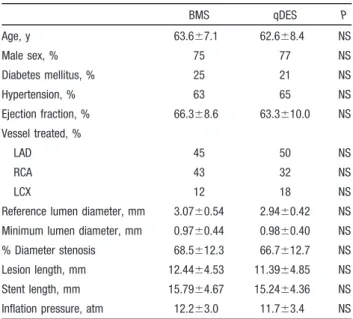
BMS qDES P
Age, y 63.6⫾7.1 62.6⫾8.4 NS
Male sex, % 75 77 NS
Diabetes mellitus, % 25 21 NS
Hypertension, % 63 65 NS
Ejection fraction, % 66.3⫾8.6 63.3⫾10.0 NS Vessel treated, %
LAD 45 50 NS
RCA 43 32 NS
LCX 12 18 NS
Reference lumen diameter, mm 3.07⫾0.54 2.94⫾0.42 NS Minimum lumen diameter, mm 0.97⫾0.44 0.98⫾0.40 NS
% Diameter stenosis 68.5⫾12.3 66.7⫾12.7 NS Lesion length, mm 12.44⫾4.53 11.39⫾4.85 NS Stent length, mm 15.79⫾4.67 15.24⫾4.36 NS Inflation pressure, atm 12.2⫾3.0 11.7⫾3.4 NS
Values are mean⫾SD or frequencies.
LAD indicates left anterior descending artery; RCA, right coronary artery; and LCX, left circumflex artery.
TABLE 2. Postprocedure IVUS Results
BMS qDES P
Reference lumen area, mm2 10.27⫾2.94 10.00⫾2.37 NS Minimum stent area, mm2 8.15⫾1.91 7.59⫾1.62 NS
% Stent expansion 84.0⫾22.1 78.7⫾17.6 NS
Stent volume index, mm3/mm 8.68⫾1.70 8.52⫾1.76 NS Incomplete stent apposition, n (%) 5 (10) 3 (6) NS
Edge tears, n (%) 6 (13) 6 (13) NS
Values are mean⫾SD or frequencies.
drug-eluting stent with BMS in a prospective, randomized, multicenter fashion.
Acute Mechanical Properties
The design of drug-eluting stents can significantly affect the pharmacokinetics as well as the mechanical scaffolding properties. Although metal stents have radial strength supe- rior to newer polymer stents, conventional metal stents had several limitations as a drug reservoir because of limited
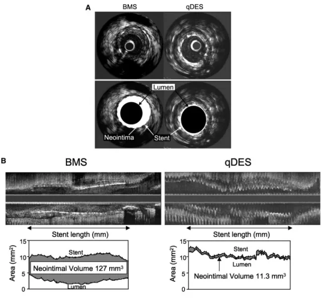
surface area and drug-binding property of the stainless steel struts. One approach to circumvent these problems is a sleeve-based drug delivery system that allows a relatively uniform application of a desired amount of drug in a Figure 1. Representative cases. A, Cross-sectional IVUS images at 6-month follow-up show abundant neointima in BMS (left) versus minimal neointima in qDES (right). B, Longitudinal IVUS images at 6-month follow-up. Large amount of in-stent neointima is observed in BMS (left), whereas qDES (right) shows distinct inhibition of neointimal growth along the entire stented segment. Both stent lengths are 17 mm.
TABLE 3. Follow-Up IVUS Results
BMS qDES P
Reference lumen area, mm2 9.20⫾3.03 9.52⫾2.82 NS Minimum lumen area, mm2 4.97⫾2.22 6.40⫾1.82 0.001 Neointimal area, mm2 3.57⫾2.19 1.07⫾0.90 ⬍0.0001 Cross-sectional narrowing, % 41.5⫾22.6 14.2⫾11.1 ⬍0.0001
Values are mean⫾SD. Figure 2. Neointimal volume index in BMS and qDES groups.
Results are reported as mean⫾SD.
controlled manner. On the other hand, these sleeves may potentially restrict initial stent expansion. Furthermore, com- plete stent apposition may be critical in antiproliferative drug-eluting stents not only for effective tissue drug uptake but also for the prevention of thrombotic events caused by delayed endothelialization, as observed in the animal models with paclitaxel-eluting stents.5In this study, however, qDES achieved an MSA and percent stent expansion comparable to those of BMS with similar lesion and procedural character- istics. Additional volumetric analysis also confirmed these findings over the entire stented segment. More importantly, no difference was observed in the occurrence of incomplete stent apposition between the two groups. Although the unique design of this drug-eluting stent may still call particular attention to major side branch protection, the technical requirement to achieve optimal stent expansion is considered comparable to conventional BMS.
Long-Term Biological Effects
In-stent restenosis is due solely to neointimal hyperplasia in response to stent-induced acute and chronic inflammation in the vessel wall. This process involves several cytokines and growth factors, inducing multiple signaling pathways to activate smooth muscle cell migration and proliferation.
Therefore, attention has long been directed at the potential of antiproliferative agents that block cell cycle progression of vascular smooth muscle cells. Among a number of antipro- liferative agents, paclitaxel and related compounds have several advantages as a candidate for local drug therapy: (1) a highly lipophilic character that promotes rapid cellular uptake by enabling easy passage through the hydrophobic barrier of cell membranes and (2) a prolonged deposition that supports a long-lasting antiproliferative action even after a brief, single-dose application at low concentrations. On the other hand, the efficacy of paclitaxel in reducing neointimal hyperplasia significantly depends on drug dose, delivery methods, and study models as well. For example, one rabbit iliac artery model demonstrated that a polymer-coated, paclitaxel-eluting stent (200 g/stent) could reduce neointi- mal hyperplasia by 59% at 28-day follow-up.5 In another study that used a pig coronary model, a paclitaxel-eluting stent with a dip-coating technique (187 g/stent) showed a 39% reduction in neointimal hyperplasia compared with
control.6On the other hand, local delivery of paclitaxel with a double-balloon local infusion catheter (10 mL; 10mol/L) failed to show any benefit after stenting in pig coronaries.17In the current study, which used a sleeve-based delivery system of 7-hexanoyltaxol, however, the favorable results of early human feasibility registries8,9 have been well replicated, achieving a 70% reduction in neointimal hyperplasia com- pared with the control BMS. Moreover, qDES showed a higher correlation coefficient between postprocedural MSA and follow-up MLA than in BMS, indicating consistent efficacy of qDES, irrespective of various patient/lesion pro- files and risk factors, and, therefore, variable degrees of biological activity in each lesion. Further follow-up will be needed to determine whether this particular type of drug- eluting stent may face late “catch-up,” as reported in animal models with other agents and delivery platforms.7
Complications Assessed by IVUS
Animal studies have demonstrated several important similar- ities in histological characteristics between antiproliferative drug-eluting stents and intracoronary radiation therapy, which raises the concern of potential negative edge effects,18 un- healed dissections,19or late stent-vessel wall malapposition.20 In this study population, however, none of those morpholog- ical complications were detected at 6-month follow-up. Nev- ertheless, significant inhibition of neointimal proliferation was accompanied with the undetectable neointimal layer (by IVUS with axial resolution 120m) in parts of qDES, which may have a relation to late thrombotic events in the stented vessels. Although it is difficult to discuss the potential relation between the undetectable neointimal layer and ad- verse follow-up events from IVUS findings, the significantly delayed healing process reported in animal studies with other drug-eluting stents5,7may underscore the need for prolonged antiplatelet therapy in this type of stent.
Future Clinical Implications
To date, several multicenter, randomized trials have been initiated worldwide, elucidating the safety and effectiveness of drug-eluting stents. The preliminary results of four trials, including RAndomized study with the sirolimus-eluting VE- locity balloon-expandable stent in the treatment of patients with de novo coronary artery Lesions (RAVEL) (rapamycin), ASian Paclitaxel-Eluting stent Clinical Trial (ASPECT), TAXUS, and European evaLUation of pacliTaxel-Eluting Stent (paclitaxel), showed minimum binary restenosis rates (0% to 14%)16 (Seung-Jung Park, Eberhard Grube, and Anthony H. Gershlick, unpublished data, 2001). Although it is difficult to compare the results of 7-hexanoyltaxol polymer sleeve stent with other coated stents directly, RAVEL and ASPECT showed a significant reduction of neointimal growth (NVI, 0.11 to 1.2 mm3/mm)16 (Myeong-Ki Hong, unpublished data, 2002), similar to the magnitude found in this study. On the other hand, the patient population of these early clinical trials may consist of relatively simple lesions or patients at low risk. With more research and advanced technologies of drugs and their delivery, a new era of drug-eluting stent may emerge in the field of real-world, complex coronary interventions, as discussed above. Further Figure 3. Relation between postprocedural MSA and follow-up
MLA. qDES showed higher correlation coefficient (r⫽0.73, P⬍0.0001) than in BMS (r⫽0.39, P⫽0.02), indicating consistent efficacy of qDES irrespective of variable degrees of biological activity in each individual lesion.
studies will be required to identify appropriate patient/lesion subsets with increased risk of restenosis that may derive true benefit from this technology, from both clinical and cost- effectiveness perspectives. In addition, whether IVUS guid- ance for these new biological treatments is advantageous will await the cumulative analysis of future trials.
Study Limitations
Several significant issues should be noted. First, enrollment in the IVUS substudy was not performed in a randomized fashion because of technical challenges during the imaging procedure. This may have created a selection bias toward larger lumens, less tortuous vessels, or lower risk patients compared with the entire population of the SCORE trial.
Despite this potential bias, baseline patient/lesion and proce- dural characteristics were identical either between the two randomized treatment groups in this substudy or between the substudy subset and overall study. Second, follow-up IVUS was not available in all patients. Although this was primarily due to several logistic reasons, one patient in the qDES group had noncardiac death (suspected pulmonary embolism) dur- ing follow-up. Third, long-term observations beyond 6 months are lacking. Fourth, IVUS data were not directly related to all follow-up events. Finally, there are some intrinsic limitations in IVUS analysis as reported previously.
Conclusions
The detailed IVUS substudy from a human, prospective, randomized multicenter trial revealed that the sleeve-based 7-hexanoyltaxol eluting stent showed comparable acute me- chanical and superior long-term biological effects to bare metal stents. Unlike another biological therapy that uses intracoronary radiation, there was no evidence of IVUS- detected adverse vessel response over the stented and adja- cent references segments. Although the long-term benefits and limitations of this technology remain to be investigated further, future directions clearly implicate a biological cellu- lar approach to compliment the conventional mechanical treatment of atherosclerotic disease.
Acknowledgments
We are grateful to Drs Mary E. Russell (Boston Scientific Corp), Martha A. Reitman, (Reitman Corp), and Yoshio Kobayashi (Car- diovascular Research Foundation) for their support.
References
1. Fischman DL, Leon MB, Baim DS, et al. A randomized comparison of coronary-stent placement and balloon angioplasty in the treatment of
coronary artery disease: Stent Restenosis Study Investigators. N Engl J Med. 1994;331:496 –501.
2. Sollott SJ, Cheng L, Pauly RR, et al. Taxol inhibits neointimal smooth muscle cell accumulation after angioplasty in the rat. J Clin Invest.
1995;95:1869 –1876.
3. Axel DI, Kunert W, Goggelmann C, et al. Paclitaxel inhibits arterial smooth muscle cell proliferation and migration in vitro and in vivo using local drug delivery. Circulation. 1997;96:636 – 645.
4. Rowinsky EK, Donehower RC. Paclitaxel (Taxol). N Engl J Med. 1995;
332:1004 –1014.
5. Drachman DE, Edelman ER, Seifert P, et al. Neointimal thickening after stent delivery of paclitaxel: change in composition and arrest of growth over six months. J Am Coll Cardiol. 2000;36:2325–2332.
6. Heldman AW, Cheng L, Jenkins GM, et al. Paclitaxel stent coating inhibits neointimal hyperplasia at 4 weeks in a porcine model of coronary restenosis. Circulation. 2001;103:2289 –2295.
7. Farb A, Heller PF, Shroff S, et al. Pathological analysis of local delivery of paclitaxel via a polymer-coated stent. Circulation. 2001;104:473– 479.
8. Honda Y, Grube E, de La Fuente LM, et al. Novel drug-delivery stent:
intravascular ultrasound observations from the first human experience with the QP2-eluting polymer stent system. Circulation. 2001;104:
380 –383.
9. de la Fuente LM, Miano J, Mrad J, et al. Initial results of the Quanam drug eluting stent (QuaDS-QP-2) Registry (BARDDS) in human subjects.
Catheter Cardiovasc Interv. 2001;53:480 – 488.
10. Potkin BN, Bartorelli AL, Gessert JM, et al. Coronary artery imaging with intravascular high-frequency ultrasound. Circulation. 1990;81:
1575–1585.
11. Okura H, Morino Y, Oshima A, et al. Preintervention arterial remodeling affects clinical outcome following stenting: an intravascular ultrasound study. J Am Coll Cardiol. 2001;37:1031–1035.
12. Prati F, Di Mario C, Moussa I, et al. In-stent neointimal proliferation correlates with the amount of residual plaque burden outside the stent: an intravascular ultrasound study. Circulation. 1999;99:1011–1014.
13. Kornowski R, Mintz GS, Kent KM, et al. Increased restenosis in diabetes mellitus after coronary interventions is due to exaggerated intimal hyper- plasia: a serial intravascular ultrasound study. Circulation. 1997;95:
1366 –1369.
14. Suzuki T, Kopia G, Hayashi S, et al. Stent-based delivery of sirolimus reduces neointimal formation in a porcine coronary model. Circulation.
2001;104:1188 –1193.
15. Sousa JE, Costa MA, Abizaid A, et al. Lack of neointimal proliferation after implantation of sirolimus-coated stents in human coronary arteries:
a quantitative coronary angiography and three-dimensional intravascular ultrasound study. Circulation. 2001;103:192–195.
16. Morice MC, Serruys PW, Sousa JE, et al. A randomized comparison of a sirolimus-eluting stent with a standard stent for coronary revasculariza- tion. N Engl J Med. 2002;346:1773–1780.
17. Oberhoff M, Herdeg C, Al Ghobainy R, et al. Local delivery of paclitaxel using the double-balloon perfusion catheter before stenting in the porcine coronary artery. Catheter Cardiovasc Interv. 2001;53:562–568.
18. Albiero R, Nishida T, Adamian M, et al. Edge restenosis after implan- tation of high activity (32)P radioactive beta-emitting stents. Circulation.
2000;101:2454 –2457.
19. Costa MA, Sabat M, van der Giessen WJ, et al. Late coronary occlusion after intracoronary brachytherapy. Circulation. 1999;100:789 –792.
20. Kozuma K, Costa MA, Sabate M, et al. Late stent malapposition occurring after intracoronary beta-irradiation detected by intravascular ultrasound. J Invasive Cardiol. 1999;11:651– 655.
for the SCORE Investigators
Pitney, Alexandra J. Lansky, Simon H. Stertzer, Paul G. Yock and Peter J. Fitzgerald Bonneau, Antonio Colombo, Carlo Di Mario, Giulio Guagliumi, Karl E. Hauptmann, Mark R.
Toru Kataoka, Eberhard Grube, Yasuhiro Honda, Yoshihiro Morino, Seung-Ho Hur, Heidi N.
QuaDS-QP2 (SCORE)
Print ISSN: 0009-7322. Online ISSN: 1524-4539
Copyright © 2002 American Heart Association, Inc. All rights reserved.
is published by the American Heart Association, 7272 Greenville Avenue, Dallas, TX 75231 Circulation
doi: 10.1161/01.CIR.0000031734.11420.1C
2002;106:1788-1793; originally published online September 9, 2002;
Circulation.
http://circ.ahajournals.org/content/106/14/1788
World Wide Web at:
The online version of this article, along with updated information and services, is located on the
http://circ.ahajournals.org//subscriptions/
is online at:
Circulation Information about subscribing to
Subscriptions:
http://www.lww.com/reprints
Information about reprints can be found online at:
Reprints:
document.
Permissions and Rights Question and Answer this process is available in the
click Request Permissions in the middle column of the Web page under Services. Further information about Office. Once the online version of the published article for which permission is being requested is located,
can be obtained via RightsLink, a service of the Copyright Clearance Center, not the Editorial Circulation
in
Requests for permissions to reproduce figures, tables, or portions of articles originally published Permissions: