Yonsei Med J http://www.eymj.org Volume 52 Number 6 November 2011 1028
Case Report
http://dx.doi.org/10.3349/ymj.2011.52.6.1028pISSN: 0513-5796, eISSN: 1976-2437 Yonsei Med J 52(6):1028-1030, 2011
A Newly Formed and Ruptured Atheromatous Plaque within Neointima after Drug-Eluting Stent Implantation:
2-Year Follow-Up Intravascular Ultrasound and Optical Coherence Tomography Studies
Chang-Myung Oh, Jeonggeun Moon, Hee Tae Yu, Ji-Yong Jang, Jung-Sun Kim, Young-Guk Ko, Donghoon Choi, Yangsoo Jang, and Myeong-Ki Hong
Division of Cardiology, Yonsei Cardiovascular Center, Yonsei University College of Medicine, Seoul, Korea.
Received: July 23, 2010 Revised: August 26, 2010 Accepted: September 2, 2010
Corresponding author: Dr. Myeong-Ki Hong, Division of Cardiology,
Severance Cardiovascular Hospital, Yonsei University College of Medicine, 50 Yonsei-ro, Seodaemun-gu, Seoul 120-752, Korea.
Tel: 82-2-2228-8458, Fax: 82-2-393-2041 E-mail: [email protected]
∙ The authors have no financial conflicts of interest.
© Copyright:
Yonsei University College of Medicine 2011 This is an Open Access article distributed under the terms of the Creative Commons Attribution Non- Commercial License (http://creativecommons.org/
licenses/by-nc/3.0) which permits unrestricted non- commercial use, distribution, and reproduction in any medium, provided the original work is properly cited.
Late stent thrombosis (LST) which is a life threatening complication has emerged as a serious problem of drug-eluting stents (DES). Several studies have suggested that incomplete neointimal coverage of stent struts contributes to LST. Progressive atherosclerosis within the neointima is an another possible cause of LST, but this phenomenon has seldom been reported in DES. We present a case of LST follow- ing DES implantation after a period of 28 months due to ruptured atheromatous plaque, despite complete neointimal coverage of stent struts proven by optical co- herence tomography.
Key Words: Drug-eluting stents, thrombosis, optical coherence tomography
INTRODUCTION
Drug eluting stent (DES) has successfully been introduced to interventional cardi- ology in order to prevent in-stent restenosis. Nevertheless, concerns about the safe- ty of DES still exist. Especially, late stent thrombosis (LST) is an unsolved prob- lem in the DES era, which was rarely seen with bare metal stents (BMS).1 The risk of LST is associated with patient and lesion characteristics, non-compliance of an- ti-platelet drugs and incomplete stent apposition, number and the length of stents, coronary dissection, etc.2,3 Incomplete neointimal coverage of stent struts is the most important predictor of LST.4,5 We report a case of LST despite complete neo- intimal coverage on well-opposed stent struts and continued dual-antiplatelet ther- apy more than 1 year.
CASE REPORT
A 57-year-old male with a history of hypertension, diabetes mellitus, normal renal function and a 60 pack years smoking received percutaneous coronary interven-
Ruptured Plaque within Neointima after DES Implantation
Yonsei Med J http://www.eymj.org Volume 52 Number 6 November 2011 1029
positive. Therefore, he underwent another follow-up angio- gram to evaluate silent ischemia in January 2010. The fol- low-up angiogram revealed 2 patent stents implanted in the right coronary artery. However, abnormal contrast filling tion at the right coronary artery with the impression of stable
angina pectoris in September 2007 (Fig. 1). Two sirolimus- eluting stents (SES) [3.0×28 mm, 2.75×28 mm (Cypher, Cordis Corp., Johnson & Johnson Co., Warren, New Jersey, USA)] were implanted at the distal part of the right coronary artery. Transthoracic echocardiography demonstrated normal left ventricular systolic function (ejection fraction=56%) with regional wall motion abnormality at the inferior wall. One- year follow-up angiogram showed that 2 stents were all patent. Intravascular ultrasound (IVUS) and optical coher- ence tomography (OCT) revealed complete neointimal coverage of stent struts (Fig. 2).
Until recently, he had good compliance with dual anti- platelet agents and other medications, and did not suffer from chest pain. However, the result of treadmill test was
Fig. 1. Initial coronary angiogram of the RCA. (A) Coronary angiogram shows significant luminal narrowing at the distal RCA. (B) After stent im- plantation, there is a minimal residual stenosis. RCA, right coronary artery.
A B
B A
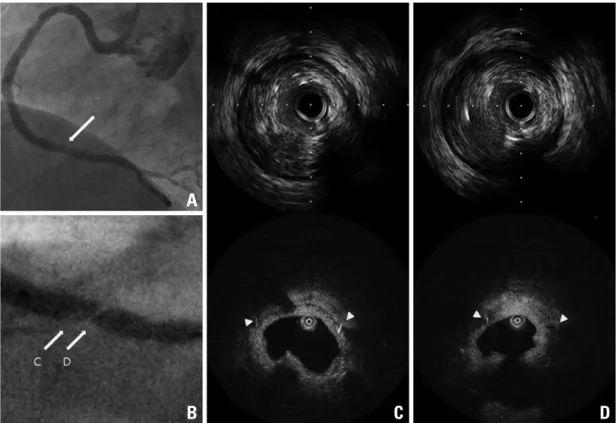
C D
Fig. 3. (A) The 2nd follow-up angiogram at 28 months after stent implantation shows abnormal contrast filling in a previously implanted stent (see arrow). (B, C and D) IVUS and OCT images reveal newly formed and ruptured atheromatous plaque within neointima.
Arrowheads indicate stent strut. IVUS , intravascular ultrasound; OCT, optical coherence tomography.
Fig. 2. Coronary angiogram and OCT findings at 12-month follow-up. (A)Coronary angiogram shows no restenosis in the stented segment.
(B, C and D)The OCT shows complete neointimal coverage of stent struts. OCT, optical coherence tomography.
A B C D
Chang-Myung Oh, et al.
Yonsei Med J http://www.eymj.org Volume 52 Number 6 November 2011 1030
curred only after 2 years. We think that early atherosclerosis is a possible mechanism in our case. This was an unexpected finding and gave us a very helpful insight for better treatment and monitoring in patients with DES implantation.
REFERENCES
1. Iakovou I, Schmidt T, Bonizzoni E, Ge L, Sangiorgi GM, Stankovic G, et al. Incidence, predictors, and outcome of throm- bosis after successful implantation of drug-eluting stents. JAMA 2005;293:2126-30.
2. Lüscher TF, Steffel J, Eberli FR, Joner M, Nakazawa G, Tanner FC, et al. Drug-eluting stent and coronary thrombosis: biological mecha- nisms and clinical implications. Circulation 2007;115:1051-8.
3. Kim U, Kim JS, Kim JS, Lee JM, Son JW, Kim J, et al. The initial extent of malapposition in ST-elevation myocardial infarction treated with drug-eluting stent: the usefulness of optical coherence tomography. Yonsei Med J 2010;51:332-8.
4. Kotani J, Awata M, Nanto S, Uematsu M, Oshima F, Minamigu- chi H, et al. Incomplete neointimal coverage of sirolimus-eluting stents: angioscopic findings. J Am Coll Cardiol 2006;47:2108-11.
5. Matsumoto D, Shite J, Shinke T, Otake H, Tanino Y, Ogasawara D, et al. Neointimal coverage of sirolimus-eluting stents at 6-month follow-up: evaluated by optical coherence tomography.
Eur Heart J 2007;28:961-7.
6. Finn AV, Joner M, Nakazawa G, Kolodgie F, Newell J, John MC, et al. Pathological correlates of late drug-eluting stent thrombosis:
strut coverage as a marker of endothelialization. Circulation 2007;115:2435-41.
7. Higo T, Ueda Y, Oyabu J, Okada K, Nishio M, Hirata A, et al.
Atherosclerotic and thrombogenic neointima formed over sirolim- us drug-eluting stent: an angioscopic study. JACC Cardiovasc Im- aging 2009;2:616-24.
8. Doyle B, Rihal CS, O’Sullivan CJ, Lennon RJ, Wiste HJ, Bell M, et al. Outcomes of stent thrombosis and restenosis during extend- ed follow-up of patients treated with bare-metal coronary stents.
Circulation 2007;116:2391-8.
9. Nakazawa G, Vorpahl M, Finn AV, Narula J, Virmani R. One step forward and two steps back with drug-eluting-stents: from pre- venting restenosis to causing late thrombosis and nouveau athero- sclerosis. JACC Cardiovasc Imaging 2009;2:625-8.
defect was observed within the previously stented segments (arrow in Fig. 3A). IVUS and OCT were performed to elu- cidate the characteristics of the abnormal contrast filling de- fect. Both studies revealed a newly formed and ruptured atheromatous plaque within the neointima over the stent struts (IVUS and OCT) (Fig. 3B, C and D). He was success- fully treated with percutaneous coronary intervention and discharged on medications of aspirin (100 mg/day), clopi- dogrel (75 mg/day), atorvastatin (10 mg/day) and atenolol (25 mg/day).
DISCUSSION
LST after DES implantation has been thought to be caused by poorly formed neointima and incomplete neointimal cov- erage over DES struts.6 In a recent study using angioscopy, however, a newly formed and thrombogenic atherosclerotic yellow neointima after DES implantation was reported.7 This phenomenon may be an another cause for late stent thrombosis after DES implantation. Stent thrombosis and myocardial infarction caused by restenosis during extended follow-up after bare metal stent implantation have been re- ported and might be explained by newly formed and pro- gressive atherosclerosis within neointima.8 However, this phenomenon has seldom been reported in DES. Atheroscle- rotic change within neointima after bare metal stent implan- tation usually occurs after 5 years.7 The intracoronary imag- es of our patient illustrate a newly formed and ruptured atheromatous plaque within neointima after DES implanta- tion in a relatively short period (less than 3 years after DES implantation). A recent pathologic study showed that ath- erosclerotic lesions progress more rapidly within SES than within BMS. In the autopsy study by Nakazawa, et al.9 the atherosclerotic change in SES was seen in more than 40 percent of cases by 9 months, but the change in BMS oc-