671
Print ISSN 1738-5520 / On-line ISSN 1738-5555 Copyright © 2011 The Korean Society of Cardiology CASE REPORT
http://dx.doi.org/10.4070/kcj.2011.41.11.671
Open Access
A Case of In-Stent Neointimal Plaque Rupture 10 Years After Bare Metal Stent Implantation: Intravascular Ultrasound
and Optical Coherence Tomographic Findings
Hyuck-Jun Yoon, MD, Seung-Ho Hur, MD, Shin-Keun Kim, RT, Hyungseop Kim, MD, Hyoung-Seob Park, MD, Yun-Kyeong Cho, MD, Chang-Wook Nam, MD, Yoon-Nyun Kim, MD, and Kwon-Bae Kim, MD
Division of Cardiology, Department of Internal Medicine, Keimyung University Dongsan Medical Center, Daegu, Korea ABSTRACT
Neointimal hyperplasia mainly develops within several months of coronary stent deployment, after which it stabilizes. Al- though it was widely accepted, particularly during the bare-metal stent (BMS) era, that in-stent restenosis (ISR) generally does not present as an acute coronary syndrome (ACS), but rather as a gradual recurrence of angina symptoms, recent data have shown that a substantial number of patients with ISR present as ACS. There has also been consistent postmortem evi- dence of plaque rupture secondary to atherosclerotic change within the neointima of a BMS. We report here a case of ACS in which intravascular ultrasound and optical coherent tomographic assessments revealed neointimal atherosclerotic change and ruptured plaque 10 years after BMS deployment. (Korean Circ J 2011;41:671-673)
KEY WORDS: Coronary restenosis; Stents; Neointima; Ultrasonography; Tomography.
Received: January 24, 2011 Revision Received: April 11, 2011 Accepted: April 19, 2011
Correspondence: Seung-Ho Hur, MD, Division of Cardiology, Dongsan Medical Center, Keimyung University, 194 Dongsan-dong, Jung-gu, Daegu 700-712, Korea
Tel: 82-53-250-7949, Fax: 82-53-250-7034 E-mail: [email protected]
• The authors have no financial conflicts of interest.
cc This is an Open Access article distributed under the terms of the Cre- ative Commons Attribution Non-Commercial License (http://creativecom- mons.org/licenses/by-nc/3.0) which permits unrestricted non-commer- cial use, distribution, and reproduction in any medium, provided the origi- nal work is properly cited.
Introduction
Several authors recently reported a substantial number of bare-metal stent (BMS)-related restenosis cases presenting as acute coronary syndromes (ACS)1)2) and several case reports providing intravascular ultrasound (IVUS) evidence of pla- que rupture at in-stent neointima.3)4) Although IVUS is widely used to evaluate plaque composition, it has inherent image qu- ality limitations when compared with optical coherence to- mography (OCT) for the assessment of soft tissue compo- nents and subtle surface changes in atherosclerotic lesions.5)6) Here, we present a patient with ACS in whom a combination of IVUS and OCT imaging showed atheromatous changes and plaque rupture of neointima 10 years after BMS implantation.
Case
A 58-year-old man was admitted with unstable angina and complaining of new onset substernal chest pain at rest. The pa- tient had a history of prior percutaneous coronary interven- tion (PCI) at this hospital due to angina pectoris ten years ago.
At that time, the coronary angiogram (CAG) showed 90%
stenosis at the proximal portion of the left anterior descend- ing coronary (LAD). A BMS (NIRTM, 3.75×16 mm, Medinol, Boston Scientific, Tel Aviv, Israel) was implanted at the pro- ximal-portion of the LAD and there was no evidence of in- stent restenosis (ISR) upon follow-up CAG nine months after index PCI. During ten years of follow-up at the local clinic, he had not complained of chest pain while on his medical treatment; however, periods of substernal chest pain at rest had recurred from one month before admission.
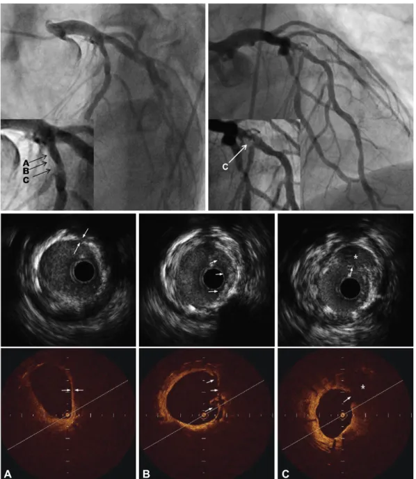
CAG on this admission revealed evidence of focal stenosis with luminal haziness on the previously stented proximal LAD and a de novo lesion at the distal left circumflex (LCX) artery (Fig. 1, upper panel). Upon quantitative coronary analysis, the previously stented lesion had a diameter stenosis of 65%.
IVUS images demonstrated a well expanded previously de- ployed BMS and remarkable neointimal proliferation caus- ing luminal narrowing (minimal luminal area=3.32 mm², pla- que burden=75.9%). Of interest was a suspicious cavitary-
672 Neointimal Plaque Rupture 10 Years After BMS Deployment
shaped ruptured plaque in the neointima (Fig. 1, middle pa- nel). For more detailed evaluation of the suggested ruptured plaque in IVUS, we conducted OCT at the ISR site, which showed evident proximally located thin fibrous cap athero- ma (TFCA) with lipid core and a ruptured plaque with tiny multiple ulcerations of neointima that were proximally lo- cated (Fig. 1, lower panel). A new-generation drug eluting stent (Xience VTM, 4.0×28 mm, Abbott, IL, USA) was suc- cessfully placed to cover the previously stented lesion, and post-stenting IVUS revealed a well expanded previous ISR
lesion (minimal stent area=7.31 mm2). A 2.75×23 mm stent was also successfully deployed in the de novo LCX lesion. At the nine months follow up, CAG showed patent stented le- sions without evidence of ISR, and the patient has been clin- ically stable for 18 months after PCI.
Discussion
Although many cardiologists are under the impression that neointimal growth after BMS implantation has benign
A B C
Fig. 1. Coronary angiogram (upper panel), intravascular ultrasound (middle panel) and optical coherence tomography (lower panel) images of the in-stent restenosis site in the proximal left anterior descending artery. A: in comparisons of IVUS and OCT images in the correspond- ing site, an atherosclerotic plaque extending from 12 to 5 o'clock contains regions consistent with fibrous tissue, and a homogenous signal- poor lesion possible representing lipid core with thin cap fibrous atheroma (TFCA) was clearly shown in OCT (arrow). Although this fibrous atheromatous lesion was also apparent in the IVUS image from the same site, it was difficult to identify the presence of TFCA and lipid core. The minimal cap thickness at the region measured 60 m by OCT. B: immediately proximal to the ruptured plaque, the OCT image showed multiple disrupted intimal flaps and a subtle ulcerated palque (arrow) that was not clearly seen in the corresponding IVUS image. C:
an obvious intimal rupture (arrow) with large cavitary change (*) was also clearly identified by OCT compard with corresponding IVUS image.
Hyuck-Jun Yoon, et al. 673
consequences, this case shows unstable clinical features and consistent neointimal atheromatous changes and plaque rup- ture findings on IVUS and OCT.
In the past, post-mortem and endarterectomy specimens had shown extensive extracellular matrix accumulation with low rates of cellular proliferation in BMS-related ISR lesions,7-9) which were believed to have benign clinical presentation as stable angina.
Although awareness of very late stent thrombosis is usually associated with the drug-eluting stent (DES) era, it is also per- tinent to the BMS era. A large retrospective study reported that the cumulative incidence of stent thrombosis after BMS implantation was 0.5% at 30 days, 0.8% at 1 year, 1.3% at 5 years, and 2.0% at 10 years.2) Another retrospective study of 1,186 cases of BMS-related ISR reported that more than one- third presented as acute myocardial infarction or unstable an- gina.1) These studies suggested that stent thrombosis and ACS long after BMS implantation might be attributable to neo- intimal plaque rupture. Recently, an interesting report on as- sessment of very late stent thrombosis after both DES and BMS using IVUS demonstrated that neointimal atheroscle- rotic changes and plaque ruptures were the main cause of BMS-related very late stent thrombosis.10) Another postmor- tem pathologic study revealed that atherosclerotic changes of neointima that occur significantly earlier and more frequent- ly with DES, were also found with BMS.11)
Despite several previous reports suggesting atheromatous changes in neointima after stenting and case reports of IVUS- documented ruptured plaque in neointima, there have been few reports of atherosclerotic progression with ruptured pla- ques of in-stent intima confirmed by both IVUS and OCT.
Because of its high resolution of approximately 10-20 µm, which is approximately 10-fold greater than that of IVUS, OCT can be a valuable tool for evaluation of intravascular plaque characterization that cannot be detected by CAG and IVUS.5)
In this case, we found a suspicious plaque rupture in the neointima using IVUS. However, OCT imaging allowed de- tection of more detailed atheromatous changes of neointima such as multiple tiny ulcerations and a TCFA with a large li- pid core, which could not be precisely discriminated in IVUS images. OCT has a higher sensitivity than IVUS for charac-
terizing lipid-rich plaques, and the higher resolution of OCT allows improved visualization of soft tissue components and detection of the cause of ACS such as plaque rupture or ero- sion when compared with IVUS images.5)6) We anticipate that neointimal atherosclerotic changes might be more easi- ly detected before plaque rupture using a combination of OCT and IVUS than IVUS alone.
In summary, this case provides in vivo evidence of athero- matous neointimal changes and ruptured plaque using com- bination of IVUS and OCT imaging modalities.
REFERENCES
1) Chen MS, John JM, Chew DP, Lee DS, Ellis SG, Bhatt DL. Bare me- tal stent restenosis is not a benign clinical entity. Am Heart J 2006;
151:1260-4.
2) Doyle B, Rihal CS, O’Sullivan CJ, et al. Outcomes of stent thrombo- sis and restenosis during extended follow-up of patients treated with bare-metal coronary stents. Circulation 2007;116:2391-8.
3) Fineschi M, Carrera A, Gori T. Atheromatous degeneration of the neo- intima in a bare metal stent: intravascular ultrasound evidence. J Cardiovasc Med (Hagerstown) 2009;10:572-3.
4) Baek EK, Park SH, Kwon KH, Shim EJ, Jo JY. A case of in-stent plaque rupture presenting as an acute myocardial infarction. Korean Circ J 2008;38:432-5.
5) Kume T, Akasaka T, Kawamoto T, et al. Assessment of coronary ar- terial plaque by optical coherence tomography. Am J Cardiol 2006;
97:1172-5.
6) Jang IK, Bouma BE, Kang DH, et al. Visualization of coronary ath- erosclerotic plaques in patients using optical coherence tomography:
comparison with intravascular ultrasound. J Am Coll Cardiol 2002;
39:604-9.
7) Komatsu R, Ueda M, Naruko T, Kojima A, Becker AE. Neointimal tissue response at sites of coronary stenting in humans: macroscopic, histological, and immunohistochemical analyses. Circulation 1998;
98:224-33.
8) Sangiorgi G, Taylor AJ, Farb A, et al. Histopathology of postpercuta- neous transluminal coronary angioplasty remodeling in human coro- nary arteries. Am Heart J 1999;138:681-7.
9) Chung IM, Gold HK, Schwartz SM, Ikari Y, Reidy MA, Wight TN.
Enhanced extracellular matrix accumulation in restenosis of coro- nary arteries after stent deployment. J Am Coll Cardiol 2002;40:
2072-81.
10) Lee CW, Kang SJ, Park DW, et al. Intravascular ultrasound findings in patients with very late stent thrombosis after either drug-eluting or bare-metal stent implantation. J Am Coll Cardiol 2010;55:1936-42.
11) Nakazawa G, Otsuka F, Nakano M, et al. The pathology of neoath- erosclerosis in human coronary implants bare-metal and drug-elut- ing stents. J Am Coll Cardiol 2011;57:1314-22.