of January 7, 2016.
This information is current as Responsive to γ -2 (Crg-2)
(Mig) and Chemokine γ
Induced by IFN-
Activity that Is Dependent on Monokine IL-2/IL-12 Produce T Cell Chemoattractant Primary Hepatocytes from Mice Treated with
G. Fenton, Ji-Ming Wang and Robert H. Wiltrout
Jong-Keuk Lee, Jeffrey Subleski, Jon M. Wigginton, Robert Jong-Wook Park, M. Eilene Gruys, Kathy McCormick,
http://www.jimmunol.org/content/166/6/3763 doi: 10.4049/jimmunol.166.6.3763
2001; 166:3763-3770; ; J Immunol
References
http://www.jimmunol.org/content/166/6/3763.full#ref-list-1
, 22 of which you can access for free at:
cites 34 articles This article
Subscriptions
http://jimmunol.org/subscriptions
is online at:
The Journal of Immunology Information about subscribing to
Permissions
http://www.aai.org/ji/copyright.html Submit copyright permission requests at:
Email Alerts
http://jimmunol.org/cgi/alerts/etoc
Receive free email-alerts when new articles cite this article. Sign up at:
Print ISSN: 0022-1767 Online ISSN: 1550-6606.
Immunologists All rights reserved.
Copyright © 2001 by The American Association of 9650 Rockville Pike, Bethesda, MD 20814-3994.
The American Association of Immunologists, Inc.,
is published twice each month by The Journal of Immunology
at Keimyung Univ Med Lib on January 7, 2016http://www.jimmunol.org/Downloaded from at Keimyung Univ Med Lib on January 7, 2016http://www.jimmunol.org/Downloaded from
Produce T Cell Chemoattractant Activity that Is Dependent on Monokine Induced by IFN- ␥ (Mig) and Chemokine Responsive to ␥-2 (Crg-2) 1
Jong-Wook Park,
2* M. Eilene Gruys,* Kathy McCormick,
†Jong-Keuk Lee,* Jeffrey Subleski,*
Jon M. Wigginton,
‡Robert G. Fenton,
§Ji-Ming Wang,
¶and Robert H. Wiltrout
3*
The IFN-␥-inducible proteins monokine induced by IFN-␥ (Mig) and chemokine responsive to ␥-2 (Crg-2) can contribute to IL-12-induced antiangiogenic and leukocyte-recruiting activities, but the extent to which leukocytes vs parenchymal cells in different organs contribute to the production of these molecules remains unclear. The results presented herein show that IFN-
␥-dependent induction of Mig and Crg-2 gene expression can occur in many nonlymphoid organs, and these genes are rapidly induced in purified hepatocytes isolated from mice treated with IL-2 plus IL-12, or from Hepa 1-6 hepatoma cells treated in vitro with IFN-␥. In addition to depending on IFN-␥, the ability of IL-12 or IL-2/IL-12 to induce Mig and Crg-2 gene expression in purified hepatocytes also is accompanied by the coordinate up-regulation of the IFN-␥ R ␣ and -chains, in the absence of IL-12R components. Supernatants of primary hepatocytes obtained from mice treated in vivo with IL-2/IL-12 or from hepatocytes treated in vitro with IFN-␥ contain increased chemotactic activity for enriched human and mouse CD3ⴙT cells, as well as mouse DX5ⴙ NK cells. The hepatocyte-derived chemotactic activity for mouse T cells but not NK cells was ablated by Abs specific for Mig and Crg-2. These results suggest that parenchymal cells in some organs may contribute substantially to initiation and/or amplification of inflammatory or antitumor responses. The Journal of Immunology, 2001, 166: 3763–3770.
I
nterleukin-12, a multifunctional cytokine produced by mac- rophages, B cell lines, and other cells, stimulates the prolif- eration of T and NK cells, enhances cell-mediated cytotox- icity, promotes the development of Th1-type responses (1–3), and mediates potent antitumor activity in numerous mouse tumor mod- els (4 – 6). IL-12 also has been reported to inhibit angiogenesis in tumor-bearing mice (7–9) and to induce production of the non- ELR CXC chemokines, such as monokine induced by IFN-␥ (Mig)4and the mouse homolog of IFN-inducible protein-10 (IP- 10, otherwise termed chemokine responsive to␥-2; Crg-2) that areactive as chemotactic factors for stimulated T cells (10 –13). These chemokines have been reported to promote damage to established tumor vasculature, cause tumor necrosis (14, 15), and contribute to the antitumor activities of IL-12 (16). Recent studies from our laboratory have demonstrated that the combination of IL-2 and IL-12 yields enhanced therapeutic efficacy in a variety of murine tumor models (17) in association with increased local expression of the Mig and Crg-2 genes (34).
The most important intermediate cytokine for induction of Mig and Crg-2 by IL-12 is IFN-␥, and leukocytes, some nonleukocytes, and tumor cells can all produce Mig and Crg-2 (12, 13, 18). We have demonstrated that both Mig and Crg-2 gene expression can be induced in several nonlymphoid organs by administration of IL-12 or IL-2/IL-12 to mice. These results raise the issue of whether all of the observed gene expression in nonlymphoid or- gans can be attributed to resident tissue macrophages and transient blood monocytes and lymphocytes, or whether parenchymal cells (PC) also contribute substantially to this effect. If PC can be in- duced to produce functional proteins that contribute to leukocyte recruitment, then these cells may represent an important innate immune component for initiating or sustaining biological activities vital to the success of cytokine therapy or may contribute to patho- genic inflammatory responses in nonlymphoid organs. In this re- port, we use the liver as a model to demonstrate that hepatocytes can produce chemotactic activity for mouse T cells that is depen- dent on Mig and Crg-2 as well as chemotactic activity for NK cells that is independent of Mig and Crg-2.
Materials and Methods
Mice
Normal male BALB/c mice were obtained from the Animal Production Area, Harlan Sprague-Dawley at National Cancer Institute-Frederick Can- cer Research and Development Center (Frederick, MD), and IFN-␥⫺/⫺
*Laboratory of Experimental Immunology, National Cancer Institute-Frederick Can- cer Research and Development Center, Frederick, MD 21702;†IRSP, Science Ap- plications International Corporation, National Cancer Institute-Frederick Cancer Re- search and Development Center, Frederick, MD 21702;‡Pediatric Oncology Branch, National Institutes of Health, Bethesda, MD;§Greenbaum Cancer Center, University of Maryland Medical School, Baltimore, MD; and¶Laboratory of Molecular Immu- noregulation, National Cancer Institute-Frederick Cancer Research and Development Center, Frederick, MD 21702
Received for publication July 24, 2000. Accepted for publication January 2, 2001.
The costs of publication of this article were defrayed in part by the payment of page charges. This article must therefore be hereby marked advertisement in accordance with 18 U.S.C. Section 1734 solely to indicate this fact.
1The project has been funded in whole or part with federal funds from the National Cancer Institute, National Institutes of Health, under Contract No. NO-56000. The content of this publication does not necessarily reflect the views or policies of the Department of Health and Human Services, nor does mention of trade names, com- mercial products, or organizations imply endorsement by the U.S. Government.
2Current address: Department of Immunology, Keimyung University School of Med- icine, No. 194 Dongsan-Dong, Jung-Gu, Taegu, Korea, 700-712. E-mail address:
3Address correspondence and reprint requests to Dr. Robert H. Wiltrout, National Cancer Institute-Frederick Cancer Research and Development Center, Building 560, Room 31-93, Frederick, MD 21702-1201. E-mail address:WiltroutR@
mail.ncifcrf.gov
4Abbreviations used in this paper: Mig, monokine induced by IFN-␥; IP-10, IFN- inducible protein-10; PC, parenchymal cells; rm, recombinant mouse; NPC, non- parenchymal cells; RPA, RNase protection assays.
Copyright © 2001 by The American Association of Immunologists 0022-1767/01/$02.00
at Keimyung Univ Med Lib on January 7, 2016http://www.jimmunol.org/Downloaded from
knockout mice were obtained from our own specific pathogen-free breed- ing colony that was initiated from mice kindly donated by Dyana Dalton (Hoffman-LaRoche, Nutley, NJ). The mice were used at 8 –10 wk of age.
Animal care was provided in accordance with procedures outlined in the
“Guide for the Care and Use of Laboratory Animals” (National Institutes of Health publication no. 86-23, 1985).
Reagents
Recombinant murine IL-12 (specific activity, 2.7⫻ 106U/mg) and IL-2 (16⫻ 106IU/mg) were generously provided by Genetics Institute (Cam- bridge, MA) and Chiron Corporation (Emeryville, CA), respectively. Re- combinant mouse (rm)IFN-␥ was kindly provided by Genentech (South San Francisco, CA).
In vivo cytokine administration
Chemokine gene expression was induced in vivo by using IL-12, IL-2, or the combination of IL-2/IL-12, which has been shown previously to induce complete regression of the Renca murine renal carcinoma in mice (17). Mice were given 300,000 IU IL-2 (or HBSS plus syngeneic normal mouse serum) i.p. on day 0, and IL-12 (0.5g) or PBS plus mouse serum was administered by daily i.p. injection on days 0 –3.
Groups of mice were euthanized for analyses on days 1, 4, and 11. In some studies, mice were treated i.p. and i.v. with 50,000 U rmIFN-␥ on days 0 –3 and tissues were harvested on day 4.
Isolation of PC and nonparenchymal cells (NPC) from the liver PC were prepared by a modification of the method described previously by Kedderis and Held (19). Briefly, after CO2inhalation and cervical dislo- cation, mice were perfused with EGTA solution at 37°C (flow rate, 5 ml/
min) for 8 min followed by a 0.33% collagenase solution for 10 –12 min.
The perfused livers were then dissociated by mild agitation, and the re- covered cell suspension was passed through a 100-m nylon filter and centrifuged at 50⫻ g for 60 s. The cell pellet was gently resuspended in ice-cold William’s media E, mixed with 45% Percoll (Pharmacia LKB Biotechnology, Uppsala, Sweden) and centrifuged again at 50⫻ g for 8 min. The viable PC, which pellet at the bottom, were resuspended in cold William’s media E and washed twice. These PC preparations then were stained with trypan blue. Microscopic and flow cytometric analyses con- firmed these preparations to be⬎90% hepatocytes. Purified PC were kept ice cold until used for RT-PCR analyses, or overnight culture in William’s media E plus 5% FBS. Where indicated, the hepatocyte cultures also in- cluded 200 U/ml rmIFN-␥. NPC containing lymphocytes, Kupffer cells, and endothelial cells were obtained by using a 30% metrizamide (Accurate Chemical and Scientific, Westbury, NY) gradient technique as described previously (20). The final preparation contains varying numbers of T, NK, or NK/T cells, and Kupffer cells, depending on the nature of preceding in vivo treatments.
Hepa 1-6 cell culture
The Hepa 1-6 cell line (obtained from the American Type Culture Collec- tion, Mannassas, VA) was cultured in DMEM containing 10% FBS. For the induction of Mig and Crg-2 gene expression, cells were treated with 200 U/ml rmIFN-␥ for 2, 6, and 24 h, harvested in Trizol solution, and then used for RT-PCR analyses.
RT-PCR
For the analysis of gene expression in various tissues and cells, total cel- lular RNA was isolated from snap-frozen tissue specimens and cultured cells by the Trizol method (Life Technologies, Gaithersburg, MD). Reverse transcription was performed in a 50-l reaction mixture containing 50 mM Tris-HCl, 75 mM KCl, 3 mM MgCl2, 10 mM DTT, 250M dATP, 250
M dCTP, 250 M dTTP, 250 M dGTP, 0.75 U/l RNase inhibitor, 5 U/l Moloney murine leukemia virus reverse transcriptase, 2.5 M oli- go(dT) primer, and 10g denatured RNA. The reverse transcription reac- tion mixture was incubated at 42°C for 60 min at 95°C for 5 min and then stored at⫺20°C until further use. The PCR was performed in a 25-l reaction mixture containing 10 mM tris-HCl, 50 mM KCl, 1.5 mM MgCl2, 200M dATP, 200 M dCTP, 200 M dTTP, 200 M dGTP, 0.6 U Taq DNA polymerase, 0.5 mM sense primer, 0.5M sense primer, 0.5 58 M anti-sense primer, and 2.5l of the products of the reverse transcription reaction. Amplification was performed in a thermocycler (Gene Amp PCR System 2400, Perkin-Elmer, Cetus, Norwalk, CT) as follows: 94°C for 3 min (1 cycle), 94°C for 30 s, 56 –57°C for 1 min, 72°C for 45 s (optimized at between 20 –28 cycles depending on the primer pair and tissue source used); and 72°C for 5 min (1 cycle). The sequence of primer pairs for the respective genes evaluated in these studies was for G3PDH: sense, GC
CACCCAGAAGACTGTGGATGGC; antisense, CATGTAGGCCATGA GGTCCACCAC; for MIG: sense; GATCAAACCTGCCTAGATCC;
antisense, GGCTGTGTAGAACACAGAGT; and for IP-10: sense, AC CATGAACCCAAGTGCTGCCGTC; antisense, GCTTCACTCCAGTT AAGGAGCCCT.
PCR products were then separated on 1% agarose gels impregnated with 0.5g/ml ethidium bromide. Density of the DNA band was measured by using the image PC program and relative intensity was calculated by fol- lowing formula; relative intensity⫽ density of target DNA band/density of G3PDH DNA band.
RNase protection assays (RPA)
Total RNA from PC was isolated by using Trizol reagent (Life Technol- ogies). RPA were performed by using RiboQuant Transcription and RPA kit (BD PharMingen, San Diego, CA) according to the manufacturer’s instructions with the exception that the Multiprobe templates (BD Phar- Mingen) and Mig probes were purified with a Tris-EDTA micro select G25 spin column (5 Prime33 Prime, Boulder, CO). The mouse Mig 416 probe was prepared by PCR cloning of Mig fragment into TOPO vector (Invitro- gen) using the following primer pairs: Mig: sense, 5⬘-GAAGTCCGCTGT TCTTTTCC; antisense, 5⬘-GATTCAGGGTGCTTGTTGGT.
The protected bands were separated on a 5% sequencing gel (National Diagnostics) and analyzed using a Beckman Phosphor imager (Beckman Coulter, Fullerton, CA).
Intracellular cytokine analysis
Purified liver leukocytes were washed in PBS plus 0.5% BSA and counted.
Intracellular labeling was performed by adding 1⫻ 106cells per well using a 96-well round-bottom micro titer plate. The wells were sealed with tape, the cells were pelleted at 400⫻ g for 2 min at 4°C, and the supernatant was removed by aspiration. Cells then were resuspended in 50 l/well PBS followed by addition of 50l/well of 8% paraformaldehyde in PBS to fix the cells. Then, the cells were incubated at 37°C for 5 min and pelleted, the supernatants were removed, and the fixed cells were washed in PBS plus 0.1% BSA, and permeablized by a wash in staining buffer (0.1% BSA⫹ 0.01 M HEPES plus 0.1% saponin in PBS). The supernatant was removed from the pelleted cells and 10l of optimally titered anti-mouse IFN-␥
(clone XMG1.2 BD, BD PharMingen) diluted in HBSS plus 0.1% BSA plus NaN3was added per well to detect intracellular IFN-␥. The wells were taped, the cells were mixed by manual shaking, incubated at 4°C for 15 min, and centrifuged at 400⫻ g for 2 min at 4°C. The supernatant was aspirated, the cells were washed twice in staining buffer, resuspended in HBSS plus 0.1% BSA plus NaN3, and transferred to 12⫻ 75-mm poly- styrene test tubes. Cells (10,000) were analyzed for the percentage of cells bearing mouse IFN-␥ by using a FACScan flow cytometer affixed with a 488-nm laser (Becton Dickinson, Mountain View, CA). Isotype-matched controls were used to quantitate nonspecific background binding that was subtracted from all results.
Purification of CD3⫹T cells or DX5⫹NK cells
T and NK cells were obtained for use as responders in microchemotaxis assays as follows: mouse and human T cells were purified by using neg- ative-selection T cell enrichment columns (R&D Systems, Minneapolis, MN) as recommended by the manufacturer. For human T cells, the starting population was Ficoll-Hypaque-separated PBL, and spleen cell prepara- tions were the source for mouse T cells. The purity of human and mouse CD3⫹ cell preparations as confirmed by flow cytometry was routinely
⬎95%. Enriched mouse NK cells were obtained from spleen cell prepa- rations as described previously (21). The resulting preparation routinely contained⬎75% DX5⫹, CD3⫺NK cells as assessed by flow cytometry.
Enriched mouse T or NK cells were cultured for 7 days in 50 U/ml IL-2 and then were used as responders in the microchemotaxis assay described below.
In vitro chemotaxis assay
T or NK cell migration was assessed by a 48-well microchemotaxis cham- ber technique. A 25-l aliquot of hepatocyte supernatant diluted in che- motaxis medium (RPMI 1640 with 1% BSA) was placed in the lower compartment and 50l of enriched T or NK cell suspensions (5 ⫻ 106 cells/ml) were placed in the upper compartment of the chamber. The two compartments were separated by a polycarbonate filter (5-m pore size;
Nucleopore, Pleasanton, CA) coated with 10g/ml fibronectin (Sigma, St.
Louis, MO) via incubation at 4°C overnight or at 37°C for 2 h. The cham- ber was incubated at 37°C for 3 h in humidified air with 5% CO2. At the end of the incubation period, the nonmigrating cells were removed from the filter that was then fixed and stained with Diff-Quik (Harleco, Gibbstown,
3764 PRIMARY HEPATOCYTES CAN PRODUCE CXC CHEMOKINES
at Keimyung Univ Med Lib on January 7, 2016http://www.jimmunol.org/Downloaded from
NJ). The number of migrated cells in three high-power fields (⫻400) was counted by light microscopy after coding the samples. From the mean (⫾ SD) value of the migration in triplicate samples, the results are ex- pressed as chemotaxis index calculated by the following formula: chemo- taxis index⫽ migration in response to chemokine/migration to control medium. For Ab neutralization, the supernatants were treated with anti- mCrg-2, anti-mMig, or a combination of the two Abs at 50g/ml each for 30 min at 37°C before chemotaxis assays. The significance of the differ- ences in cell migration between test and control groups was compared by Student’s t test.
Results
IFN-␥-dependent induction of Mig and Crg-2 genes by IL-12 ⫾ IL-2 in vivo
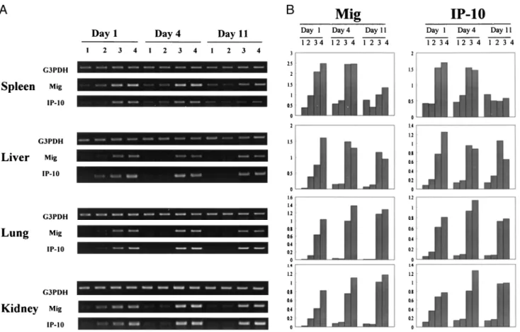
Initial studies demonstrated by RT-PCR analysis that IL-12 or IL- 2/IL-12 efficiently induced expression of both the Mig and Crg-2 genes in various organs within 24 h, with maximal expression in this setting for 4 days and enhanced gene expression still evident on day 11, whereas IL-2 alone was relatively ineffective in induc- ing the expression of either gene at any time point (Fig. 1A). Den- sitometric analysis of the intensity of the induced bands (Fig. 1B) suggested that the IL-2/IL-12 regimen was more efficient for in- ducing the expression of both of these genes by day 1 than was IL-12 alone, and therefore the IL-2/IL-12 regimen was selected for further study. Because both Mig and Crg-2 are known to be IFN-
␥-inducible genes (12) and the IL-2/IL-12 regimen enhances the expression of IFN-␥ (34), studies were performed to determine whether IFN-␥ administration could substitute for IL-2/IL-12 for the induction of Mig and Crg-2 gene expression in parenchymal tissue sites. The data presented in Fig. 2 demonstrates that the daily administration of 50,000 U rmIFN-␥ for 4 days clearly induced
expression of both genes in the spleen, liver, and lung, with min- imal induction of these genes also seen in the kidney. Based on these results, we speculated that at least some of the Mig and Crg-2 gene induction by IL-2/IL-12 was IFN-␥ dependent and the studies presented in Fig. 3 were designed to investigate this issue. In this experiment, IL-2/IL-12 was injected into wild-type and IFN-␥⫺/⫺
mice and gene expression was assessed on day 4. The results show that IL-2/IL-12 induced expression of both Mig and Crg-2 genes in
FIGURE 1. Induction of Mig and Crg-2 genes by IL-2, IL-12, or IL-2/IL-12 in mouse lymphoid and nonlymphoid tissues. BALB/c mice were treated with HBSS vehicle (lane 1), IL-2 (lane 2), IL-12 (lane 3), or IL-2/IL-12 (lane 4) and spleen, kidney, lung, and liver were harvested at days 1, 4, and 11 for analysis of Mig and Crg-2 gene expression by RT-PCR (A), with the intensity of each band relative to the expression of the G3PDH band presented by densitometric analysis (B).
FIGURE 2. Comparison of IL-2/IL-12 with rmIFN-␥ for ability to in- duce Crg-2 and Mig gene expression in various tissues. BALB/c mice were injected with IL-2/IL-12 50,000 U rmIFN-␥ by both the i.p. and i.v. routes each day for 4 days. Spleen, livers, lung, and kidney tissues were then harvested for analysis of Mig and IP-10 gene expression by RT-PCR.
at Keimyung Univ Med Lib on January 7, 2016http://www.jimmunol.org/Downloaded from
wild-type mice but not in IFN-␥⫺/⫺mice. These data demonstrate that all of the gene expression for IL-2/IL-12 in these organs is IFN-␥ dependent, irrespective of whether it may occur in tissue- associated leukocytes or PC.
Contribution of PC to Mig and Crg-2 gene expression after treatment with IL-12 or IL-2/IL-12
The results shown in Figs. 1–3 demonstrate that administration of IL-2⫾ IL-12 induced Mig and Crg-2 gene expression in a variety of nonlymphoid organs, and that this induction occurs via an IFN-
␥-dependent mechanism. However, it is unclear to what extent transiting or resident leukocytes (such as macrophages) vs tissue PC may contribute to this gene expression, and ultimately to the biological effects induced IL-2⫾ IL-12. Therefore, the liver was used as a model organ to study induction of the Mig and Crg-2 genes in highly purified PC (⬎90% hepatocytes) and NPC (⬎94%
leukocytes and endothelial cells). RT-PCR analyses showed that IL-2/IL-12 induced both Mig and Crg-2 gene expression at day 4 in PC as well as NPC (Fig. 4A) and subsequent RPA analyses confirmed this finding in PC (Fig. 4B). It should be noted that low levels of Mig and Crg-2 gene expression can sometimes be ob- served by RT-PCR analysis in unstimulated hepatocytes. In such cases, as shown in Fig. 4A, the levels of expression can fall below detection by RPA (Fig. 4B), suggesting that expression of these genes in unstimulated hepatocytes is generally minimal. To further confirm the ability of hepatocyte-lineage cells to express the Mig and Crg-2 genes, the hepatoma line (Hepa 1-6) was treated with
100 U/ml IFN-␥ for 2, 6, and 24 h, and RT-PCR analysis showed that the Mig and Crg-2 genes also were rapidly up-regulated in these cells (Fig. 4C). Thus, both PC and PC-derived cell lines can be induced to express the Mig and Crg-2 genes in response to IL-2/IL-12 treatment in vivo or by exposure to IFN-␥ in vitro, suggesting that the up-regulation of these chemokine genes in PC could contribute to local cytokine-induced inflammatory and anti- tumor effects.
Local production of IFN-␥ and expression of TH1cytokine receptors on purified hepatocytes
The data presented above demonstrates that the induction of the Mig and Crg-2 chemokine genes in lymphoid and nonlymphoid tissues by IL-2/IL-12 is critically dependent on induction of IFN-␥ gene expression (Fig. 3) and that direct administration of IFN-␥ also induces a similar pattern of expression of these genes in vivo (Fig. 2). We speculated that these IFN-␥-dependent effects on hep- atocyte-derived Mig and Crg-2 could simply result from increased production of the ligand (IFN-␥) that is clearly required (Fig. 3), or that the effects of increased levels of IFN-␥ could be further mag- nified by a coordinate induction of IFN-␥R components on the hepatocytes. Increased local production of IFN-␥ was detected by day 2 in NPC isolated from mice treated with IL-2/IL-12 (Fig. 5).
Intracellular staining of NPC with an Ab specific for mouse IFN-␥ revealed a 3-fold increase in the percentage of liver-associated leukocytes synthesizing IFN-␥. These results formally document an enhanced local production of the IFN-␥ ligand in the liver. In conjunction with these studies, RPA analysis of TH1cytokine re- ceptor expression presented in Fig. 6 demonstrates that the genes for both the␣- and -chains of the IFN-␥R are constitutively ex- pressed in freshly isolated hepatocytes, and expression of both chains is further increased by in vivo administration of IL-12 alone and even more so by IL-2/IL-12. In contrast, expression of genes encoding the respective components of the IL-2 or IL-12 receptors was not detected in these hepatocyte preparations. Therefore, this data suggests that the IL-2/IL-12-induced IFN-␥-dependent in- crease in Mig and Crg-2 gene expression by hepatocytes results exclusively from induction of IFN-␥ and not from direct stimula- tion of hepatocytes by IL-2 or IL-12. Furthermore, this effect may be potentiated by concurrent increases in the expression of the IFN-␥R on hepatocytes. In addition, there is very strong induction of the gene for the p55 chain of the TNFR by IL-2 ⫾ IL-12.
Because TNF-␣ has been reported to synergize with IFN-␥ for CXC chemokine induction in human liver (22), it is possible that FIGURE 3. Induction of Mig and Crg-2 genes by IL-2/IL-12 in wild-
type vs IFN-␥⫺/⫺ mice. Wild-type BALB/c and IFN-␥⫺/⫺ mice were treated with vehicle (lane 1) or IL-2/IL-12 (lane 2) for 4 days as described in Materials and Methods. Spleen, liver, lung, and kidney tissues were then harvested for analysis of Mig and IP-10 gene expression by RT-PCR.
FIGURE 4. Induction of Mig and Crg-2 genes in PC, NPC, or a hepatocyte cell line. A, Mice were treated with vehicle (lane 1) or IL-2/IL-12 by using the 4-day protocol outlined in Materials and Meth- ods (lane 2), and PC and NPC were obtained on day 4 for analysis of Mig and Crg-2 gene expression by RT-PCR. B, Mice were treated with vehicle (lane 1) or IL-2/IL-12 (lane 2) and PC were obtained for analysis of Mig, Crg-2, and L32 control gene ex- pression in hepatocytes by RPA at 24 h. C, Hepa 1-6 cells were cultured with 100 U/ml IFN-␥ for 2, 6, and 24 h before analysis of Mig and Crg-2 gene expression by RT-PCR.
3766 PRIMARY HEPATOCYTES CAN PRODUCE CXC CHEMOKINES
at Keimyung Univ Med Lib on January 7, 2016http://www.jimmunol.org/Downloaded from
modulation of TNFR components also may contribute to enhanced expression of the Mig and Crg-2 genes.
Hepatocyte-derived chemotactic activity for T cells, but not NK cells, is mediated by a combination of Mig and Crg-2
The data presented above demonstrates that administration of IL- 2/IL-12 can rapidly enhance expression of the Crg-2 and Mig genes in hepatocytes, suggesting that stimulated hepatocytes might produce functional chemotactic activity for activated T cells. To test this hypothesis, purified hepatocytes from normal mice or mice treated with IL-2/IL-12 were cultured in media or 200 U/ml rmIFN-␥ for 24 h, and supernatants were tested in vitro for their ability to chemoattract purified, IL-2-activated human CD3⫹ T cells that were placed in the upper compartments. The results of these studies demonstrated that culture supernatants from hepato- cytes obtained from IL-2/IL-12-treated mice did induce significant
chemotactic activity for these purified CD3⫹T cells (Fig. 7). Su- pernatants from hepatocytes of untreated mice did not contain de- tectable chemotactic activity unless they were pretreated in vitro with 200 U/ml IFN-␥. The level of chemotactic activity produced by IL-2/IL-12-derived hepatocytes was equivalent to that exhibited by 1 ng/ml of recombinant human IP-10.
The data shown in Fig. 7 confirmed the presence of T cell che- moattractant activity in the supernatants of hepatocytes obtained from mice treated with IL-2/IL-12 but did not specifically address the role of Crg-2 or Mig protein in this process. Therefore, the study shown in Fig. 7, with mouse T lymphocytes and/or enriched NK cells was performed to define the role of Mig or Crg-2 in hepatocyte-derived chemotactic activity. Both highly purified IL- 2-activated CD3⫹T cells (Fig. 8A) or enriched DX5⫹NK cells (Fig. 8B) were chemoattracted to supernatants derived from ap- propriately induced hepatocytes. Specifically, the in vivo treatment
FIGURE 6. Receptor gene profiles of hepato- cytes treated with HBSS vehicle, IL-2, IL-12, and IL-2/IL-12. A, BALB/c mice were treated with HBSS vehicle (lane 1), IL-2 (lane 2), IL-12 (lane 3), or IL-2/IL-12 (lane 4) for 24 h. Hepa- tocyte RNA was harvested and analyzed for cy- tokine receptor genes by RPA. B, Receptor mes- sage of IFN-␥ and TNF-␣ from A was quantitated by using a phosphor imager. Signal strength was normalized to control L32 gene expression.
FIGURE 5. Detection of intracellular IFN-␥ in liver-associated NPC. BALB/c mice were injected i.p with HBSS (A) or 300,000 IU IL-2 on day 0 and 0.5g of IL-12 on days 0 and 1 (B). On day 2, mice were euthanized and NPC were obtained as described in Materials and Methods. Intracellular IFN-␥
was detected in these purified liver leukocytes with anti-mouse IFN-␥ (solid line) and isotype matched control Ab (dashed line). The percentage of liver-associated leukocytes containing IFN-␥ (shaded area) was assessed by flow cytometric analysis by subtracting the percentage staining with the isotype control from the percentage staining with the anti-IFN-␥
at Keimyung Univ Med Lib on January 7, 2016http://www.jimmunol.org/Downloaded from
of mice with IL-2/IL-12 or in vitro treatment of hepatocytes from normal mice with IFN-␥ was able to induce the production of chemoattractant activity for these purified T cells, and in vitro treatment of normal hepatocytes with IFN-␥ was also able to in- duce chemoattractant activity for DX5⫹NK cells. The hepatocyte- derived chemoattractant activity for mouse CD3⫹T cells was par- tially neutralized by Abs specific for Crg-2 ( p⬍ 0.05) or Mig and totally ablated by a combination of both Abs. Interestingly, the IFN-␥-induced, hepatocyte-derived chemotactic activity for puri- fied IL-2-activated NK cells (Fig. 7B) was not significantly re- duced by treatment with these Abs.
Discussion
The CXC chemokines Crg-2 and Mig have been associated with the antitumor activity of IL-12-based therapies for a variety of tumors in mice (9, 16, 23, 24), and these genes can be induced in many organ sites in response to infectious agents (12, 25). A com- mon theme during most cytokine-induced antitumor and antimi- crobial responses is the development of local inflammation with infiltration of activated T and NK cells. Because Mig and Crg-2 are potent chemoattractants for activated lymphocytes (18), a better insight into which cells produce them and how they are regulated in different organ compartments could be very useful in under- standing the critical events that initiate immune and inflammatory responses in different tissues. This insight could be of value both for enhancing the initiation of desirable immune responses as well as for developing new strategies to curtail unwanted pathological activities. We have demonstrated previously that the IL-2/IL-12 combination induces greater antitumor effects against a variety of transplantable and spontaneous tumors in mice than does IL-12 or IL-2 alone (17, 34), and this enhanced activity is routinely asso- ciated with increased expression of IFN-␥ and the IFN-␥-inducible genes Mig and Crg-2 (34). However, the relative roles of PC, NPC, and blood-borne leukocytes in these processes remain ill-defined.
By using the IL-2/IL-12 regimen as a model, our results show the induction of the Mig and Crg-2 genes and biologically active che-
moattractant activities for T vs NK cells by liver-derived PC (hepatocytes).
The liver is an interesting model in which to study the expres- sion and function of chemokine genes because enriched popula- tions of hepatic PC and NPC can be easily obtained (19, 20), and there is considerable information available about inflammatory, anti-microbial, and antitumor responses mediated by T, NK/T, and NK cells in the liver. The leukocyte composition of the liver can vary considerably in response to infection or cytokine therapies.
We have demonstrated that IL-12 and IL-2 have distinct effects on leukocyte recruitment to the liver (26, 27), whereas the combina- tion of IL-2 plus IL-12 has enhanced antitumor effects (17). Our studies show that the ability of IL-2/IL-12 to induce Mig and Crg-2 expression in a variety of organs is IFN-␥ dependent, which ex- tends previous conclusions for a variety of infections (25) or sys- temic administration of IFN-␥ (28). However, it should be noted that the endogenous IFN-␥-dependent induction of Mig and Crg-2 genes by IL-2/IL-12 can be easily and reproducibly detected, whereas the ability of exogenously administered IFN-␥ to induce these genes seems less effective. This may be a result of higher and/or more sustained levels of IFN-␥ occurring in the liver due to local production than can be achieved by exogenous administra- tion of IFN-␥ protein. Alternatively, maximal IFN-␥-dependent gene induction may occur in concert with expression of other gene products such as TNF-␣. To address this possiblity, studies are planned that use mice deficient in TNF-␣, lymphotoxin, or both.
Other studies have shown that some chemokine genes can be in- duced by LPS in Kupffer cell-depleted livers, implying gene ex- pression by nonmacrophages (29). Chemokine gene expression also has been documented in transformed mouse hepatocytes (30, 31). In our studies, low-level expression of Mig and Crg-2 genes can be sometimes detected by RT-PCR but not by RPA, suggesting that under homeostatic conditions, the expression of these genes in hepatocytes is minimal to negative. Our studies demonstrate for the first time that IL-2/IL-12 efficiently induces gene expression of Mig and Crg-2 in purified primary hepatocytes and reveals that all of the detectable expression of these genes in the liver and other nonlymphoid organs in response to IL-2/IL-12 is IFN-␥ dependent.
In addition, the amplification of Mig and Crg-2 genes in hepato- cytes by IL-12 or IL-2/IL-12 also may relate to the ability of these cytokines to up-regulate IFN-␥R components on those hepato- cytes. The ligand-binding␣-chain and the accessory -chain of the IFN-␥R are constitutively expressed at low to moderate levels on many cell types and can be regulated in some cell types by external stimuli (32). Thus, IL-12 and IL-2/IL-12 might increase the ex- pression of Mig and Crg-2 genes via the coordinate induction of both IFN-␥ from leukocytes as well as its receptor on the target cell (hepatocytes) population. It should be noted that the lack of expression of either IL-2 or IL-12 receptor gene components in hepatocytes strongly argues against any direct effects of IL-2 or IL-12 on hepatocytes. However, it remains possible that additional stimuli may contribute to induction of Mig and Crg-2 in this model. In particular, it is intriguing that both the p55 and p75 components of the TNFR are constitutively expressed on hepato- cytes and that IL-12⫾ IL-2 strongly up-regulate the p55 chain.
Because a previous study (22) has demonstrated that TNF-␣ syn- ergizes with IFN-␥ for CXC chemokine induction in human liver, a similar role for the TNF/TNFR system could play a role in our studies. Further studies with TNFR or TNF-␣⫺/⫺ mice will be required to define contributions of TNF-␣ to Mig and Crg-2 gene induction in vivo.
Because we have previously demonstrated that the administra- tion of either IL-2 or IL-12 to mice results in a rapid recruitment FIGURE 7. Chemotactic activity of hepatocyte-derived supernatants for
human CD3⫹T cells. Purified hepatocyte preparations were obtained from mice treated on days 0 to 3 with vehicles or IL-2/IL-12 as described in Materials and Methods. These cells were cultured for 20 h with or without 200 U/ml IFN-␥ and the supernatants assessed at a 1:3 dilution for their ability to attract purified human CD3⫹T cells. SDF-1␣ and recombinant human IP-10 were used as positive chemoattractant controls.ⴱ, p ⬍ 0.05, significantly greater than the normal hepatocyte control.
3768 PRIMARY HEPATOCYTES CAN PRODUCE CXC CHEMOKINES
at Keimyung Univ Med Lib on January 7, 2016http://www.jimmunol.org/Downloaded from
of T lymphocytes and NK cells to the liver, and the IFN-␥-induc- ible Mig and Crg-2 genes are rapidly induced in the liver by the IL-2/IL-12 combination, we speculated that culture supernatants from purified primary hepatocytes obtained from these mice would have chemotactic activity for T and NK cells. The results demon- strate for the first time that IL-2/IL-12 treatment in vivo, as well as IFN-␥ treatment in vitro, induces hepatocytes to produce chemo- tactic activity for human T cells as well as mouse T and/or NK cells. The hepatocyte-derived chemoattractant activity for T cells is mediated by a combination of Mig and Crg-2, whereas the che- moattractant activity for NK cells is largely independent of Mig and Crg-2. This latter finding may be consistent with a recent report by Salazar-Mather et al. (33) who demonstrated that murine CMV-induced NK cell infiltration into the liver was dependent on a CC chemokine, MIP-1␣. Therefore, T cell recruitment to the liver in the setting of IL-2/IL-12 therapy may be initially induced by Mig/Crg-2, whereas NK cell recruitment may be regulated via Mip-1␣ and/or other as yet unidentified factors.
Overall, our results demonstrate for the first time that purified primary mouse hepatocytes can be induced by IL-2/IL-12 to ex- press the genes for Mig, Crg-2, and IFN-␥ R, and to produce Mig and Crg-2 at levels sufficient to chemoattract T cells in vitro.
Therefore, these results suggest that expression of these genes by
primary PC in vivo may constitute an important component of the innate immune response in the initiation and/or progression of im- mune responses against cancer or infectious agents in nonlym- phoid organs.
Acknowledgments
We thank W. Gong for excellent technical assistance and Dr. Sara D. Held (Chemical Industry Institute of Technology, Research Triangle Park, NC) for her invaluable advice on the procedure for obtaining purified mouse hepatocytes. We also thank Susan Charbonneau and Joyce Vincent for excellent secretarial and editorial assistance.
References
1. Trinchieri, G. 1998. Interleukin-12: a cytokine at the interface of inflammation and immunity. Adv. Immunol. 70:83.
2. Zeh, H. J., S. Hurd, W. J. Storkus, and M. T. Lotze. 1993. Interleukin-12 pro- motes the proliferation and cytolytic maturation of immune effectors: implica- tions for the immunotherapy of cancer. J. Immunother. 14:155.
3. Manetti, R., P. Parronchi, M. G. Giudizi, M. P. Piccinni, E. Maggi, G. Trinchieri, and S. Romagnani. 1993. Natural killer cell stimulatory factor (interleukin 12 [IL-12]) induces T helper type 1 (Th1)-specific immune responses and inhibits the development of IL-4-producing Th cells. J. Exp. Med. 177:1199.
4. Brunda, M. J., L. Luistro, R. R. Warrier, R. B. Wright, B. R. Hubbard, M. Murphy, S. F. Wolf, and M. K. Gately. 1994. Antitumor and antimetastatic activity of interleukin 12 against murine tumors. J. Exp. Med. 178:1223.
FIGURE 8. Chemotactic activity of hepatocyte- derived supernatants for purified mouse CD3⫹ T cells (A) and purified mouse NK cells (B). Purified hepatocyte preparations were obtained from normal or IL-2/IL-12 pretreated mice as described in Mate- rials and Methods. These cells were cultured for 20 h with or without 200 U/ml rmIFN-␥ and super- natants obtained. Some supernatants were also incu- bated with Abs specific for the Mig and Crg-2 pro- teins. Supernatants then were tested for their ability to chemoattract highly enriched T or NK cell pop- ulations as described in Materials and Methods.ⴱ, p⬍ 0.05, significantly less than IL-2/IL-12 positive control;ⴱⴱ, p ⬍ 0.05, significantly greater than con- trol medium.
at Keimyung Univ Med Lib on January 7, 2016http://www.jimmunol.org/Downloaded from
5. Brunda, M. J., L. Luistro, J. A. Hendrzak, M. Fountoulakis, G. Garotta, and M. K. Gately. 1995. Role of interferon-␥ in mediating the antitumor efficacy of interleukin-12. J. Immunother. Emphasis Tumor Immunol. 17:71.
6. Nastala, C. L., H. D. Edington, T. G. McKinney, H. Tahara, M. A. Nalesnik, M. J. Brunda, M. K. Gately, S. F. Wolf, R. D. Schreiber, W. J. Storkus, and M. T. Lotze. 1994. Recombinant IL-2 administration induces tumor regression in association with IFN-␥ production. J. Immunol. 153:1697.
7. Voest, E. E., B. M. Kenyon, M. S. O’Reilly, G. Truitt, R. J. D’Amato, and J. Folkman. 1995. Inhibition of angiogenesis in vivo by interleukin 12. J. Natl.
Cancer Inst. 87:581.
8. Angiolillo, A. L., C. Sgadari, and G. Tosato. 1996. A role for the interferon- inducible protein 10 in inhibition of angiogenesis by interleukin-12. Ann. NY Acad. Sci. 795:158.
9. Sgadari, C., A. L. Angiolillo, and G. Tosato. 1996. Inhibition of angiogenesis by interleukin-12 is mediated by the interferon-inducible protein 10. Blood 87:3877.
10. Kanegane, C., C. Sgadari, H. Kanegane, J. Teruya-Fieldstein, L.Yao, G. Gupta, J. M. Farber, F. Liao, L. Liu, and G. Tosato. 1998. Contribution of CXC che- mokines IP-10 and Mig to the antitumor effects of IL-12. J. Leukocyte Biol.
64:384.
11. Loetscher, M., B. Gerber, P. Loetscher, S. A. Jones, L. Piali, I. Clark-Lewis, M. Baggiolini, and B. Moser. 1996. Chemokine receptor specific for IP 10 and Mig: structure, function, and expression in activated T-lymphocytes. J. Exp.
Med.184: 963.
12. Farber, J. M. 1997. Mig and IP-10: CXC chemokines that target lymphocytes.
J. Leukocyte Biol. 61:246.
13. Kim, C. H., and H. E. Broxmeyer. 1999. Chemokines: signal lamps for trafficking of T and B cells for development and effector function. J. Leukocyte Biol. 65:6.
14. Arenberg, D. A., S. C. Kunkel, P. J. Palverini, S. B. Morris, M. D. Bardick, M. C. Glass, D. T. Taub, M. D. Iannettoni, R. I. Whyte, and R. M. Strieter. 1996.
Interferon-inducible protein 10 (IP-10) is an angiostatic factor that inhibits human non-small cell lung cancer (NSCLC) tumorigenesis and spontaneous metastases.
J. Exp. Med. 184:981.
15. Sgadari, C., A. L. Angiolillo, B. W. Cherney, S. E. Pike, J. M. Farber, L. G. Koniaris, P. Vanguri, P. R. Burd, N. Sheikh, G. Gupta, et al. 1996. Inter- feron-inducible protein-10 identified as a mediator of tumor necrosis in vivo.
Proc. Natl. Acad. Sci. USA 93:3791.
16. Tannenbaum, C. S., R. Tubbs, D. Armstrong, J. H. Finke, R. M. Bukowski, and T. A. Hamilton. 1998. The CXC chemokines IP-10 and Mig are necessary for IL-12-mediated regression of the mouse Renca tumor. J. Immunol. 161:927.
17. Wigginton, J. M., K. L. Komschlies, T. C. Back, J. L. Franco, M. J. Brunda, and R. H. Wiltrout. 1996. Administration of interleukin 12 with pulse interleukin 2 and the rapid and complete eradication of murine renal carcinoma. J. Natl. Can- cer Inst. 88:38.
18. Sonouchi, K., T. A. Hamilton, C. S. Tannenbaum, R. R. Tubbs, R. Bukowski, and J. H. Finke. 1994. Chemokine gene expression in the murine renal carcinoma, Renca, following treatment in vivo with interferon-␣ and interleukin-2.
Am. J. Pathol. 144:747.
19. Kedderis, G. L., and S. D. Held. 1996. Prediction of Furan pharmacokinetics from hepatocyte studies: comparison of bioactivation and hepatic dosimetry in rats, mice, and humans. Toxicol. Appl. Pharmacol. 140:124.
20. Fogler, W. E., K. Volker, K. L. McCormick, M. Watanabe, J. R. Ortaldo, and R. H. Wiltrout. 1996. NK cell infiltration into lung, liver, and subcutaneous B16 melanoma is mediated by VCAM-1/VLA-4 interaction. J. Immunol. 156:4707.
21. Mason, L., S. L. Giardina, T. Hecht, J. Ortaldo, and B. J. Mathieson. 1988.
LGL-1: a non-polymorphic antigen expressed on a major population of mouse natural killer cells. J. Immunol. 140:440.
22. Shields, P. L., C. M. Morland, M. Salmon, S. Qin, S. G. Hubscher, and D. H. Adams. 1999. Chemokine and chemokine receptor interactions provide a mechanism for selective T cell recruitment to specific liver compartments within hepatitis C-infected liver. J. Immunol. 163:6236.
23. Cavallo, F., E. DiCarlo, M. Butera, R. Verrua, M. P. Columbo, P. Musiani, and G. Forni. 1999. Immune events associated with the cure of established tumors and spontaneous metastasis by local and systemic interleukin 12. Cancer Res. 59:414.
24. Coughlin, C. M., K. E. Salhany, M. S. Gee, D. C. LaTemple, S. Kotenko, X. Ma, G. Gri, M. Wysocka, J. E. Kim, L. Liu, et al. 1998. Tumor cell responses to IFN␥ affect tumorigenicity and response to IL-12 therapy and antiangiogenesis. Immu- nity 9:25.
25. Amichay, D., R. T. Gazzinelli, G. Karupiah, T. R. Moench, A. Sheri, and J. M. Farber. 1996. The gene for chemokinesMig and Crg-2 are induced in protozoan and viral infections in response to IFN-␥ with patterns of tissue ex- pression that suggest nonredundant roles in vivo. J. Immunol. 157:4511.
26. Fogler, W. E., K. Volker, M. Watanabe, J. M. Wigginton, P. Roessler, M. J. Brunda, J. R. Ortaldo, and R. H. Wiltrout. 1998. Recruitment of hepatic NK cells by IL-12 is dependent on IFN-␥ and VCAM-1 and is rapidly down-regulated by a mechanism involving T cells and expression of Fas. J. Immunol. 161:6014.
27. Wiltrout, R. H. 2000. Regulation and antimetastatic functions of liver-associated natural killer cells. Immunol. Rev. 174:63.
28. Narumi, S., L. M. Wyner, M. H. Stoler, C. S. Tannenbaum, and T. A. Hamilton.
1992. Tissue specific expression of murine IP-10 mRNA following systemic treatment with interferon-␥. J. Leukocyte Biol. 52:27.
29. Kopydlowski, K. M., C. A. Salkowski, M. J. Cody, N. van Rooijen, J. Major, T. A. Hamilton, and S. N. Vogel. 1999. Regulation of macrophage chemokine expression by lipopolysaccharide in vitro and in vivo. J. Immunol. 163:1537.
30. Barsig, J., I. E. Flesch, and S. H. Kaufmann. 1998. Macrophages and hepatic cells as chemokine producers in murine listeriosis. Immunobiology 199:87.
31. Dong, W., P. P. Simeonova, R. Galluci, J. Mathesan, R. Fannin, P. Montuschi, L. Flood, and M. I. Luster. 1998. Cytokine expression in hepatocytes: role of oxidant stress. J. Interferon Cytokine Res. 18:629.
32. Bach, E. A., M. Aguet, and R. D. Schreiber. 1997. The IFN-␥ receptor: a para- digm for cytokine receptor signaling. Annu. Rev. Immunol. 15:563.
33. Salazar-Mather, T. P., J. S. Orange, and C. A. Biron. 1998. Early murine cyto- megalovirus (MCMV) infection induces liver natural killer (NK) cell inflamma- tion and protection through macrophage inflammatory protein 1␣ (MIP-1␣)-de- pendent pathways. J. Exp. Med. 187:1.
34. Wigginton, J. M., J.-W. Park, M. E. Gruys, H. A. Young, C. L. Jorcyk, T. C. Back, M. J. Brunda, R. B. Strieter, J. E. Green, and R. H. Wiltrout. 2001.
Complete regression of established spontaneous mammary carcinoma, and the therapeutic prevention of genetically programmed neoplastic transition by IL-12/
pulse IL-2: induction of local T cell infiltration, Fas/Fas ligand gene expression and mammary epithelial apoptosis. J. Immunol. 166:1156.
3770 PRIMARY HEPATOCYTES CAN PRODUCE CXC CHEMOKINES
at Keimyung Univ Med Lib on January 7, 2016http://www.jimmunol.org/Downloaded from